Epithelial Cellular Adhesion Molecule (EpCAM), also known as KS1/4, gp40, GA733-2, 17-1A, and TROP-1, is a 40 kDa transmembrane glycoprotein composed of a 242 amino acid (aa) extracellular domain with two epidermal-growth-factor-like (EGF-like) repeats within the cysteine-rich N-terminal region, a 23 aa transmembrane domain, and a 26 aa cytoplasmic domain. Human and mouse EpCAM share 82% aa sequence identity. In human, EpCAM also shares 49% aa sequence homology with TROP-2/EGP-1. During embryonic development, EpCAM is detected in fetal lung, kidney, liver, pancreas, skin, and germ cells. In adults, human EpCAM is detected in basolateral cell membranes of all simple, pseudo-stratified, and transitional epithelia, but is not detected in normal squamous stratified epithelia, mesenchymal tissue, muscular tissue, neuro-endocrine tissue, or lymphoid tissue (1). EpCAM expression has been found to increase in actively proliferating epithelia tissues and during adult liver regeneration (1, 2). EpCAM expression is also found to increase in human malignant neoplasias, with most carcinoma expressing EpCAM including those of arising from squamous epithelia (1). EpCAM has been shown function as a homophilic Ca2+ independent adhesion molecule (3). Homophilic adhesion via EpCAM requires the interaction of both EGF-like repeats, with the first EGF-like repeat mediating reciprocal interaction between EpCAM molecules on opposing cells, while the second repeat is involved in lateral interaction of EpCAM. Lateral interaction of EpCAM lead to the formation of dimers and tetramers (4). During homophilic adhesion the cytoplasmic tail of EpCAM interacts with the actin cytoskeleton via a direct association alpha -actinin (5).

Key Product Details
Species Reactivity
Validated:
Human
Cited:
Human, Mouse
Applications
Validated:
Immunohistochemistry, Western Blot, Flow Cytometry, Immunocytochemistry, Simple Western, CyTOF-ready
Cited:
Immunohistochemistry, Western Blot, Flow Cytometry, Immunocytochemistry, Adhesion Assay, Bioassay, Capture Assay, Cell Selection, CTC, CyTof, ELISA Capture, FACS
Label
Unconjugated
Antibody Source
Polyclonal Goat IgG
Loading...
Product Specifications
Immunogen
Mouse myeloma cell line NS0-derived recombinant human EpCAM/TROP-1, extracellular domain
Gln24-Lys265
Accession # CAA32870
Gln24-Lys265
Accession # CAA32870
Specificity
Detects human EpCAM/TROP-1 in direct ELISAs and Western blots. In direct ELISAs, less than 1% cross-reactivity with recombinant human TROP-2 and approximately 26% cross-reactivity with recombinant mouse EpCAM/TROP-1 is observed.
Clonality
Polyclonal
Host
Goat
Isotype
IgG
Scientific Data Images for Human EpCAM/TROP1 Antibody
Detection of Human EpCAM/TROP‑1 by Western Blot.
Western blot shows lysates of HCT-116 human colorectal carcinoma cell line, MCF-7 human breast cancer cell line, and BG01V human embryonic stem cells. PVDF membrane was probed with 0.2 µg/mL of Goat Anti-Human EpCAM/TROP-1 Antigen Affinity-purified Polyclonal Antibody (Catalog # AF960) followed by HRP-conjugated Anti-Goat IgG Secondary Antibody (HAF017). A specific band was detected for EpCAM/TROP-1 at approximately 40 kDa (as indicated). This experiment was conducted under reducing conditions and using Immunoblot Buffer Group 1.
Detection of EpCAM/TROP‑1 in HT‑29 Human Cell Line by Flow Cytometry.
HT-29 human colon adenocarcinoma cell line was stained with Goat Anti-Human EpCAM/TROP-1 Antigen Affinity-purified Polyclonal Antibody (Catalog # AF960, filled histogram) or isotype control antibody (AB-108-C, open histogram), followed by Phycoerythrin-conjugated Anti-Goat IgG Secondary Antibody (F0107).
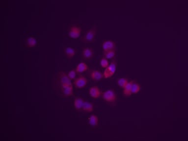
EpCAM/TROP‑1 in BG01V Human Embryonic Stem Cells.
EpCAM/TROP-1 was detected in immersion fixed BG01V human embryonic stem cells using Goat Anti-Human EpCAM/TROP-1 Antigen Affinity-purified Polyclonal Antibody (Catalog # AF960) at 10 µg/mL for 3 hours at room temperature. Cells were stained using the NorthernLights™ 557-conjugated Anti-Goat IgG Secondary Antibody (red; Catalog # NL001) and counterstained with DAPI (blue). Specific staining was localized to cytoplasm and cell membrane. View our protocol for Fluorescent ICC Staining of Cells on Coverslips.
EpCAM/TROP‑1 in Human Colon.
EpCAM/TROP-1 was detected in immersion fixed paraffin-embedded sections of human colon using Goat Anti-Human EpCAM/TROP-1 Antigen Affinity-purified Polyclonal Antibody (Catalog # AF960) at 0.3 µg/mL for 1 hour at room temperature followed by incubation with the Anti-Goat IgG VisUCyte™ HRP Polymer Antibody (VC004). Before incubation with the primary antibody, tissue was subjected to heat-induced epitope retrieval using Antigen Retrieval Reagent-Basic (Catalog # CTS013). Tissue was stained using DAB (brown) and counterstained with hematoxylin (blue). Specific staining was localized to cytoplasm and plasma membrane. View our protocol for IHC Staining with VisUCyte HRP Polymer Detection Reagents.
EpCAM/TROP‑1 in Human Colon.
EpCAM/TROP-1 was detected in immersion fixed paraffin-embedded sections of human colon using Goat Anti-Human EpCAM/TROP-1 Antigen Affinity-purified Polyclonal Antibody (Catalog # AF960) at 0.3 µg/mL for 1 hour at room temperature followed by incubation with the Anti-Goat IgG VisUCyte™ HRP Polymer Antibody (VC004). Before incubation with the primary antibody, tissue was subjected to heat-induced epitope retrieval using Antigen Retrieval Reagent-Basic (CTS013). Tissue was stained using DAB (brown) and counterstained with hematoxylin (blue). Specific staining was localized to cytoplasm and plasma membrane. View our protocol for IHC Staining with VisUCyte HRP Polymer Detection Reagents.
Detection of Human EpCAM/TROP‑1 by Simple WesternTM.
Simple Western lane view shows lysates of A549 human lung carcinoma cell line exosomes, HT‑29 human colon adenocarcinoma cell line exosomes, and COLO 205 human colorectal adenocarcinoma cell line whole cell lysate (WCL), loaded at 0.2 mg/mL. A specific band was detected for EpCAM/TROP‑1 at approximately 52 kDa (as indicated) using 10 µg/mL of Goat Anti-Human EpCAM/TROP‑1 Antigen Affinity-purified Polyclonal Antibody (Catalog # AF960). This experiment was conducted under reducing conditions and using the 12-230 kDa separation system. and Jurkat Human Acute T Cell Leukemia Cell Line (Negative) Cells.")
Detection of EpCAM/TROP‑1 in HT-29 Human Colon Adenocarcinoma Cell Line (Positive) and Jurkat Human Acute T Cell Leukemia Cell Line (Negative) Cells.
EpCAM/TROP‑1 was detected in immersion fixed HT‑29 human colon adenocarcinoma cell line (positive) and Jurkat human acute T cell leukemia cell line (negative) cells using Goat Anti-Human EpCAM/TROP‑1 Antigen Affinity-purified Polyclonal Antibody (Catalog # AF960) at 5 µg/mL for 3 hours at room temperature. Cells were stained using the NorthernLights™ 557-conjugated Anti-Goat IgG Secondary Antibody (red; Catalog # NL001) and counterstained with DAPI (blue). Specific staining was localized to cell surface. View our protocol for Fluorescent ICC Staining of Cells on Coverslips.
Human EpCAM / TROP1 ELISA Standard Curve
Recombinant HumanEpCAM/TROP‑1 Fc Chimera (Catalog # 960-EP) was serially diluted and captured by Mouse Anti-Human
EpCAM/TROP‑1 Monoclonal Antibody (Catalog # MAB9601) coated on a Clear Polystyrene Microplate (Catalog # DY990). Goat Anti-Human
EpCAM/TROP‑1 Antigen Affinity-purified Polyclonal Antibody (Catalog # AF960) was biotinylated and incubated with the protein captured on the plate. Detection of the standard curve was achieved by incubating Streptavidin-HRP (Catalog # DY998)
Applications for Human EpCAM/TROP1 Antibody
Application
Recommended Usage
CyTOF-ready
Ready to be labeled using established conjugation methods. No BSA or other carrier proteins that could interfere with conjugation.
Flow Cytometry
0.25 µg/106 cells
Sample: HT‑29 human colon adenocarcinoma cell line
Sample: HT‑29 human colon adenocarcinoma cell line
Immunocytochemistry
5-15 µg/mL
Sample: Immersion fixed BG01V human embryonic stem cells, HT-29 Human Colon Adenocarcinoma Cell Line and Jurkat Human Acute T Cell Leukemia Cell Line cells.
Sample: Immersion fixed BG01V human embryonic stem cells, HT-29 Human Colon Adenocarcinoma Cell Line and Jurkat Human Acute T Cell Leukemia Cell Line cells.
Immunohistochemistry
0.3-15 µg/mL
Sample: Immersion fixed paraffin-embedded sections of human colon
Sample: Immersion fixed paraffin-embedded sections of human colon
Simple Western
10 µg/mL
Sample: A549 human lung carcinoma cell line exosomes, HT‑29 human colon adenocarcinoma cell line exosomes, and COLO 205 human colorectal adenocarcinoma cell line whole cell lysate (WCL)
Sample: A549 human lung carcinoma cell line exosomes, HT‑29 human colon adenocarcinoma cell line exosomes, and COLO 205 human colorectal adenocarcinoma cell line whole cell lysate (WCL)
Western Blot
0.2 µg/mL
Sample: HCT‑116 human colorectal carcinoma cell line, MCF‑7 human breast cancer cell line, and BG01V human embryonic stem cells
Sample: HCT‑116 human colorectal carcinoma cell line, MCF‑7 human breast cancer cell line, and BG01V human embryonic stem cells
Reviewed Applications
Read 1 review rated 5 using AF960 in the following applications:
Flow Cytometry Panel Builder
Bio-Techne Knows Flow Cytometry
Save time and reduce costly mistakes by quickly finding compatible reagents using the Panel Builder Tool.
Advanced Features
- Spectra Viewer - Custom analysis of spectra from multiple fluorochromes
- Spillover Popups - Visualize the spectra of individual fluorochromes
- Antigen Density Selector - Match fluorochrome brightness with antigen density

Formulation, Preparation, and Storage
Purification
Antigen Affinity-purified
Reconstitution
Reconstitute at 0.2 mg/mL in sterile PBS. For liquid material, refer to CoA for concentration.
Loading...
Formulation
Lyophilized from a 0.2 μm filtered solution in PBS with Trehalose. See Certificate of Analysis for details.
*Small pack size (-SP) is supplied either lyophilized or as a 0.2 µm filtered solution in PBS.
*Small pack size (-SP) is supplied either lyophilized or as a 0.2 µm filtered solution in PBS.
Shipping
Lyophilized product is shipped at ambient temperature. Liquid small pack size (-SP) is shipped with polar packs. Upon receipt, store immediately at the temperature recommended below.
Stability & Storage
Use a manual defrost freezer and avoid repeated freeze-thaw cycles.
- 12 months from date of receipt, -20 to -70 °C as supplied.
- 1 month, 2 to 8 °C under sterile conditions after reconstitution.
- 6 months, -20 to -70 °C under sterile conditions after reconstitution.
Calculators
Background: EpCAM/TROP1
References
- Balzar, M. et al. (1999) J. Mol. Med. 77:699.
- Boer, C.J. et al. (1999) J. Pathol. 188:201.
- Litvinow, S.V. et al. (1994) J. Cell Biol. 125:437.
- Balzar, M. et al. (2001) Mol. Cell. Biol. 21:2570.
- Balzar, M. et al. (1998) Mol. Cell. Biol. 18:4388.
Long Name
Epithelial Cell Adhesion Molecule
Alternate Names
17-1A, CD326, GA733-2, gp40, KS1/4, M4S1, TACSTD1, TROP1
Gene Symbol
EPCAM
UniProt
Additional EpCAM/TROP1 Products
Product Documents for Human EpCAM/TROP1 Antibody
Certificate of Analysis
To download a Certificate of Analysis, please enter a lot or batch number in the search box below.
Note: Certificate of Analysis not available for kit components.
Product Specific Notices for Human EpCAM/TROP1 Antibody
For research use only
Citations for Human EpCAM/TROP1 Antibody
 Powered by Bioz
Customer Reviews for Human EpCAM/TROP1 Antibody (1)
5 out of 5
1 Customer Rating
Have you used Human EpCAM/TROP1 Antibody?
Submit a review and receive an Amazon gift card!
$25/€18/£15/$25CAN/¥2500 Yen for a review with an image
$10/€7/£6/$10CAN/¥1110 Yen for a review without an image
Submit a review
Customer Images
Showing
1
-
1 of
1 review
Showing All
Filter By:
-
Application: ImmunocytochemistrySample Tested: MDA MB 231 cellsSpecies: HumanVerified Customer | Posted 11/16/2023MDA-MB 231 cells stained with AF960 antibody (red)

There are no reviews that match your criteria.
Protocols
Find general support by application which include: protocols, troubleshooting, illustrated assays, videos and webinars.
- 7-Amino Actinomycin D (7-AAD) Cell Viability Flow Cytometry Protocol
- Antigen Retrieval Protocol (PIER)
- Antigen Retrieval for Frozen Sections Protocol
- Appropriate Fixation of IHC/ICC Samples
- Cellular Response to Hypoxia Protocols
- Chromogenic IHC Staining of Formalin-Fixed Paraffin-Embedded (FFPE) Tissue Protocol
- Chromogenic Immunohistochemistry Staining of Frozen Tissue
- ClariTSA™ Fluorophore Kits
- Detection & Visualization of Antibody Binding
- Extracellular Membrane Flow Cytometry Protocol
- Flow Cytometry Protocol for Cell Surface Markers
- Flow Cytometry Protocol for Staining Membrane Associated Proteins
- Flow Cytometry Staining Protocols
- Flow Cytometry Troubleshooting Guide
- Fluorescent IHC Staining of Frozen Tissue Protocol
- Graphic Protocol for Heat-induced Epitope Retrieval
- Graphic Protocol for the Preparation and Fluorescent IHC Staining of Frozen Tissue Sections
- Graphic Protocol for the Preparation and Fluorescent IHC Staining of Paraffin-embedded Tissue Sections
- Graphic Protocol for the Preparation of Gelatin-coated Slides for Histological Tissue Sections
- ICC Cell Smear Protocol for Suspension Cells
- ICC Immunocytochemistry Protocol Videos
- ICC for Adherent Cells
- IHC Sample Preparation (Frozen sections vs Paraffin)
- Immunocytochemistry (ICC) Protocol
- Immunocytochemistry Troubleshooting
- Immunofluorescence of Organoids Embedded in Cultrex Basement Membrane Extract
- Immunofluorescent IHC Staining of Formalin-Fixed Paraffin-Embedded (FFPE) Tissue Protocol
- Immunohistochemistry (IHC) and Immunocytochemistry (ICC) Protocols
- Immunohistochemistry Frozen Troubleshooting
- Immunohistochemistry Paraffin Troubleshooting
- Intracellular Flow Cytometry Protocol Using Alcohol (Methanol)
- Intracellular Flow Cytometry Protocol Using Detergents
- Intracellular Nuclear Staining Flow Cytometry Protocol Using Detergents
- Intracellular Staining Flow Cytometry Protocol Using Alcohol Permeabilization
- Intracellular Staining Flow Cytometry Protocol Using Detergents to Permeabilize Cells
- Preparing Samples for IHC/ICC Experiments
- Preventing Non-Specific Staining (Non-Specific Binding)
- Primary Antibody Selection & Optimization
- Propidium Iodide Cell Viability Flow Cytometry Protocol
- Protocol for Heat-Induced Epitope Retrieval (HIER)
- Protocol for Liperfluo
- Protocol for Making a 4% Formaldehyde Solution in PBS
- Protocol for VisUCyte™ HRP Polymer Detection Reagent
- Protocol for the Characterization of Human Th22 Cells
- Protocol for the Characterization of Human Th9 Cells
- Protocol for the Fluorescent ICC Staining of Cell Smears - Graphic
- Protocol for the Fluorescent ICC Staining of Cultured Cells on Coverslips - Graphic
- Protocol for the Preparation & Fixation of Cells on Coverslips
- Protocol for the Preparation and Chromogenic IHC Staining of Frozen Tissue Sections
- Protocol for the Preparation and Chromogenic IHC Staining of Frozen Tissue Sections - Graphic
- Protocol for the Preparation and Chromogenic IHC Staining of Paraffin-embedded Tissue Sections
- Protocol for the Preparation and Chromogenic IHC Staining of Paraffin-embedded Tissue Sections - Graphic
- Protocol for the Preparation and Fluorescent ICC Staining of Cells on Coverslips
- Protocol for the Preparation and Fluorescent ICC Staining of Non-adherent Cells
- Protocol for the Preparation and Fluorescent ICC Staining of Stem Cells on Coverslips
- Protocol for the Preparation and Fluorescent IHC Staining of Frozen Tissue Sections
- Protocol for the Preparation and Fluorescent IHC Staining of Paraffin-embedded Tissue Sections
- Protocol for the Preparation of Gelatin-coated Slides for Histological Tissue Sections
- Protocol for the Preparation of a Cell Smear for Non-adherent Cell ICC - Graphic
- Protocol: Annexin V and PI Staining by Flow Cytometry
- Protocol: Annexin V and PI Staining for Apoptosis by Flow Cytometry
- R&D Systems Quality Control Western Blot Protocol
- TUNEL and Active Caspase-3 Detection by IHC/ICC Protocol
- The Importance of IHC/ICC Controls
- Troubleshooting Guide: Fluorokine Flow Cytometry Kits
- Troubleshooting Guide: Immunohistochemistry
- Troubleshooting Guide: Western Blot Figures
- Western Blot Conditions
- Western Blot Protocol
- Western Blot Protocol for Cell Lysates
- Western Blot Troubleshooting
- Western Blot Troubleshooting Guide
- View all Protocols, Troubleshooting, Illustrated assays and Webinars

