PRAS40 (Proline-rich AKT1 substrate 1; also Akt1S1 and p39) is a 40-42 kDa cytoplasmic phosphoprotein that lacks generally recognized structural motifs. It is widely expressed and is considered to be key regulator of mTORC1 (mTOR plus raptor and G beta L), a complex through which Akt signals into the cell. Through phosphorylation, mTORC1 activity is upregulated by PRAS40. In particular, nonphosphorylated PRAS40 binds to and serves as a negative regulator of mTORC1 activity. Upon Insulin signaling, PRAS40 is phosphorylated on Thr246, Ser221 and Ser183. This causes it to bind 14-3-3 and results in its dissociation from mTORC1, freeing up mTOR to regulate (positively or negatively) protein synthesis. Human PRAS40 is 256 amino acids (aa) in length. It contains one extended Pro-rich region (aa 35-96) plus at least nine utilized Ser/Thr phosphorylation sites. There is one alternative start site at Met131. Over aa 119-256, human PRAS40 shares 93% aa identity with mouse PRAS40.
phospho-PRAS40 (T246) Antibody (760502)
R&D Systems | Catalog # MAB6890
 by Western Blot.")
Key Product Details
Validated by
Biological Validation
Species Reactivity
Validated:
Human, Mouse
Cited:
Human
Applications
Validated:
Western Blot, Immunocytochemistry
Cited:
Western Blot
Label
Unconjugated
Antibody Source
Monoclonal Mouse IgG2A Clone # 760502
Loading...
Product Specifications
Immunogen
Phosphopeptide containing the human PRAS40 T246 site
Specificity
Detects human and mouse PRAS40 when phosphorylated at T246
Clonality
Monoclonal
Host
Mouse
Isotype
IgG2A
Scientific Data Images for phospho-PRAS40 (T246) Antibody (760502)
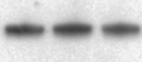
Detection of Human and Mouse Phospho-PRAS40 (T246) by Western Blot.
Western blot shows lysates of MCF-7 human breast cancer cell line and NIH-3T3 mouse embryonic fibroblast cell line untreated (-) or treated (+) with 100 ng/mL Recombinant Human IGF-I (Catalog # 291-G1) for 20 minutes and 10 ng/mL Recombinant Human PDGF-BB (Catalog # 220-BB) for 5 minutes. PVDF membrane was probed with 0.5 µg/mL of Mouse Anti-Human Phospho-PRAS40 (T246) Monoclonal Antibody (Catalog # MAB6890) followed by HRP-conjugated Anti-Mouse IgG Secondary Antibody (Catalog # HAF018). A specific band was detected for Phospho-PRAS40 (T246) at approximately 40 kDa (as indicated). This experiment was conducted under reducing conditions and using Immunoblot Buffer Group 1. in MCF‑7 Human Cell Line.")
Phospho-PRAS40 (T246) in MCF‑7 Human Cell Line.
PRAS40 phosphorylated at T246 was detected in immersion fixed MCF-7 human breast cancer cell line using Mouse Anti-Human/Mouse Phospho-PRAS40 (T246) Monoclonal Antibody (Catalog # MAB6890) at 25 µg/mL for 3 hours at room temperature. Cells were stained using the NorthernLights™ 557-conjugated Anti-Mouse IgG Secondary Antibody (red; Catalog # NL007) and counterstained with DAPI (blue). Specific staining was localized to cytoplasm. View our protocol for Fluorescent ICC Staining of Cells on Coverslips.
Detection of Human PRAS40 by Western Blot
PIK3R2 depletion, but not PI3K inhibitors, induces stable PI3K pathway inhibitionA. Tumor xenografts were obtained using H226, CaLu-1 and H520 lung SQCC cells expressing inducible PIK3R1 or PIK3R2 shRNA. When tumors reached ~50 mm3, mice were treated with doxycycline in drinking water (2 mg/ml); they were sacrificed when tumors began to diminish. Normalized tumor extracts were examined by WB with indicated antibodies. B. H226, CaLu-1 and H520 cells were cultured in exponential growth were incubated with: vehicle (DMSO, 1:103 V:V), Ly294002 (5 μM), PIK75 (200 nM), TGX221 (30 μM), or PIK75 (200 nM) plus TGX221 (30 μM) for the last 1 h of culture or during 48 h; extracts were tested in WB. Mr indicates relative mobility. In both A and B, the pAkt or pPRAS40 signal was measured and normalized to the loading control (Akt or tubulin, respectively), and compared to that in controls (DMSO, considered 100%). Graphs show percentage of signal compared to maximal as mean ± SD at 48h of treatment. Arrows on the right side of bars show the percentage of signal compared to maximal detected after 1h of treatment.* P <0.05; **P <0.01; unpaired Student's t test with Welch correction. Image collected and cropped by CiteAb from the following publication (https://www.oncotarget.com/lookup/doi/10.18632/oncotarget.13195), licensed under a CC-BY license. Not internally tested by R&D Systems.Applications for phospho-PRAS40 (T246) Antibody (760502)
Application
Recommended Usage
Immunocytochemistry
8-25 µg/mL
Sample: Immersion fixed MCF‑7 human breast cancer cell line
Sample: Immersion fixed MCF‑7 human breast cancer cell line
Western Blot
0.5 µg/mL
Sample: MCF‑7 human breast cancer cell line and NIH‑3T3 mouse embryonic fibroblast cell line treated with Recombinant Human IGF‑I (Catalog # 291-G1) and Recombinant Human PDGF‑BB (Catalog # 220-BB)
Sample: MCF‑7 human breast cancer cell line and NIH‑3T3 mouse embryonic fibroblast cell line treated with Recombinant Human IGF‑I (Catalog # 291-G1) and Recombinant Human PDGF‑BB (Catalog # 220-BB)
Reviewed Applications
Read 1 review rated 5 using MAB6890 in the following applications:
Formulation, Preparation, and Storage
Purification
Protein A or G purified from hybridoma culture supernatant
Reconstitution
Sterile PBS to a final concentration of 0.5 mg/mL. For liquid material, refer to CoA for concentration.
Loading...
Formulation
Lyophilized from a 0.2 μm filtered solution in PBS with Trehalose. *Small pack size (SP) is supplied either lyophilized or as a 0.2 µm filtered solution in PBS.
Shipping
Lyophilized product is shipped at ambient temperature. Liquid small pack size (-SP) is shipped with polar packs. Upon receipt, store immediately at the temperature recommended below.
Stability & Storage
Use a manual defrost freezer and avoid repeated freeze-thaw cycles.
- 12 months from date of receipt, -20 to -70 °C as supplied.
- 1 month, 2 to 8 °C under sterile conditions after reconstitution.
- 6 months, -20 to -70 °C under sterile conditions after reconstitution.
Calculators
Background: PRAS40
Long Name
40 kDa Proline-rich Akt1 Substrate
Alternate Names
AKT1S1, Lobe
Gene Symbol
AKT1S1
Additional PRAS40 Products
Product Documents for phospho-PRAS40 (T246) Antibody (760502)
Certificate of Analysis
To download a Certificate of Analysis, please enter a lot or batch number in the search box below.
Note: Certificate of Analysis not available for kit components.
Product Specific Notices for phospho-PRAS40 (T246) Antibody (760502)
For research use only
Related Research Areas
Citations for phospho-PRAS40 (T246) Antibody (760502)
 Powered by Bioz
Customer Reviews for phospho-PRAS40 (T246) Antibody (760502) (1)
5 out of 5
1 Customer Rating
Have you used phospho-PRAS40 (T246) Antibody (760502)?
Submit a review and receive an Amazon gift card!
$25/€18/£15/$25CAN/¥2500 Yen for a review with an image
$10/€7/£6/$10CAN/¥1110 Yen for a review without an image
Submit a review
Customer Images
Showing
1
-
1 of
1 review
Showing All
Filter By:
-
Application: Western BlotSample Tested: MCF-7 human breast cancer cell lineSpecies: HumanVerified Customer | Posted 12/23/2021

There are no reviews that match your criteria.
Protocols
Find general support by application which include: protocols, troubleshooting, illustrated assays, videos and webinars.
- Appropriate Fixation of IHC/ICC Samples
- Cellular Response to Hypoxia Protocols
- ClariTSA™ Fluorophore Kits
- Detection & Visualization of Antibody Binding
- ICC Cell Smear Protocol for Suspension Cells
- ICC Immunocytochemistry Protocol Videos
- ICC for Adherent Cells
- Immunocytochemistry (ICC) Protocol
- Immunocytochemistry Troubleshooting
- Immunofluorescence of Organoids Embedded in Cultrex Basement Membrane Extract
- Immunohistochemistry (IHC) and Immunocytochemistry (ICC) Protocols
- Preparing Samples for IHC/ICC Experiments
- Preventing Non-Specific Staining (Non-Specific Binding)
- Primary Antibody Selection & Optimization
- Protocol for VisUCyte™ HRP Polymer Detection Reagent
- Protocol for the Fluorescent ICC Staining of Cell Smears - Graphic
- Protocol for the Fluorescent ICC Staining of Cultured Cells on Coverslips - Graphic
- Protocol for the Preparation and Fluorescent ICC Staining of Cells on Coverslips
- Protocol for the Preparation and Fluorescent ICC Staining of Non-adherent Cells
- Protocol for the Preparation and Fluorescent ICC Staining of Stem Cells on Coverslips
- Protocol for the Preparation of a Cell Smear for Non-adherent Cell ICC - Graphic
- R&D Systems Quality Control Western Blot Protocol
- TUNEL and Active Caspase-3 Detection by IHC/ICC Protocol
- The Importance of IHC/ICC Controls
- Troubleshooting Guide: Western Blot Figures
- Western Blot Conditions
- Western Blot Protocol
- Western Blot Protocol for Cell Lysates
- Western Blot Troubleshooting
- Western Blot Troubleshooting Guide
- View all Protocols, Troubleshooting, Illustrated assays and Webinars
Loading...
Associated Pathways


