Bcl-2 is a member of a family of proteins that regulates outer mitochondrial membrane permeability (1, 2). Bcl-2 is an anti-apoptotic member that prevents release of cytochrome c from the mitochondria intermembrane space into the cytosol. Bcl-2 is present on the outer mitochondrial membrane and is also found on other membranes in some cell types. Natural Bcl-2 contains a carboxyl-terminal mitochondria targeting sequence. Recombinant Bcl-2, missing the mitochondrial targeting sequence, maintains its ability to neutralize pro-apoptotic Bcl-2 family members. Neutralization by Bcl-2 appears to be through binding the BH3 region of pro-apoptotic Bcl-2 family members. This activity does not require the mitochondrial targeting sequence.

Key Product Details
Validated by
Knockout/Knockdown, Biological Validation
Species Reactivity
Validated:
Human, Mouse, Rat
Cited:
Human, Mouse, Rat
Applications
Validated:
Knockout Validated, Immunohistochemistry, Western Blot
Cited:
Western Blot, Cell Culture
Label
Unconjugated
Antibody Source
Monoclonal Mouse IgG2B Clone # 625509
Loading...
Product Specifications
Immunogen
E. coli-derived recombinant human Bcl-2
Ala2-Asp211
Accession # P10415
Ala2-Asp211
Accession # P10415
Specificity
Detects human Bcl-2 in direct ELISAs and Western blots. In direct ELISAs, approximately 25‑40% cross‑reactivity with recombinant mouse (rm) Bcl‑2 is observed and no cross-reactivity with recombinant human BCL2L12 is observed. In Western blots, 100% cross-reactivity with rmBcl-2 is observed.
Clonality
Monoclonal
Host
Mouse
Isotype
IgG2B
Scientific Data Images for Bcl-2 Antibody (625509)
Detection of Human, Mouse, and Rat Bcl‑2 by Western Blot.
Western blot shows lysates of THP-1 human acute monocytic leukemia cell line, KG-1 human acute myelogenous leukemia cell line, CTLL-2 mouse cytotoxic T cell line, and NRK rat normal kidney cell line. PVDF Membrane was probed with 0.1 µg/mL of Mouse Anti-Human/Mouse/Rat Bcl-2 Monoclonal Antibody (Catalog # MAB8272) followed by HRP-conjugated Anti-Mouse IgG Secondary Antibody (Catalog # HAF007). A specific band was detected for Bcl-2 at approximately 24 kDa (as indicated). This experiment was conducted under reducing conditions and using Immunoblot Buffer Group 1.
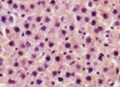
Bcl‑2 in Human Lymphoma.
Bcl-2 was detected in immersion fixed paraffin-embedded sections of human lymphoma using Mouse Anti-Human/Mouse/Rat Bcl-2 Monoclonal Antibody (Catalog # MAB8272) at 15 µg/mL overnight at 4 °C. Before incubation with the primary antibody, tissue was subjected to heat-induced epitope retrieval using Antigen Retrieval Reagent-Basic (Catalog # CTS013). Tissue was stained using the Anti-Mouse HRP-DAB Cell & Tissue Staining Kit (brown; Catalog # CTS002) and counterstained with hematoxylin (blue). Specific staining was localized to cytoplasm and nuclei. View our protocol for Chromogenic IHC Staining of Paraffin-embedded Tissue Sections.
Western Blot Shows Human Bcl‑2 Specificity by Using Knockout Cell Line.
Western blot shows lysates of HeLa human cervical epithelial carcinoma parental cell line and Bcl-2 knockout HeLa cell line (KO). PVDF membrane was probed with 0.1 µg/mL of Mouse Anti-Human/Mouse/Rat Bcl-2 Monoclonal Antibody (Catalog # MAB8272) followed by HRP-conjugated Anti-Mouse IgG Secondary Antibody (Catalog # HAF018). A specific band was detected for Bcl-2 at approximately 24 kDa (as indicated) in the parental HeLa cell line, but is not detectable in knockout HeLa cell line. GAPDH (Catalog # MAB5718) is shown as a loading control. This experiment was conducted under reducing conditions and using Immunoblot Buffer Group 1.
Detection of Human Bcl-2 by Western Blot
Cholesterol reversed metformin-mediated anticancer potential.Cells were treated with metformin (2 mM) alone and in combination with metformin (2 mM) plus cholesterol (10 μg/ml). (A) Cell viability (MTT) assay was performed to check the reverse effect of cholesterol on metformin- inhibited cell viability. Bars are representing the relative cell viability after 24 hr of treatment. Values represent mean ± SEM of triplicate measurements, **p <.001 vs. control and *p <.001 vs. metformin. (B) Scratch assay was performed. MDA-MB-231 cell monolayers were scratched and incubated with metformin (2 mM) alone and in combination with metformin (2 mM) plus cholesterol (10 μg/ml). The cell monolayers were photographed, and represented photos at 0 hr and 24 hr for control, metformin and metformin plus cholesterol cells were shown here. (C) The unfilled gap areas between two ends of the scratch were measured and subsequently plotted. Values represent mean ± SEM of triplicate measurements, **p <.01 vs. control at 0 hr and **p <.01 vs. metformin at 24 hr. (D) Colony formation assay was performed. After 24 hr of cell seeding, cells were treated with metformin (1mM) alone and in combination with metformin (1mM) plus cholesterol (5 μg/ml). After 5 days, formed colonies were stained with crystal violet. Photos of the wells were taken by a camera. (E) Soft agar assay of MDA-MB-231 cells in presence and absence of metformin (1mM) alone and in combination with metformin (1mM) plus cholesterol (5 μg/ml). Day 9 pictures were shown in the figure. Arrows depict the spheres. (F, G and H) RT-PCR analysis was performed using total RNA isolated from metformin, metformin (1mM) plus cholesterol treated and untreated cells, and gene specific primers. Cholesterol treatment reverses the metformin inhibited genes expressions (Bcl-xL [F], Zeb1 [G] and BMI1 [H]) (compare lane 2 vs. lane 3). (I and J) Western blot analysis was performed using total proteins from metformin, metformin (1mM) plus cholesteroApplications for Bcl-2 Antibody (625509)
Application
Recommended Usage
Immunohistochemistry
8-25 µg/mL
Sample: Immersion fixed paraffin-embedded sections of human lymphoma
Sample: Immersion fixed paraffin-embedded sections of human lymphoma
Knockout Validated
Bcl‑2
is specifically detected in HeLa human cervical epithelial carcinoma parental cell line but is not detectable in
Bcl‑2 knockout HeLa cell line.
Western Blot
0.1 µg/mL
Sample: THP‑1 human acute monocytic leukemia cell line, KG‑1 human acute myelogenous leukemia cell line, CTLL‑2 mouse cytotoxic T cell line, and NRK rat normal kidney cell line
Sample: THP‑1 human acute monocytic leukemia cell line, KG‑1 human acute myelogenous leukemia cell line, CTLL‑2 mouse cytotoxic T cell line, and NRK rat normal kidney cell line
Reviewed Applications
Read 1 review rated 5 using MAB8272 in the following applications:
Formulation, Preparation, and Storage
Purification
Protein A or G purified from hybridoma culture supernatant
Reconstitution
Sterile PBS to a final concentration of 0.5 mg/mL. For liquid material, refer to CoA for concentration.
Loading...
Formulation
Lyophilized from a 0.2 μm filtered solution in PBS with Trehalose. *Small pack size (SP) is supplied either lyophilized or as a 0.2 µm filtered solution in PBS.
Shipping
Lyophilized product is shipped at ambient temperature. Liquid small pack size (-SP) is shipped with polar packs. Upon receipt, store immediately at the temperature recommended below.
Stability & Storage
Use a manual defrost freezer and avoid repeated freeze-thaw cycles.
- 12 months from date of receipt, -20 to -70 °C as supplied.
- 1 month, 2 to 8 °C under sterile conditions after reconstitution.
- 6 months, -20 to -70 °C under sterile conditions after reconstitution.
Calculators
Background: Bcl-2
References
- Gross, A. et al. (1999) Genes and Develop. 13:1899.
- Kroemer, G. (1997) Nature Med. 3:614.
Long Name
B Cell Lymphoma/Leukemia 2
Alternate Names
Bcl2
Gene Symbol
BCL2
UniProt
Additional Bcl-2 Products
Product Documents for Bcl-2 Antibody (625509)
Certificate of Analysis
To download a Certificate of Analysis, please enter a lot or batch number in the search box below.
Note: Certificate of Analysis not available for kit components.
Product Specific Notices for Bcl-2 Antibody (625509)
For research use only
Related Research Areas
Citations for Bcl-2 Antibody (625509)
 Powered by Bioz
Customer Reviews for Bcl-2 Antibody (625509) (1)
5 out of 5
1 Customer Rating
Have you used Bcl-2 Antibody (625509)?
Submit a review and receive an Amazon gift card!
$25/€18/£15/$25CAN/¥2500 Yen for a review with an image
$10/€7/£6/$10CAN/¥1110 Yen for a review without an image
Submit a review
Customer Images
Showing
1
-
1 of
1 review
Showing All
Filter By:
-
Application: Immunohistochemistry-FrozenSample Tested: LiverSpecies: MouseVerified Customer | Posted 08/20/2021Bcl-2 Antibody, brown staining

There are no reviews that match your criteria.
Protocols
Find general support by application which include: protocols, troubleshooting, illustrated assays, videos and webinars.
- Antigen Retrieval Protocol (PIER)
- Antigen Retrieval for Frozen Sections Protocol
- Appropriate Fixation of IHC/ICC Samples
- Cellular Response to Hypoxia Protocols
- Chromogenic IHC Staining of Formalin-Fixed Paraffin-Embedded (FFPE) Tissue Protocol
- Chromogenic Immunohistochemistry Staining of Frozen Tissue
- ClariTSA™ Fluorophore Kits
- Detection & Visualization of Antibody Binding
- Fluorescent IHC Staining of Frozen Tissue Protocol
- Graphic Protocol for Heat-induced Epitope Retrieval
- Graphic Protocol for the Preparation and Fluorescent IHC Staining of Frozen Tissue Sections
- Graphic Protocol for the Preparation and Fluorescent IHC Staining of Paraffin-embedded Tissue Sections
- Graphic Protocol for the Preparation of Gelatin-coated Slides for Histological Tissue Sections
- IHC Sample Preparation (Frozen sections vs Paraffin)
- Immunofluorescent IHC Staining of Formalin-Fixed Paraffin-Embedded (FFPE) Tissue Protocol
- Immunohistochemistry (IHC) and Immunocytochemistry (ICC) Protocols
- Immunohistochemistry Frozen Troubleshooting
- Immunohistochemistry Paraffin Troubleshooting
- Preparing Samples for IHC/ICC Experiments
- Preventing Non-Specific Staining (Non-Specific Binding)
- Primary Antibody Selection & Optimization
- Protocol for Heat-Induced Epitope Retrieval (HIER)
- Protocol for Making a 4% Formaldehyde Solution in PBS
- Protocol for VisUCyte™ HRP Polymer Detection Reagent
- Protocol for the Preparation & Fixation of Cells on Coverslips
- Protocol for the Preparation and Chromogenic IHC Staining of Frozen Tissue Sections
- Protocol for the Preparation and Chromogenic IHC Staining of Frozen Tissue Sections - Graphic
- Protocol for the Preparation and Chromogenic IHC Staining of Paraffin-embedded Tissue Sections
- Protocol for the Preparation and Chromogenic IHC Staining of Paraffin-embedded Tissue Sections - Graphic
- Protocol for the Preparation and Fluorescent IHC Staining of Frozen Tissue Sections
- Protocol for the Preparation and Fluorescent IHC Staining of Paraffin-embedded Tissue Sections
- Protocol for the Preparation of Gelatin-coated Slides for Histological Tissue Sections
- R&D Systems Quality Control Western Blot Protocol
- TUNEL and Active Caspase-3 Detection by IHC/ICC Protocol
- The Importance of IHC/ICC Controls
- Troubleshooting Guide: Immunohistochemistry
- Troubleshooting Guide: Western Blot Figures
- Western Blot Conditions
- Western Blot Protocol
- Western Blot Protocol for Cell Lysates
- Western Blot Troubleshooting
- Western Blot Troubleshooting Guide
- View all Protocols, Troubleshooting, Illustrated assays and Webinars


