
Key Product Details
Validated by
Knockout/Knockdown
Species Reactivity
Validated:
Human, Mouse, Rat
Cited:
Human, Mouse
Applications
Validated:
Knockout Validated, Western Blot, Immunocytochemistry
Cited:
Western Blot, ELISA Development
Label
Unconjugated
Antibody Source
Monoclonal Mouse IgG2B Clone # 252320
Loading...
Product Specifications
Immunogen
E. coli-derived recombinant human JNK2 isoform 2
Specificity
Detects human, mouse, and rat JNK2, expressed as p46 JNK (isoforms 2 and/or 3, both 382 aa) and p54 JNK (isoforms 1 and/or 4, both 424 aa) in Western blots. Does not detect recombinant JNK1 or JNK3.
Clonality
Monoclonal
Host
Mouse
Isotype
IgG2B
Scientific Data Images for JNK2 Antibody (252320)
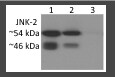
Detection of Human, Mouse, and Rat JNK2 by Western Blot.
Western blot shows lysates of HeLa human cervical epithelial carcinoma cell line and C2C12 mouse myoblast cell line. PVDF membrane was probed with 0.2 µg/mL Mouse Anti-Human/Mouse/Rat JNK2 Monoclonal Antibody (Catalog # MAB1846) followed by HRP-conjugated Anti-Mouse IgG Secondary Antibody (Catalog # HAF007). For additional reference, recombinant human JNK1, JNK2, and JNK3 (1 ng/lane) were included. Specific bands for JNK2 were detected at approximately 46 and 54 kDa (as indicated). This experiment was conducted under reducing conditions and using Immunoblot Buffer Group 1.
JNK2 in HeLa Human Cell Line.
JNK2 was detected in immersion fixed HeLa human cervical epithelial carcinoma cell line using Mouse Anti-Human/Mouse/Rat JNK2 Monoclonal Antibody (Catalog # MAB1846) at 25 µg/mL for 3 hours at room temperature. Cells were stained using the NorthernLights™ 557-conjugated Anti-Mouse IgG Secondary Antibody (red; Catalog # NL007) and counterstained with DAPI (blue). Specific staining was localized to cytoplasm. View our protocol for Fluorescent ICC Staining of Cells on Coverslips.
Western Blot Shows Human JNK2 Specificity by Using Knockout Cell Line.
Western blot shows lysates of HEK293T human embryonic kidney parental cell line and JNK2 knockout HEK293T cell line (KO). PVDF membrane was probed with 0.2 µg/mL of Mouse Anti-Human/Mouse/Rat JNK2 Monoclonal Antibody (Catalog # MAB1846) followed by HRP-conjugated Anti-Mouse IgG Secondary Antibody (Catalog # HAF018). Specific bands were detected for JNK2 at approximately 45 and 54 kDa (as indicated) in the parental HEK293T cell line, but is not detectable in knockout HEK293T cell line. GAPDH (Catalog # MAB5718) is shown as a loading control. This experiment was conducted under reducing conditions and using Immunoblot Buffer Group 1.
JNK2 in C2C12 Mouse Cell Line.
JNK2 was detected in immersion fixed C2C12 mouse myoblast cell line using Mouse Anti-Human/Mouse/Rat JNK2 Monoclonal Antibody (Catalog # MAB1846) at 25 µg/mL for 3 hours at room temperature. Cells were stained using the NorthernLights™ 557-conjugated Anti-Mouse IgG Secondary Antibody (red; NL007) and counterstained with DAPI (blue). Specific staining was localized to cytoplasm. Staining was performed using our protocol for Fluorescent ICC Staining of Non-adherent Cells.Applications for JNK2 Antibody (252320)
Application
Recommended Usage
Immunocytochemistry
8-25 µg/mL
Sample: Immersion fixed HeLa human cervical epithelial carcinoma cell line, MCF-7 human breast cancer cell line, and C2C12 mouse myoblast cell line
Sample: Immersion fixed HeLa human cervical epithelial carcinoma cell line, MCF-7 human breast cancer cell line, and C2C12 mouse myoblast cell line
Knockout Validated
JNK2
is specifically detected in HEK293T human embryonic kidney parental cell line but is not detectable in
JNK2 knockout HEK293T cell line.
Western Blot
0.2 µg/mL
Sample: HeLa human cervical epithelial carcinoma cell line and C2C12 mouse myoblast cell line
Sample: HeLa human cervical epithelial carcinoma cell line and C2C12 mouse myoblast cell line
Reviewed Applications
Read 2 reviews rated 5 using MAB1846 in the following applications:
Formulation, Preparation, and Storage
Purification
Protein A or G purified from hybridoma culture supernatant
Reconstitution
Reconstitute at 0.5 mg/mL in sterile PBS. For liquid material, refer to CoA for concentration.
Loading...
Formulation
Lyophilized from a 0.2 μm filtered solution in PBS with Trehalose. *Small pack size (SP) is supplied either lyophilized or as a 0.2 µm filtered solution in PBS.
Shipping
Lyophilized product is shipped at ambient temperature. Liquid small pack size (-SP) is shipped with polar packs. Upon receipt, store immediately at the temperature recommended below.
Stability & Storage
Use a manual defrost freezer and avoid repeated freeze-thaw cycles.
- 12 months from date of receipt, -20 to -70 °C as supplied.
- 1 month, 2 to 8 °C under sterile conditions after reconstitution.
- 6 months, -20 to -70 °C under sterile conditions after reconstitution.
Calculators
Background: JNK2
Long Name
C-Jun N-terminal Kinase 2
Alternate Names
MAPK9, SAPK1 alpha
Gene Symbol
MAPK9
Additional JNK2 Products
Product Documents for JNK2 Antibody (252320)
Certificate of Analysis
To download a Certificate of Analysis, please enter a lot or batch number in the search box below.
Note: Certificate of Analysis not available for kit components.
Product Specific Notices for JNK2 Antibody (252320)
For research use only
Citations for JNK2 Antibody (252320)
 Powered by Bioz
Customer Reviews for JNK2 Antibody (252320) (2)
5 out of 5
2 Customer Ratings
Have you used JNK2 Antibody (252320)?
Submit a review and receive an Amazon gift card!
$25/€18/£15/$25CAN/¥2500 Yen for a review with an image
$10/€7/£6/$10CAN/¥1110 Yen for a review without an image
Submit a review
Customer Images
Showing
1
-
2 of
2 reviews
Showing All
Filter By:
-
Application: Western BlotSample Tested: RetinaSpecies: Rat and MouseVerified Customer | Posted 12/31/2016Retina samples from rat (1), wild-type mouse (2), and JNK2 ko mouse (3) probed for JNK2. Major band at 54 kDa and additional band at 46 kDa are as expected from prior publications. Absence of bands in lanes with JNK2 ko sample demonstrates specificity.JNK2 Ab was used at 1:2000 over night, followed by peroxidase conjugated donkey anti mouse secondary antibody and chemiluminescence detection.

-
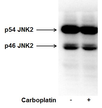
Application: Western BlotSample Tested: Human cancer cell whole cell lysateSpecies: HumanVerified Customer | Posted 10/04/2015JNK2 expression in MDA-MB-231 in response to carboplatin treatment.
There are no reviews that match your criteria.
Protocols
Find general support by application which include: protocols, troubleshooting, illustrated assays, videos and webinars.
- Appropriate Fixation of IHC/ICC Samples
- Cellular Response to Hypoxia Protocols
- ClariTSA™ Fluorophore Kits
- Detection & Visualization of Antibody Binding
- ICC Cell Smear Protocol for Suspension Cells
- ICC Immunocytochemistry Protocol Videos
- ICC for Adherent Cells
- Immunocytochemistry (ICC) Protocol
- Immunocytochemistry Troubleshooting
- Immunofluorescence of Organoids Embedded in Cultrex Basement Membrane Extract
- Immunohistochemistry (IHC) and Immunocytochemistry (ICC) Protocols
- Preparing Samples for IHC/ICC Experiments
- Preventing Non-Specific Staining (Non-Specific Binding)
- Primary Antibody Selection & Optimization
- Protocol for VisUCyte™ HRP Polymer Detection Reagent
- Protocol for the Fluorescent ICC Staining of Cell Smears - Graphic
- Protocol for the Fluorescent ICC Staining of Cultured Cells on Coverslips - Graphic
- Protocol for the Preparation and Fluorescent ICC Staining of Cells on Coverslips
- Protocol for the Preparation and Fluorescent ICC Staining of Non-adherent Cells
- Protocol for the Preparation and Fluorescent ICC Staining of Stem Cells on Coverslips
- Protocol for the Preparation of a Cell Smear for Non-adherent Cell ICC - Graphic
- R&D Systems Quality Control Western Blot Protocol
- TUNEL and Active Caspase-3 Detection by IHC/ICC Protocol
- The Importance of IHC/ICC Controls
- Troubleshooting Guide: Western Blot Figures
- Western Blot Conditions
- Western Blot Protocol
- Western Blot Protocol for Cell Lysates
- Western Blot Troubleshooting
- Western Blot Troubleshooting Guide
- View all Protocols, Troubleshooting, Illustrated assays and Webinars
Loading...
Associated Pathways


TGF-beta Signaling Pathways
 Toll-Like Receptor Signaling Pathways
 Wnt Signaling Pathways: beta-Catenin-dependent Wnt Signaling
