
Key Product Details
Validated by
Knockout/Knockdown
Species Reactivity
Validated:
Human, Mouse, Rat
Cited:
Human, Mouse, Xenograft
Applications
Validated:
Knockout Validated, Immunohistochemistry, Western Blot, Simple Western
Cited:
Immunohistochemistry, Immunohistochemistry-Paraffin, Western Blot, Flow Cytometry, Simple Western
Label
Unconjugated
Antibody Source
Polyclonal Rabbit IgG
Loading...
Product Specifications
Immunogen
E. coli-derived recombinant human p38 alpha
Met1-Ser360
Accession # Q16539
Met1-Ser360
Accession # Q16539
Specificity
Detects human, mouse and rat p38 alpha. Does not detect recombinant human p38 beta, p38 gamma or p38 delta.
Clonality
Polyclonal
Host
Rabbit
Isotype
IgG
Scientific Data Images for p38 alpha Antibody
Detection of Human, Mouse, and Rat p38 alpha by Western Blot.
Western blot shows lysates of HepG2 human hepatocellular carcinoma cell line, HeLa human cervical epithelial carcinoma cell line, NIH-3T3 mouse embryonic fibroblast cell line, and PC-12 rat adrenal pheochromocytoma cell line. PVDF membrane was probed with 0.5 µg/mL Rabbit Anti-Human/Mouse/Rat p38a Antigen Affinity-purified Polyclonal Antibody (Catalog # AF8691) followed by HRP-conjugated Anti-Rabbit IgG Secondary Antibody (HAF008). A specific band for p38a was detected at approximately 40 kDa (as indicated). This experiment was conducted under reducing conditions and using Immunoblot Buffer Group 1.
Detection of Human p38 alpha by Western Blot.
Western blot shows recombinant human p38 beta, p38 gamma, p38 delta and Recombinant Human Active p38 alpha (5477-KS) (2 ng/lane). PVDF membrane was probed with 0.5 µg/mL Rabbit Anti-Human/Mouse/Rat p38 alpha Antigen Affinity-purified Polyclonal Antibody (Catalog # AF8691) followed by HRP-conjugated Anti-Rabbit IgG Secondary Antibody (HAF008). This experiment was conducted under reducing conditions and using Immunoblot Buffer Group 1.
Orexin B, p38 alpha, & Integrin beta 1 in Mouse Brainstem.
Orexin B, p38a, and Integrin beta 1 were detected in perfusion fixed frozen sections of mouse brainstem using Human Orexin B Monoclonal Antibody (red; MAB734), Rabbit Anti-Human/Mouse/Rat p38a Antigen Affinity-purified Polyclonal Antibody (green; Catalog # AF8691), and Mouse Integrin beta 1 Antigen Affinity-purified Polyclonal Antibody (blue; AF2405). Tissue was incubated with primary antibodies overnight at 4 °C. Tissue was stained using NorthernLights™ 493-conjugated Anti-Rabbit IgG Secondary Antibody (green; NL006), NorthernLights 557-conjugated Anti-Mouse IgG Secondary Antibody (red; NL007), and NorthernLights 637-conjugated Anti-Goat IgG Secondary Antibody (blue; NL002). The image of Integrin beta 1 is pseudo-colored for presentation. View our protocol for Fluorescent IHC Staining of Frozen Tissue Sections.
Detection of Human and Mouse p38 alpha by Simple WesternTM.
Simple Western lane view shows lysates of HeLa human cervical epithelial carcinoma cell line and NIH‑3T3 mouse embryonic fibroblast cell line, loaded at 0.2 mg/mL. A specific band was detected for p38 alpha at approximately 43 kDa (as indicated) using 5 µg/mL of Rabbit Anti-Human/Mouse/Rat p38 alpha Antigen Affinity-purified Polyclonal Antibody (Catalog # AF8691). This experiment was conducted under reducing conditions and using the 12-230 kDa separation system.
Western Blot Shows Human p38 alpha Specificity by Using Knockout Cell Line.
Western blot shows lysates of HEK293T human embryonic kidney parental cell line and p38a knockout HEK293T cell line (KO). PVDF membrane was probed with 0.5 µg/mL of Rabbit Anti-Human/Mouse/Rat p38a Antigen Affinity-purified Polyclonal Antibody (Catalog # AF8691) followed by HRP-conjugated Anti-Rabbit IgG Secondary Antibody (HAF008). A specific band was detected for p38a at approximately 38 kDa (as indicated) in the parental HEK293T cell line, but is not detectable in knockout HEK293T cell line. GAPDH (AF5718) is shown as a loading control. This experiment was conducted under reducing conditions and using Immunoblot Buffer Group 1.
Detection of p38 alpha in Mouse Brain Cerebellum.
p38 alpha was detected in perfusion fixed paraffin-embedded sections of Mouse Brain Cerebellum using Rabbit Anti-Human/Mouse/Rat p38 alpha Antigen Affinity-purified Polyclonal Antibody (Catalog # AF8691) at 15 µg/mL for 1 hour at room temperature followed by incubation with the Anti-Rabbit IgG VisUCyte™ HRP Polymer Antibody (Catalog # VC003). Before incubation with the primary antibody, tissue was subjected to heat-induced epitope retrieval using VisUCyte Antigen Retrieval Reagent-Basic (Catalog # VCTS021). Tissue was stained using DAB (brown) and counterstained with hematoxylin (blue). Specific staining was localized to cytoplasm in Purkinje neurons. View our protocol for IHC Staining with VisUCyte HRP Polymer Detection Reagents.
Detection of Human p38 alpha by Western Blot
Pharmacological and genetic inhibition of the p38 MAPK pathway and its impact on human myotube formation in vitro.(A) Luminometric analysis of cell lysates generated from co-cultures initiated with 105 myoblastFLPe and 105 myoblastGS.Luc cells and exposed for 3 days to differentiation medium (white bar), to differentiation medium supplemented with 0.1, 0.5 and 2.5 µM SB 203580 (black bars) or to differentiation medium containing a final concentration of vehicle equivalent to that applied to co-cultures incubated with 2.5 µM SB 203580 (grey bar). Cumulative data are presented as means ± standard deviations (n = 3). RLU, relative light units. (B) Western blot analysis of p38 alpha levels in protein lysates of parental myoblasts and myoblastsGS.Luc (mock) and of myoblasts and myoblastsGS.Luc stably transduced with shRNA modules designed to down-regulate expression of eGFP (sh.eGFP), hif1 alpha (sh.hif1 alpha ) and human p38 alpha (sh.p38 alpha.35, sh.p38 alpha.33, sh.p38 alpha.36 and sh.p38 alpha.34). The alpha - and beta -tubulins served as loading control. (C) Diagram outlining the experimental set-up applied to investigate the impact of post-transcriptional down-regulation of p38 alpha expression on human myocyte fusion (see text for details). (D) Quantification through chemiluminescence of myoblast fusion activity in co-cultures consisting of a 1∶1 mixture of FLPe- and GS.Luc-encoding myoblasts either not transduced (none) or stably transduced with shRNAs sh.p38 alpha.33, sh.p38 alpha.36 or sh.hif1 alpha. Data were derived from a minimum of 3 and a maximum of 6 different experiments and are presented as means ± standard error of the mean. RLU, relative light units. Image collected and cropped by CiteAb from the following open publication (https://pubmed.ncbi.nlm.nih.gov/20532169), licensed under a CC-BY license. Not internally tested by R&D Systems.Applications for p38 alpha Antibody
Application
Recommended Usage
Immunohistochemistry
5-15 µg/mL
Sample: Perfusion fixed frozen sections of mouse brainstem and perfusion fixed paraffin-embedded sections of Mouse Brain Cerebellum
Sample: Perfusion fixed frozen sections of mouse brainstem and perfusion fixed paraffin-embedded sections of Mouse Brain Cerebellum
Knockout Validated
p38 alpha
is specifically detected in HEK293T human embryonic kidney parental cell line but is not detectable in
p38 alpha knockout HEK293T cell line.
Simple Western
5 µg/mL
Sample: HeLa human cervical epithelial carcinoma cell line and NIH‑3T3 mouse embryonic fibroblast cell line
Sample: HeLa human cervical epithelial carcinoma cell line and NIH‑3T3 mouse embryonic fibroblast cell line
Western Blot
0.5 µg/mL
Sample: HepG2 human hepatocellular carcinoma cell line, HeLa human cervical epithelial carcinoma cell line, NIH-3T3 mouse embryonic fibroblast cell line, and PC-12 rat adrenal pheochromocytoma cell line
Sample: HepG2 human hepatocellular carcinoma cell line, HeLa human cervical epithelial carcinoma cell line, NIH-3T3 mouse embryonic fibroblast cell line, and PC-12 rat adrenal pheochromocytoma cell line
Reviewed Applications
Read 3 reviews rated 5 using AF8691 in the following applications:
Formulation, Preparation, and Storage
Purification
Antigen and protein A Affinity-purified
Reconstitution
Reconstitute at 0.2 mg/mL in sterile PBS. For liquid material, refer to CoA for concentration.
Loading...
Formulation
Lyophilized from a 0.2 μm filtered solution in PBS with Trehalose. *Small pack size (SP) is supplied either lyophilized or as a 0.2 µm filtered solution in PBS.
Shipping
Lyophilized product is shipped at ambient temperature. Liquid small pack size (-SP) is shipped with polar packs. Upon receipt, store immediately at the temperature recommended below.
Stability & Storage
Use a manual defrost freezer and avoid repeated freeze-thaw cycles.
- 12 months from date of receipt, -20 to -70 °C as supplied.
- 1 month, 2 to 8 °C under sterile conditions after reconstitution.
- 6 months, -20 to -70 °C under sterile conditions after reconstitution.
Calculators
Background: p38 alpha
Alternate Names
MAPK14, SAPK2a
Gene Symbol
MAPK14
UniProt
Additional p38 alpha Products
Product Documents for p38 alpha Antibody
Certificate of Analysis
To download a Certificate of Analysis, please enter a lot or batch number in the search box below.
Note: Certificate of Analysis not available for kit components.
Product Specific Notices for p38 alpha Antibody
For research use only
Citations for p38 alpha Antibody
 Powered by Bioz
Customer Reviews for p38 alpha Antibody (3)
5 out of 5
3 Customer Ratings
Have you used p38 alpha Antibody?
Submit a review and receive an Amazon gift card!
$25/€18/£15/$25CAN/¥2500 Yen for a review with an image
$10/€7/£6/$10CAN/¥1110 Yen for a review without an image
Submit a review
Customer Images
Showing
1
-
3 of
3 reviews
Showing All
Filter By:
-
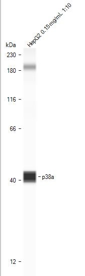
Application: Simple WesternSample Tested: HepG2 human hepatocellular carcinoma cell lineSpecies: HumanVerified Customer | Posted 05/17/2022Very good detection peaks while using Simple Western, as indicated.

-
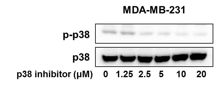
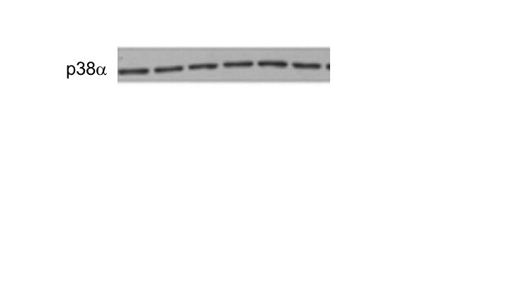
Application: Western BlotSample Tested: Human breast cancer cellsSpecies: HumanVerified Customer | Posted 10/24/2018Human breast cancer cell line MDA-MB-231 was treated with different doses of p38 inhibitor and the expression of phospho-p38 and total p38 were detected by western blot.
-
Application: Western BlotSample Tested: HEK293 human embryonic kidney cell lineSpecies: HumanVerified Customer | Posted 07/17/2018
There are no reviews that match your criteria.
Protocols
Find general support by application which include: protocols, troubleshooting, illustrated assays, videos and webinars.
- Antigen Retrieval Protocol (PIER)
- Antigen Retrieval for Frozen Sections Protocol
- Appropriate Fixation of IHC/ICC Samples
- Cellular Response to Hypoxia Protocols
- Chromogenic IHC Staining of Formalin-Fixed Paraffin-Embedded (FFPE) Tissue Protocol
- Chromogenic Immunohistochemistry Staining of Frozen Tissue
- ClariTSA™ Fluorophore Kits
- Detection & Visualization of Antibody Binding
- Fluorescent IHC Staining of Frozen Tissue Protocol
- Graphic Protocol for Heat-induced Epitope Retrieval
- Graphic Protocol for the Preparation and Fluorescent IHC Staining of Frozen Tissue Sections
- Graphic Protocol for the Preparation and Fluorescent IHC Staining of Paraffin-embedded Tissue Sections
- Graphic Protocol for the Preparation of Gelatin-coated Slides for Histological Tissue Sections
- IHC Sample Preparation (Frozen sections vs Paraffin)
- Immunofluorescent IHC Staining of Formalin-Fixed Paraffin-Embedded (FFPE) Tissue Protocol
- Immunohistochemistry (IHC) and Immunocytochemistry (ICC) Protocols
- Immunohistochemistry Frozen Troubleshooting
- Immunohistochemistry Paraffin Troubleshooting
- Preparing Samples for IHC/ICC Experiments
- Preventing Non-Specific Staining (Non-Specific Binding)
- Primary Antibody Selection & Optimization
- Protocol for Heat-Induced Epitope Retrieval (HIER)
- Protocol for Making a 4% Formaldehyde Solution in PBS
- Protocol for VisUCyte™ HRP Polymer Detection Reagent
- Protocol for the Preparation & Fixation of Cells on Coverslips
- Protocol for the Preparation and Chromogenic IHC Staining of Frozen Tissue Sections
- Protocol for the Preparation and Chromogenic IHC Staining of Frozen Tissue Sections - Graphic
- Protocol for the Preparation and Chromogenic IHC Staining of Paraffin-embedded Tissue Sections
- Protocol for the Preparation and Chromogenic IHC Staining of Paraffin-embedded Tissue Sections - Graphic
- Protocol for the Preparation and Fluorescent IHC Staining of Frozen Tissue Sections
- Protocol for the Preparation and Fluorescent IHC Staining of Paraffin-embedded Tissue Sections
- Protocol for the Preparation of Gelatin-coated Slides for Histological Tissue Sections
- R&D Systems Quality Control Western Blot Protocol
- TUNEL and Active Caspase-3 Detection by IHC/ICC Protocol
- The Importance of IHC/ICC Controls
- Troubleshooting Guide: Immunohistochemistry
- Troubleshooting Guide: Western Blot Figures
- Western Blot Conditions
- Western Blot Protocol
- Western Blot Protocol for Cell Lysates
- Western Blot Troubleshooting
- Western Blot Troubleshooting Guide
- View all Protocols, Troubleshooting, Illustrated assays and Webinars
Loading...
Associated Pathways


Pathogen or Damage-activated C-Type Lectin Receptor Signaling Pathways
 TGF-beta Signaling Pathways
 Toll-Like Receptor Signaling Pathways
 VEGF - VEGF R2 Signaling Pathways
