
Key Product Details
Species Reactivity
Validated:
Human, Mouse, Rat
Cited:
Human, Mouse
Applications
Validated:
Immunohistochemistry, Western Blot, Simple Western
Cited:
Western Blot
Label
Unconjugated
Antibody Source
Monoclonal Mouse IgG2A Clone # 563002
Loading...
Product Specifications
Immunogen
E. coli-derived recombinant human Raf-1
Asn189-Thr353
Accession # P04049
Asn189-Thr353
Accession # P04049
Specificity
Detects human, mouse, and rat Raf-1 at 74 kDa in Western blots.
Clonality
Monoclonal
Host
Mouse
Isotype
IgG2A
Scientific Data Images for Raf-1 Antibody (563002)
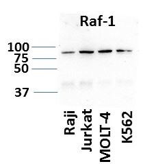
Detection of Human, Mouse, and Rat Raf‑1 by Western Blot.
Western blot shows lysates of MCF-7 human breast cancer cell line, HepG2 human hepatocellular carcinoma cell line, A431 human epithelial carcinoma cell line, L-929 mouse fibroblast cell line, and NRK rat normal kidney cell line. PVDF Membrane was probed with 1 µg/mL of Mouse Anti-Human/Mouse/Rat Raf-1 Monoclonal Antibody (Catalog # MAB4540) followed by HRP-conjugated Anti-Mouse IgG Secondary Antibody (Catalog # HAF007). A specific band was detected for Raf-1 at approximately 74 kDa (as indicated). This experiment was conducted under reducing conditions and using Immunoblot Buffer Group 3.
Raf‑1 in Human Liver.
Raf-1 was detected in immersion fixed paraffin-embedded sections of human liver array using Mouse Anti-Human/Mouse/Rat Raf-1 Monoclonal Antibody (Catalog # MAB4540) at 15 µg/mL overnight at 4 °C. Tissue was stained using the Anti-Mouse HRP-DAB Cell & Tissue Staining Kit (brown; Catalog # CTS002) and counterstained with hematoxylin (blue). Lower panel shows a lack of labeling if primary antibodies are omitted and tissue is stained only with secondary antibody followed by incubation with detection reagents. View our protocol for Chromogenic IHC Staining of Paraffin-embedded Tissue Sections.
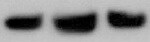
Detection of Human Raf‑1 by Simple WesternTM.
Simple Western lane view shows lysates of MCF‑7 human breast cancer cell line, loaded at 0.2 mg/mL. A specific band was detected for Raf‑1 at approximately 75 kDa (as indicated) using 10 µg/mL of Mouse Anti-Human/Mouse/Rat Raf‑1 Monoclonal Antibody (Catalog # MAB4540). This experiment was conducted under reducing conditions and using the 12-230 kDa separation system.Applications for Raf-1 Antibody (563002)
Application
Recommended Usage
Immunohistochemistry
8-25 µg/mL
Sample: Immersion-fixed paraffin-embedded sections of human liver and human liver cancer tissue
Sample: Immersion-fixed paraffin-embedded sections of human liver and human liver cancer tissue
Simple Western
10 µg/mL
Sample: MCF‑7 human breast cancer cell line
Sample: MCF‑7 human breast cancer cell line
Western Blot
1 µg/mL
Sample: MCF‑7 human breast cancer cell line, HepG2 human hepatocellular carcinoma cell line, A431 human epithelial carcinoma cell line, L‑929 mouse fibroblast cell line, and NRK rat normal kidney cell line
Sample: MCF‑7 human breast cancer cell line, HepG2 human hepatocellular carcinoma cell line, A431 human epithelial carcinoma cell line, L‑929 mouse fibroblast cell line, and NRK rat normal kidney cell line
Reviewed Applications
Read 3 reviews rated 4.3 using MAB4540 in the following applications:
Formulation, Preparation, and Storage
Purification
Protein A or G purified from hybridoma culture supernatant
Reconstitution
Reconstitute at 0.5 mg/mL in sterile PBS. For liquid material, refer to CoA for concentration.
Loading...
Formulation
Lyophilized from a 0.2 μm filtered solution in PBS with Trehalose. *Small pack size (SP) is supplied either lyophilized or as a 0.2 µm filtered solution in PBS.
Shipping
Lyophilized product is shipped at ambient temperature. Liquid small pack size (-SP) is shipped with polar packs. Upon receipt, store immediately at the temperature recommended below.
Stability & Storage
Use a manual defrost freezer and avoid repeated freeze-thaw cycles.
- 12 months from date of receipt, -20 to -70 °C as supplied.
- 1 month, 2 to 8 °C under sterile conditions after reconstitution.
- 6 months, -20 to -70 °C under sterile conditions after reconstitution.
Calculators
Background: Raf-1
Long Name
v-raf-1Murine Leukemia Viral Oncogene Homolog 1
Alternate Names
c-Raf-1, Raf1, v-Raf-1
Gene Symbol
RAF1
UniProt
Additional Raf-1 Products
Product Documents for Raf-1 Antibody (563002)
Certificate of Analysis
To download a Certificate of Analysis, please enter a lot or batch number in the search box below.
Note: Certificate of Analysis not available for kit components.
Product Specific Notices for Raf-1 Antibody (563002)
For research use only
Related Research Areas
Citations for Raf-1 Antibody (563002)
 Powered by Bioz
Customer Reviews for Raf-1 Antibody (563002) (3)
4.3 out of 5
3 Customer Ratings
Have you used Raf-1 Antibody (563002)?
Submit a review and receive an Amazon gift card!
$25/€18/£15/$25CAN/¥2500 Yen for a review with an image
$10/€7/£6/$10CAN/¥1110 Yen for a review without an image
Submit a review
Customer Images
Showing
1
-
3 of
3 reviews
Showing All
Filter By:
-
Application: Western BlotSample Tested: Lung cellsSpecies: MouseVerified Customer | Posted 10/26/2021

-
Application: Western BlotSample Tested: Hematological cancer cell lines lysatesSpecies: HumanVerified Customer | Posted 11/18/201930 ug of protein per sample. 1:500 dilution of primary Ab.
-
Application: Western BlotSample Tested: Lung single-cell suspensionSpecies: MouseVerified Customer | Posted 06/06/2019
There are no reviews that match your criteria.
Protocols
Find general support by application which include: protocols, troubleshooting, illustrated assays, videos and webinars.
- Antigen Retrieval Protocol (PIER)
- Antigen Retrieval for Frozen Sections Protocol
- Appropriate Fixation of IHC/ICC Samples
- Cellular Response to Hypoxia Protocols
- Chromogenic IHC Staining of Formalin-Fixed Paraffin-Embedded (FFPE) Tissue Protocol
- Chromogenic Immunohistochemistry Staining of Frozen Tissue
- ClariTSA™ Fluorophore Kits
- Detection & Visualization of Antibody Binding
- Fluorescent IHC Staining of Frozen Tissue Protocol
- Graphic Protocol for Heat-induced Epitope Retrieval
- Graphic Protocol for the Preparation and Fluorescent IHC Staining of Frozen Tissue Sections
- Graphic Protocol for the Preparation and Fluorescent IHC Staining of Paraffin-embedded Tissue Sections
- Graphic Protocol for the Preparation of Gelatin-coated Slides for Histological Tissue Sections
- IHC Sample Preparation (Frozen sections vs Paraffin)
- Immunofluorescent IHC Staining of Formalin-Fixed Paraffin-Embedded (FFPE) Tissue Protocol
- Immunohistochemistry (IHC) and Immunocytochemistry (ICC) Protocols
- Immunohistochemistry Frozen Troubleshooting
- Immunohistochemistry Paraffin Troubleshooting
- Preparing Samples for IHC/ICC Experiments
- Preventing Non-Specific Staining (Non-Specific Binding)
- Primary Antibody Selection & Optimization
- Protocol for Heat-Induced Epitope Retrieval (HIER)
- Protocol for Making a 4% Formaldehyde Solution in PBS
- Protocol for VisUCyte™ HRP Polymer Detection Reagent
- Protocol for the Preparation & Fixation of Cells on Coverslips
- Protocol for the Preparation and Chromogenic IHC Staining of Frozen Tissue Sections
- Protocol for the Preparation and Chromogenic IHC Staining of Frozen Tissue Sections - Graphic
- Protocol for the Preparation and Chromogenic IHC Staining of Paraffin-embedded Tissue Sections
- Protocol for the Preparation and Chromogenic IHC Staining of Paraffin-embedded Tissue Sections - Graphic
- Protocol for the Preparation and Fluorescent IHC Staining of Frozen Tissue Sections
- Protocol for the Preparation and Fluorescent IHC Staining of Paraffin-embedded Tissue Sections
- Protocol for the Preparation of Gelatin-coated Slides for Histological Tissue Sections
- R&D Systems Quality Control Western Blot Protocol
- TUNEL and Active Caspase-3 Detection by IHC/ICC Protocol
- The Importance of IHC/ICC Controls
- Troubleshooting Guide: Immunohistochemistry
- Troubleshooting Guide: Western Blot Figures
- Western Blot Conditions
- Western Blot Protocol
- Western Blot Protocol for Cell Lysates
- Western Blot Troubleshooting
- Western Blot Troubleshooting Guide
- View all Protocols, Troubleshooting, Illustrated assays and Webinars
Loading...
Associated Pathways


IL-9 Signaling Pathways
 IL-15 Signaling Pathways
 IL-21 Signaling Pathways
 MAPK Signaling: Oxidative Stress Pathway
 MAPK Signaling Pathway: Mitogen Stimulation Pathway
 mTOR Signaling Pathway
 Pathogen or Damage-activated C-Type Lectin Receptor Signaling Pathways
 TGF-beta Signaling Pathways
 VEGF - VEGF R2 Signaling Pathways
