Myeloperoxidase (MPO) is a hemeprotein that belongs to the XPO subfamily of the heme peroxidase superfamily. MPO is synthesized as a preproprotein that undergoes proteolytic processing to generate a disulfide-linked heterodimer of the N-terminal beta -subunit (12 kDa) and C-terminal alpha subunit (60 kDa). Active MPO is a tetramer of two beta -subunits and two alpha -subunits that are also disulfide-linked through the two alpha -subunits. MPO is stored in granules and is an abundant protein in neutrophils and monocytes. MPO is released upon activation to catalyze the formation of powerful oxidants such as hypochlorous acid, which kills microbes. Unprocessed pro-MPO can also be released. Human and mouse MPO share 87% amino acid sequence identity.
Human Myeloperoxidase/MPO Antibody (392105)
R&D Systems | Catalog # MAB3174

Key Product Details
Species Reactivity
Validated:
Human
Cited:
Human, Mouse, Gerbil
Applications
Validated:
Immunohistochemistry, Western Blot, Immunocytochemistry
Cited:
Immunohistochemistry, Immunohistochemistry-Paraffin, Western Blot, ELISA Capture
Label
Unconjugated
Antibody Source
Monoclonal Mouse IgG2B Clone # 392105
Loading...
Product Specifications
Immunogen
E. coli-derived recombinant human Myeloperoxidase/MPO
Val279-Ser745
Accession # P05164
Val279-Ser745
Accession # P05164
Specificity
Detects human Myeloperoxidase/MPO in Western blots. No cross-reactivity with recombinant human Eosinophil Peroxidase is observed.
Clonality
Monoclonal
Host
Mouse
Isotype
IgG2B
Scientific Data Images for Human Myeloperoxidase/MPO Antibody (392105)
Detection of Human Myeloperoxidase/MPO by Western Blot.
Western blot shows lysates of HL-60 human acute promyelocytic leukemia cell line and human neutrophil. PVDF membrane was probed with 1 µg/mL of Mouse Anti-Human Myeloperoxidase/MPO Monoclonal Antibody (Catalog # MAB3174) followed by HRP-conjugated Anti-Mouse IgG Secondary Antibody (Catalog # HAF007). A specific band was detected for Myeloperoxidase/MPO at approximately 65 kDa (as indicated). This experiment was conducted under reducing conditions and using Immunoblot Buffer Group 2.
Myeloperoxidase/MPO in MOLT-4 Human Cell Line.
Myeloperoxidase/ MPO was detected in immersion fixed MOLT-4 human acute lymphoblastic leukemia cell line using 8 µg/mL Mouse Anti-Human Myeloperoxidase/ MPO Monoclonal Antibody (Catalog # MAB3174) for 3 hours at room temperature. Cells were stained (red) and counter-stained (green). View our protocol for Fluorescent ICC Staining of Cells on Coverslips.
Myeloperoxidase/MPO in HL‑60 Human Cell Line.
Myeloperoxidase/MPO was detected in immersion fixed HL-60 human acute promyelocytic leukemia cell line using Mouse Anti-Human Myeloperoxidase/MPO Mono-clonal Antibody (Catalog # MAB3174) at 10 µg/mL for 3 hours at room temperature. Cells were stained using the NorthernLights™ 557-conju-gated Anti-Mouse IgG Secondary Antibody (yellow; Catalog # NL007) and counterstained with DAPI (blue). View our protocol for Fluorescent ICC Staining of Non-adherent Cells.
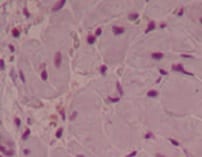
Myeloperoxidase/MPO in Human Spleen.
Myeloperoxidase/MPO was detected in immersion fixed paraffin-embedded sections of human spleen using 15 µg/mL Mouse Anti-Human Myeloperoxidase/MPO Monoclonal Antibody (Catalog # MAB3174) overnight at 4 °C. Before incubation with the primary antibody tissue was subjected to heat-induced epitope retrieval using Antigen Retrieval Reagent-Basic (Catalog # CTS013). Tissue was stained with the Anti-Mouse HRP-DAB Cell & Tissue Staining Kit (brown; Catalog # CTS002) and counterstained with hematoxylin (blue). View our protocol for Chromogenic IHC Staining of Paraffin-embedded Tissue Sections.
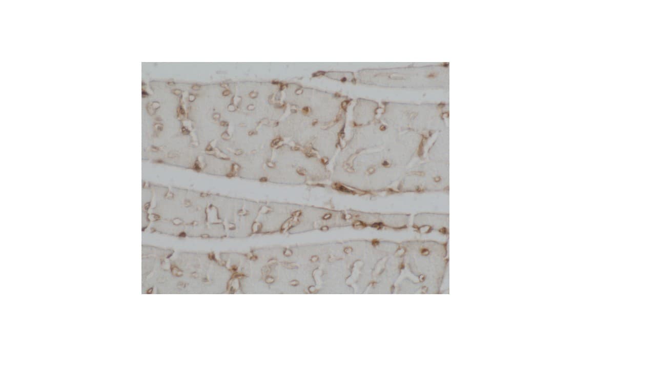
Myeloperoxidase/MPO in Mouse Spleen.
Myeloperoxidase/MPO was detected in immersion fixed paraffin-embedded sections of mouse spleen using Mouse Anti-Human Myeloperoxidase/MPO Monoclonal Antibody (Catalog # MAB3174) at 10 µg/mL for 1 hour at room temperature followed by incubation with the Anti-Mouse IgG VisUCyte™ HRP Polymer Antibody (VC001). Before incubation with the primary antibody, tissue was subjected to heat-induced epitope retrieval using Antigen Retrieval Reagent-Basic (CTS013). Tissue was stained using DAB (brown) and counterstained with hematoxylin (blue). Specific staining was localized to cytoplasm in lymphocytes. Staining was performed using our protocol for IHC Staining with VisUCyte HRP Polymer Detection Reagents.Applications for Human Myeloperoxidase/MPO Antibody (392105)
Application
Recommended Usage
Immunocytochemistry
8-25 µg/mL
Sample: Immersion fixed MOLT-4 human acute lymphoblastic leukemia cell line and HL-60 human acute promyelocytic leukemia cell line
Sample: Immersion fixed MOLT-4 human acute lymphoblastic leukemia cell line and HL-60 human acute promyelocytic leukemia cell line
Immunohistochemistry
8-25 µg/mL
Sample: Immersion fixed paraffin-embedded sections of human spleen and mouse spleen
Sample: Immersion fixed paraffin-embedded sections of human spleen and mouse spleen
Western Blot
1 µg/mL
Sample: HL-60 human acute promyelocytic leukemia cell line and human neutrophil
Sample: HL-60 human acute promyelocytic leukemia cell line and human neutrophil
Reviewed Applications
Read 5 reviews rated 3.4 using MAB3174 in the following applications:
Formulation, Preparation, and Storage
Purification
Protein A or G purified from hybridoma culture supernatant
Reconstitution
Reconstitute at 0.5 mg/mL in sterile PBS. For liquid material, refer to CoA for concentration.
Loading...
Formulation
Lyophilized from a 0.2 μm filtered solution in PBS with Trehalose. *Small pack size (SP) is supplied either lyophilized or as a 0.2 µm filtered solution in PBS.
Shipping
Lyophilized product is shipped at ambient temperature. Liquid small pack size (-SP) is shipped with polar packs. Upon receipt, store immediately at the temperature recommended below.
Stability & Storage
Use a manual defrost freezer and avoid repeated freeze-thaw cycles.
- 12 months from date of receipt, -20 to -70 °C as supplied.
- 1 month, 2 to 8 °C under sterile conditions after reconstitution.
- 6 months, -20 to -70 °C under sterile conditions after reconstitution.
Calculators
Background: Myeloperoxidase/MPO
Alternate Names
MPO
Gene Symbol
MPO
UniProt
Additional Myeloperoxidase/MPO Products
Product Documents for Human Myeloperoxidase/MPO Antibody (392105)
Certificate of Analysis
To download a Certificate of Analysis, please enter a lot or batch number in the search box below.
Note: Certificate of Analysis not available for kit components.
Product Specific Notices for Human Myeloperoxidase/MPO Antibody (392105)
For research use only
Related Research Areas
Citations for Human Myeloperoxidase/MPO Antibody (392105)
 Powered by Bioz
Customer Reviews for Human Myeloperoxidase/MPO Antibody (392105) (5)
3.4 out of 5
5 Customer Ratings
Have you used Human Myeloperoxidase/MPO Antibody (392105)?
Submit a review and receive an Amazon gift card!
$25/€18/£15/$25CAN/¥2500 Yen for a review with an image
$10/€7/£6/$10CAN/¥1110 Yen for a review without an image
Submit a review
Customer Images
Showing
1
-
5 of
5 reviews
Showing All
Filter By:
-
Application: ImmunohistochemistrySample Tested: MyocardiaSpecies: HumanVerified Customer | Posted 10/10/2021

-
Application: ELISASample Tested: EDTA PlasmaSpecies: HumanVerified Customer | Posted 12/20/2017
-
Application: Immunocytochemistry/ImmunofluorescenceSample Tested: Peripheral blood neutrophilsSpecies: HumanVerified Customer | Posted 09/08/2016This antibody does not stain Neutrophil Extracellular Traps (NETs) on coverslips, whereas it does stain intracellular MPO. This raises questions about its specificity. This antibody is also not reactive in a sandwich ELISA to detect NETs, despite the fact that MPO is known to be a very abundant protein on NETs... On western blot it does detect a protein at the right molecular weight already at low concentrations.Bio-Techne ResponseR&D Systems Technical Service is investigating.
-
Application: ImmunohistochemistrySample Tested: Adult heartSpecies: MouseVerified Customer | Posted 09/02/2016Used at a dilution of 1:50. Non-R-D secondary, biotinylated at a dilution of 1:200
-
Application: Western BlotSample Tested: Colon tissueSpecies: MouseVerified Customer | Posted 03/30/2016None
There are no reviews that match your criteria.
Protocols
Find general support by application which include: protocols, troubleshooting, illustrated assays, videos and webinars.
- Antigen Retrieval Protocol (PIER)
- Antigen Retrieval for Frozen Sections Protocol
- Appropriate Fixation of IHC/ICC Samples
- Cellular Response to Hypoxia Protocols
- Chromogenic IHC Staining of Formalin-Fixed Paraffin-Embedded (FFPE) Tissue Protocol
- Chromogenic Immunohistochemistry Staining of Frozen Tissue
- ClariTSA™ Fluorophore Kits
- Detection & Visualization of Antibody Binding
- Fluorescent IHC Staining of Frozen Tissue Protocol
- Graphic Protocol for Heat-induced Epitope Retrieval
- Graphic Protocol for the Preparation and Fluorescent IHC Staining of Frozen Tissue Sections
- Graphic Protocol for the Preparation and Fluorescent IHC Staining of Paraffin-embedded Tissue Sections
- Graphic Protocol for the Preparation of Gelatin-coated Slides for Histological Tissue Sections
- ICC Cell Smear Protocol for Suspension Cells
- ICC Immunocytochemistry Protocol Videos
- ICC for Adherent Cells
- IHC Sample Preparation (Frozen sections vs Paraffin)
- Immunocytochemistry (ICC) Protocol
- Immunocytochemistry Troubleshooting
- Immunofluorescence of Organoids Embedded in Cultrex Basement Membrane Extract
- Immunofluorescent IHC Staining of Formalin-Fixed Paraffin-Embedded (FFPE) Tissue Protocol
- Immunohistochemistry (IHC) and Immunocytochemistry (ICC) Protocols
- Immunohistochemistry Frozen Troubleshooting
- Immunohistochemistry Paraffin Troubleshooting
- Preparing Samples for IHC/ICC Experiments
- Preventing Non-Specific Staining (Non-Specific Binding)
- Primary Antibody Selection & Optimization
- Protocol for Heat-Induced Epitope Retrieval (HIER)
- Protocol for Making a 4% Formaldehyde Solution in PBS
- Protocol for VisUCyte™ HRP Polymer Detection Reagent
- Protocol for the Fluorescent ICC Staining of Cell Smears - Graphic
- Protocol for the Fluorescent ICC Staining of Cultured Cells on Coverslips - Graphic
- Protocol for the Preparation & Fixation of Cells on Coverslips
- Protocol for the Preparation and Chromogenic IHC Staining of Frozen Tissue Sections
- Protocol for the Preparation and Chromogenic IHC Staining of Frozen Tissue Sections - Graphic
- Protocol for the Preparation and Chromogenic IHC Staining of Paraffin-embedded Tissue Sections
- Protocol for the Preparation and Chromogenic IHC Staining of Paraffin-embedded Tissue Sections - Graphic
- Protocol for the Preparation and Fluorescent ICC Staining of Cells on Coverslips
- Protocol for the Preparation and Fluorescent ICC Staining of Non-adherent Cells
- Protocol for the Preparation and Fluorescent ICC Staining of Stem Cells on Coverslips
- Protocol for the Preparation and Fluorescent IHC Staining of Frozen Tissue Sections
- Protocol for the Preparation and Fluorescent IHC Staining of Paraffin-embedded Tissue Sections
- Protocol for the Preparation of Gelatin-coated Slides for Histological Tissue Sections
- Protocol for the Preparation of a Cell Smear for Non-adherent Cell ICC - Graphic
- R&D Systems Quality Control Western Blot Protocol
- TUNEL and Active Caspase-3 Detection by IHC/ICC Protocol
- The Importance of IHC/ICC Controls
- Troubleshooting Guide: Immunohistochemistry
- Troubleshooting Guide: Western Blot Figures
- Western Blot Conditions
- Western Blot Protocol
- Western Blot Protocol for Cell Lysates
- Western Blot Troubleshooting
- Western Blot Troubleshooting Guide
- View all Protocols, Troubleshooting, Illustrated assays and Webinars
Loading...
Associated Pathways
