Glycogen Synthase Kinase-3 (GSK-3) is a serine/threonine kinase initially identified asan inhibitor of glycogen synthase. Two isoforms (GSK-3 alpha and GSK-3 beta ) share 85% amino acid identity. GSK-3 beta, inhibited by phosphorylation at S9 by Akt, is involved in energy metabolism, body pattern formation, and neuronal cell development.
Human phospho-GSK-3 beta (S9) Antibody (609739)
R&D Systems | Catalog # MAB25062
 by Western Blot.")
Key Product Details
Validated by
Biological Validation
Species Reactivity
Validated:
Human
Cited:
Human
Applications
Validated:
Western Blot, Intracellular Staining by Flow Cytometry, Immunocytochemistry, CyTOF-ready
Cited:
Immunocytochemistry
Label
Unconjugated
Antibody Source
Monoclonal Mouse IgG1 Clone # 609739
Loading...
Product Specifications
Immunogen
Phosphopeptide containing the human GSK-3 beta S9 site
Specificity
Detects human GSK-3 beta when phosphorylated at S9.
Clonality
Monoclonal
Host
Mouse
Isotype
IgG1
Scientific Data Images for Human phospho-GSK-3 beta (S9) Antibody (609739)
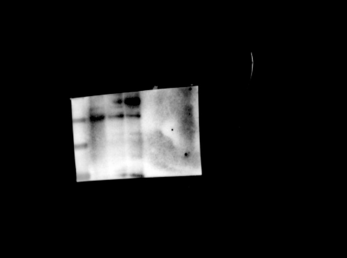
Detection of Human Phospho-GSK‑3 beta (S9) by Western Blot.
Western blot shows lysates of HeLa human cervical epithelial carcinoma cell line untreated (-) or treated (+) with 200 nM PMA for 20 minutes. PVDF Membrane was probed with 1 µg/mL of Human Phospho-GSK-3 beta (S9) Monoclonal Antibody (Catalog # MAB25062) followed by HRP-conjugated Anti-Mouse IgG Secondary Antibody (Catalog # HAF007). A specific band was detected for Phospho-GSK-3 beta (S9) at approximately 46 kDa (as indicated). This experiment was conducted under reducing conditions and using Immunoblot Buffer Group 1. in HeLa Human Cell Line.")
Phospho-GSK‑3 beta (S9) in HeLa Human Cell Line.
GSK-3 beta phosphorylated at S9 was detected in immersion fixed HeLa human cervical epithelial carcinoma cells, unstimulated (lower panel) or stimulated (upper panel) with PMA, using Mouse Anti-Human Phospho-GSK-3 beta (S9) Monoclonal Antibody (Catalog # MAB25062) at 10 µg/mL for 3 hours at room temperature. Cells were stained using the NorthernLights™ 557-conjugated Anti-Mouse IgG Secondary Antibody (red; Catalog # NL007) and counterstained with DAPI (blue). Specific staining was localized to cytoplasm. View our protocol for Fluorescent ICC Staining of Cells on Coverslips. in HeLa Human Cell Line by Flow Cytometry.")
Detection of Phospho-GSK-3 beta (S9) in HeLa Human Cell Line by Flow Cytometry.
HeLa human cervical epithelial carcinoma cell line untreated (open histogram) or treated with 120 ng/mL PMA for 15 minutes (filled histogram) was stained with Human Phospho-GSK-3 beta (S9) Monoclonal Antibody (Catalog # MAB25062) or isotype control antibody (Catalog # MAB002, not shown), followed by Fluorescein-conjugated Anti-Mouse IgG Secondary Antibody (Catalog # F0103B). To facilitate intracellular staining, cells were fixed with paraformaldehyde and permeabilized with saponin.Applications for Human phospho-GSK-3 beta (S9) Antibody (609739)
Application
Recommended Usage
CyTOF-ready
Ready to be labeled using established conjugation methods. No BSA or other carrier proteins that could interfere with conjugation.
Immunocytochemistry
8-25 µg/mL
Sample: Immersion fixed HeLa human cervical epithelial carcinoma cell line stimulated with PMA
Sample: Immersion fixed HeLa human cervical epithelial carcinoma cell line stimulated with PMA
Intracellular Staining by Flow Cytometry
2.5 µg/106 cells
Sample: HeLa human cervical epithelial carcinoma cell line treated with PMA, fixed with paraformaldehyde and permeabilized with saponin
Sample: HeLa human cervical epithelial carcinoma cell line treated with PMA, fixed with paraformaldehyde and permeabilized with saponin
Western Blot
1 µg/mL
Sample: HeLa human cervical epithelial carcinoma cell line treated with PMA
Sample: HeLa human cervical epithelial carcinoma cell line treated with PMA
Reviewed Applications
Read 3 reviews rated 3.7 using MAB25062 in the following applications:
Flow Cytometry Panel Builder
Bio-Techne Knows Flow Cytometry
Save time and reduce costly mistakes by quickly finding compatible reagents using the Panel Builder Tool.
Advanced Features
- Spectra Viewer - Custom analysis of spectra from multiple fluorochromes
- Spillover Popups - Visualize the spectra of individual fluorochromes
- Antigen Density Selector - Match fluorochrome brightness with antigen density

Formulation, Preparation, and Storage
Purification
Protein A or G purified from hybridoma culture supernatant
Reconstitution
Sterile PBS to a final concentration of 0.5 mg/mL. For liquid material, refer to CoA for concentration.
Loading...
Formulation
Lyophilized from a 0.2 μm filtered solution in PBS with Trehalose. *Small pack size (SP) is supplied either lyophilized or as a 0.2 µm filtered solution in PBS.
Shipping
Lyophilized product is shipped at ambient temperature. Liquid small pack size (-SP) is shipped with polar packs. Upon receipt, store immediately at the temperature recommended below.
Stability & Storage
Use a manual defrost freezer and avoid repeated freeze-thaw cycles.
- 12 months from date of receipt, -20 to -70 °C as supplied.
- 1 month, 2 to 8 °C under sterile conditions after reconstitution.
- 6 months, -20 to -70 °C under sterile conditions after reconstitution.
Calculators
Background: GSK-3 beta
Long Name
Glycogen Synthase Kinase 3 beta
Alternate Names
GSK3 beta, GSK3B
Gene Symbol
GSK3B
Additional GSK-3 beta Products
Product Documents for Human phospho-GSK-3 beta (S9) Antibody (609739)
Certificate of Analysis
To download a Certificate of Analysis, please enter a lot or batch number in the search box below.
Note: Certificate of Analysis not available for kit components.
Product Specific Notices for Human phospho-GSK-3 beta (S9) Antibody (609739)
For research use only
Related Research Areas
Customer Reviews for Human phospho-GSK-3 beta (S9) Antibody (609739) (3)
3.7 out of 5
3 Customer Ratings
Have you used Human phospho-GSK-3 beta (S9) Antibody (609739)?
Submit a review and receive an Amazon gift card!
$25/€18/£15/$25CAN/¥2500 Yen for a review with an image
$10/€7/£6/$10CAN/¥1110 Yen for a review without an image
Submit a review
Customer Images
Showing
1
-
3 of
3 reviews
Showing All
Filter By:
-
Application: Western BlotSample Tested: MDA-MB-231 human breast cancer cell lineSpecies: HumanVerified Customer | Posted 05/28/2026Unspecific bands in MDA-MB-23110 ug of total protein from MDA-MB-231 cells was loaded and antibody used at 1ug/mL as recommended

-
Application: Western BlotSample Tested: HCT-116 human colorectal carcinoma cell lineSpecies: HumanVerified Customer | Posted 05/28/2026Unspecific bands in HCT116 cells total proteinTotal protein from HCT116 Cells. Followed recommended dilution
-
Application: Western BlotSample Tested: MDA-MB-231 human breast cancer cell lineSpecies: HumanVerified Customer | Posted 04/18/2022
There are no reviews that match your criteria.
Protocols
Find general support by application which include: protocols, troubleshooting, illustrated assays, videos and webinars.
- 7-Amino Actinomycin D (7-AAD) Cell Viability Flow Cytometry Protocol
- Appropriate Fixation of IHC/ICC Samples
- Cellular Response to Hypoxia Protocols
- ClariTSA™ Fluorophore Kits
- Detection & Visualization of Antibody Binding
- Extracellular Membrane Flow Cytometry Protocol
- Flow Cytometry Protocol for Cell Surface Markers
- Flow Cytometry Protocol for Staining Membrane Associated Proteins
- Flow Cytometry Staining Protocols
- Flow Cytometry Troubleshooting Guide
- ICC Cell Smear Protocol for Suspension Cells
- ICC Immunocytochemistry Protocol Videos
- ICC for Adherent Cells
- Immunocytochemistry (ICC) Protocol
- Immunocytochemistry Troubleshooting
- Immunofluorescence of Organoids Embedded in Cultrex Basement Membrane Extract
- Immunohistochemistry (IHC) and Immunocytochemistry (ICC) Protocols
- Intracellular Flow Cytometry Protocol Using Alcohol (Methanol)
- Intracellular Flow Cytometry Protocol Using Detergents
- Intracellular Nuclear Staining Flow Cytometry Protocol Using Detergents
- Intracellular Staining Flow Cytometry Protocol Using Alcohol Permeabilization
- Intracellular Staining Flow Cytometry Protocol Using Detergents to Permeabilize Cells
- Preparing Samples for IHC/ICC Experiments
- Preventing Non-Specific Staining (Non-Specific Binding)
- Primary Antibody Selection & Optimization
- Propidium Iodide Cell Viability Flow Cytometry Protocol
- Protocol for Liperfluo
- Protocol for VisUCyte™ HRP Polymer Detection Reagent
- Protocol for the Characterization of Human Th22 Cells
- Protocol for the Characterization of Human Th9 Cells
- Protocol for the Fluorescent ICC Staining of Cell Smears - Graphic
- Protocol for the Fluorescent ICC Staining of Cultured Cells on Coverslips - Graphic
- Protocol for the Preparation and Fluorescent ICC Staining of Cells on Coverslips
- Protocol for the Preparation and Fluorescent ICC Staining of Non-adherent Cells
- Protocol for the Preparation and Fluorescent ICC Staining of Stem Cells on Coverslips
- Protocol for the Preparation of a Cell Smear for Non-adherent Cell ICC - Graphic
- Protocol: Annexin V and PI Staining by Flow Cytometry
- Protocol: Annexin V and PI Staining for Apoptosis by Flow Cytometry
- R&D Systems Quality Control Western Blot Protocol
- TUNEL and Active Caspase-3 Detection by IHC/ICC Protocol
- The Importance of IHC/ICC Controls
- Troubleshooting Guide: Fluorokine Flow Cytometry Kits
- Troubleshooting Guide: Western Blot Figures
- Western Blot Conditions
- Western Blot Protocol
- Western Blot Protocol for Cell Lysates
- Western Blot Troubleshooting
- Western Blot Troubleshooting Guide
- View all Protocols, Troubleshooting, Illustrated assays and Webinars
Loading...
Associated Pathways


IL-7 Signaling Pathways
 IL-9 Signaling Pathways
 IL-15 Signaling Pathways
 IL-17 Family Signaling Pathways
 IL-21 Signaling Pathways
 mTOR Signaling Pathway
 Wnt Signaling Pathways: beta-Catenin-dependent Wnt Signaling
