Interleukin-2 (IL-2) is a cytokine that stimulates the growth and differentiation of B cells, T cells, NK cells, and monocyte/macrophages. It functions through the heterotrimeric IL-2 receptor comprising alpha, beta, and gamma chains.

Key Product Details
Species Reactivity
Validated:
Human, Primate
Cited:
Human, Mouse, Primate - Papio anubis (Olive Baboon), Transgenic Mouse, Xenograft
Applications
Validated:
ELISA Capture (Matched Antibody Pair), Immunocytochemistry
Cited:
Neutralization, ELISA Capture, ELISA Detection, ELISA Development, ELISA Development (Capture), In vivo assay, Luminex Development, Stimulation
Label
Unconjugated
Antibody Source
Monoclonal Mouse IgG2A Clone # 5355
Loading...
Product Specifications
Immunogen
E. coli-derived recombinant human IL-2
Ala21-Thr153
Accession # NP_000577
Ala21-Thr153
Accession # NP_000577
Specificity
Detects human and primate IL-2 in ELISAs. In sandwich immunoassays, no cross-reactivity with recombinant human (rh) IL-1 alpha, rhIL-1 beta, rhIL-3, rhIL-4, rhIL‑6, rhIL-7, rhIL-8, recombinant mouse (rm) IL-1 beta, rmIL-2, rmIL-3, rmIL-4, rmIL-5, rmIL-6, or rmIL-7 is observed.
Clonality
Monoclonal
Host
Mouse
Isotype
IgG2A
Scientific Data Images for IL-2 Antibody (5355)
IL‑2 in Human PBMCs.
IL-2 was detected in immersion fixed human peripheral blood mononuclear cells (PBMCs) stimulated with PMA and ionomycin using 10 µg/mL Mouse Anti-Human/Primate IL-2 Monoclonal Antibody (Catalog # MAB602) for 3 hours at room temperature. Cells were stained with the NorthernLights™ 557-conjugated Anti-Mouse IgG Secondary Antibody (red; Catalog # NL007) and counterstained with DAPI (blue). View our protocol for Fluorescent ICC Staining of Non-adherent Cells.
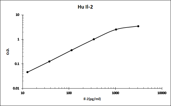
Human IL-2 ELISA Standard Curve
Recombinant Human IL‑2 (Catalog # 202-IL) was serially diluted and captured by Mouse Anti-Human/Primate IL‑2 Monoclonal Antibody (Catalog # MAB602) coated on a Clear Polystyrene Microplate (Catalog # DY990). Goat Anti-Human IL‑2 Antigen Affinity-purified Polyclonal Antibody (Catalog # AF-202-NA) was biotinylated and incubated with the protein captured on the plate. Detection of the standard curve was achieved by incubating Streptavidin-HRP (Catalog # DY998)Applications for IL-2 Antibody (5355)
Application
Recommended Usage
Immunocytochemistry
8-25 µg/mL
Sample: Immersion fixed human peripheral blood mononuclear cells (PBMCs) stimulated with PMA and ionomycin
Sample: Immersion fixed human peripheral blood mononuclear cells (PBMCs) stimulated with PMA and ionomycin
Human/Primate IL-2 Sandwich Immunoassay
Please Note: Optimal dilutions of this antibody should be experimentally determined.
Reviewed Applications
Read 2 reviews rated 5 using MAB602 in the following applications:
Formulation, Preparation, and Storage
Purification
Protein A or G purified from hybridoma culture supernatant
Reconstitution
Reconstitute at 0.5 mg/mL in sterile PBS. For liquid material, refer to CoA for concentration.
Loading...
Formulation
Lyophilized from a 0.2 μm filtered solution in PBS with Trehalose. *Small pack size (SP) is supplied either lyophilized or as a 0.2 µm filtered solution in PBS.
Shipping
Lyophilized product is shipped at ambient temperature. Liquid small pack size (-SP) is shipped with polar packs. Upon receipt, store immediately at the temperature recommended below.
Stability & Storage
Use a manual defrost freezer and avoid repeated freeze-thaw cycles.
- 12 months from date of receipt, -20 to -70 °C as supplied.
- 1 month, 2 to 8 °C under sterile conditions after reconstitution.
- 6 months, -20 to -70 °C under sterile conditions after reconstitution.
Calculators
Background: IL-2
Long Name
Interleukin 2
Alternate Names
Aldesleukin, IL2, Proleukin, TCGF
Entrez Gene IDs
Gene Symbol
IL2
UniProt
Additional IL-2 Products
Product Documents for IL-2 Antibody (5355)
Certificate of Analysis
To download a Certificate of Analysis, please enter a lot or batch number in the search box below.
Note: Certificate of Analysis not available for kit components.
Product Specific Notices for IL-2 Antibody (5355)
For research use only
Related Research Areas
Citations for IL-2 Antibody (5355)
 Powered by Bioz
Customer Reviews for IL-2 Antibody (5355) (2)
5 out of 5
2 Customer Ratings
Have you used IL-2 Antibody (5355)?
Submit a review and receive an Amazon gift card!
$25/€18/£15/$25CAN/¥2500 Yen for a review with an image
$10/€7/£6/$10CAN/¥1110 Yen for a review without an image
Submit a review
Customer Images
Showing
1
-
2 of
2 reviews
Showing All
Filter By:
-
Application: ELISASample Tested: Serum and PlasmaSpecies: HumanVerified Customer | Posted 08/20/2021

-
Application: ELISASample Tested: SerumSpecies: Cynomolgus MonkeyVerified Customer | Posted 12/12/2020
There are no reviews that match your criteria.
Protocols
Find general support by application which include: protocols, troubleshooting, illustrated assays, videos and webinars.
- Appropriate Fixation of IHC/ICC Samples
- Cellular Response to Hypoxia Protocols
- ClariTSA™ Fluorophore Kits
- Detection & Visualization of Antibody Binding
- ICC Cell Smear Protocol for Suspension Cells
- ICC Immunocytochemistry Protocol Videos
- ICC for Adherent Cells
- Immunocytochemistry (ICC) Protocol
- Immunocytochemistry Troubleshooting
- Immunofluorescence of Organoids Embedded in Cultrex Basement Membrane Extract
- Immunohistochemistry (IHC) and Immunocytochemistry (ICC) Protocols
- Preparing Samples for IHC/ICC Experiments
- Preventing Non-Specific Staining (Non-Specific Binding)
- Primary Antibody Selection & Optimization
- Protocol for VisUCyte™ HRP Polymer Detection Reagent
- Protocol for the Fluorescent ICC Staining of Cell Smears - Graphic
- Protocol for the Fluorescent ICC Staining of Cultured Cells on Coverslips - Graphic
- Protocol for the Preparation and Fluorescent ICC Staining of Cells on Coverslips
- Protocol for the Preparation and Fluorescent ICC Staining of Non-adherent Cells
- Protocol for the Preparation and Fluorescent ICC Staining of Stem Cells on Coverslips
- Protocol for the Preparation of a Cell Smear for Non-adherent Cell ICC - Graphic
- TUNEL and Active Caspase-3 Detection by IHC/ICC Protocol
- The Importance of IHC/ICC Controls
- View all Protocols, Troubleshooting, Illustrated assays and Webinars
Loading...
Associated Pathways


Innate Lymphoid Cell Differentiation Pathways
 Jak/STAT Signaling Pathway
 Th1 Differentiation Pathway
 Th2 Differentiation Pathway
