Signal Transduction and Activator of Transcription 5B (STAT5b) is a member of the Jak/STAT signal transduction pathway and is activated by a variety of cytokines (IL-22, IL-6, IFN alpha ). Upon cytokine activation, STAT5b is phosphorylated and translocates to the nucleus where it binds to the DNA consensus site TTCN3GAA and activates transcription of specific genes. STAT5b shares 93% amino acid identity with STAT5a. STAT5b is involved in a wide array of biological processes ranging from regulating apoptosis to adult mammary gland proliferation, differentiation and survival.

Key Product Details
Validated by
Knockout/Knockdown
Species Reactivity
Human
Applications
Knockout Validated, Western Blot, Intracellular Staining by Flow Cytometry, Simple Western, Immunoprecipitation, CyTOF-ready
Label
Unconjugated
Antibody Source
Monoclonal Mouse IgG2B Clone # 389215
Loading...
Product Specifications
Immunogen
Human STAT5b synthetic peptide
MDSQWIPHAQS
Accession # NP_036580
MDSQWIPHAQS
Accession # NP_036580
Specificity
Detects human STAT5b in Western blots.
Clonality
Monoclonal
Host
Mouse
Isotype
IgG2B
Scientific Data Images for Human STAT5b Antibody (389215)
Detection of Human STAT5b by Western Blot.
Western blot shows lysates of HeLa human cervical epithelial carcinoma cell line, Daudi human Burkitt's lymphoma cell line, and K562 human chronic myelogenous leukemia cell line. PVDF membrane was probed with 1 µg/mL of Mouse Anti-Human STAT5b Monoclonal Antibody (Catalog # MAB1584) followed by HRP-conjugated Anti-Mouse IgG Secondary Antibody (HAF018). A specific band was detected for STAT5b at approximately 90 kDa (as indicated). This experiment was conducted under reducing conditions and using Immunoblot Buffer Group 1.
Detection of STAT5b in Jurkat Human Cell Line by Flow Cytometry.
Jurkat human acute T cell leukemia cell line was stained with Mouse Anti-Human STAT5b Monoclonal Antibody (Catalog # MAB1584, filled histogram) or isotype control antibody (MAB0041, open histogram), followed by Allophycocyanin-conjugated Anti-Mouse IgG F(ab')2Secondary Antibody (F0101B).
Detection of Human STAT5b by Simple WesternTM.
Simple Western lane view shows lysates of HeLa human cervical epithelial carcinoma cell line, loaded at 0.2 mg/mL. A specific band was detected for STAT5b at approximately 91 kDa (as indicated) using 20 µg/mL of Mouse Anti-Human STAT5b Monoclonal Antibody (Catalog # MAB1584). This experiment was conducted under reducing conditions and using the 12-230 kDa separation system.
Western Blot Shows Human STAT5b Specificity by Using Knockout Cell Line.
Western blot shows lysates of HeLa human cervical epithelial carcinoma parental cell line and STAT5b knockout HeLa cell line (KO). PVDF membrane was probed with 1 µg/mL of Mouse Anti-Human STAT5b Monoclonal Antibody (Catalog # MAB1584) followed by HRP-conjugated Anti-Mouse IgG Secondary Antibody (HAF018). A specific band was detected for STAT5b at approximately 90 kDa (as indicated) in the parental HeLa cell line, but is not detectable in knockout HeLa cell line. GAPDH (Catalog # MAB5718) is shown as a loading control. This experiment was conducted under reducing conditions and using Immunoblot Buffer Group 1.
Detection of Stat5b by Immunoprecipitation.
Immunoprecipitation was performed on cell lysate of HAP1 human near-haploid cell line using 2.0 μg of Mouse Anti-Human Stat5b Monoclonal Antibody (Catalog # MAB1584) pre-coupled to protein G or protein A beads. Immunoprecipitated Stat5b was detected with a Rabbit Anti-Stat5b antibody. The Ponceau stained transfers of each blot are shown. SM=10% starting material; UB=10% unbound fraction; IP=immunoprecipitated. Image, protocol, and testing courtesy of YCharOS Inc. (ycharos.com).Applications for Human STAT5b Antibody (389215)
Application
Recommended Usage
CyTOF-ready
Ready to be labeled using established conjugation methods. No BSA or other carrier proteins that could interfere with conjugation.
Immunoprecipitation
2 µg/1 mg cell lysate
Sample: Cell lysate of HAP1 human near-haploid cell line
Sample: Cell lysate of HAP1 human near-haploid cell line
Intracellular Staining by Flow Cytometry
2.5 µg/106 cells
Sample: Jurkat human acute T cell leukemia cell line fixed with paraformaldehyde and permeabilized with methanol
Sample: Jurkat human acute T cell leukemia cell line fixed with paraformaldehyde and permeabilized with methanol
Knockout Validated
STAT5b
is specifically detected in HeLa human cervical epithelial carcinoma parental cell line but is not detectable in
STAT5b knockout HeLa cell line.
Simple Western
20 µg/mL
Sample: HeLa human cervical epithelial carcinoma cell line
Sample: HeLa human cervical epithelial carcinoma cell line
Western Blot
1 µg/mL
Sample: HeLa human cervical epithelial carcinoma cell line, Daudi human Burkitt's lymphoma cell line, and K562 human chronic myelogenous leukemia cell line
Sample: HeLa human cervical epithelial carcinoma cell line, Daudi human Burkitt's lymphoma cell line, and K562 human chronic myelogenous leukemia cell line
Reviewed Applications
Read 1 review rated 3 using MAB1584 in the following applications:
Flow Cytometry Panel Builder
Bio-Techne Knows Flow Cytometry
Save time and reduce costly mistakes by quickly finding compatible reagents using the Panel Builder Tool.
Advanced Features
- Spectra Viewer - Custom analysis of spectra from multiple fluorochromes
- Spillover Popups - Visualize the spectra of individual fluorochromes
- Antigen Density Selector - Match fluorochrome brightness with antigen density

Formulation, Preparation, and Storage
Purification
Protein A or G purified from hybridoma culture supernatant
Reconstitution
Reconstitute at 0.5 mg/mL in sterile PBS. For liquid material, refer to CoA for concentration.
Loading...
Formulation
Lyophilized from a 0.2 μm filtered solution in PBS with Trehalose. *Small pack size (SP) is supplied either lyophilized or as a 0.2 µm filtered solution in PBS.
Shipping
Lyophilized product is shipped at ambient temperature. Liquid small pack size (-SP) is shipped with polar packs. Upon receipt, store immediately at the temperature recommended below.
Stability & Storage
Use a manual defrost freezer and avoid repeated freeze-thaw cycles.
- 12 months from date of receipt, -20 to -70 °C as supplied.
- 1 month, 2 to 8 °C under sterile conditions after reconstitution.
- 6 months, -20 to -70 °C under sterile conditions after reconstitution.
Calculators
Background: STAT5b
Long Name
Signal Transducer and Activator of Transcription 5b
Alternate Names
signal transducer and activator of transcription 5B, STAT5, transcription factor STAT5B
Gene Symbol
STAT5B
UniProt
Additional STAT5b Products
Product Documents for Human STAT5b Antibody (389215)
Certificate of Analysis
To download a Certificate of Analysis, please enter a lot or batch number in the search box below.
Note: Certificate of Analysis not available for kit components.
Product Specific Notices for Human STAT5b Antibody (389215)
For research use only
Citations for Human STAT5b Antibody (389215)
 Powered by Bioz
Customer Reviews for Human STAT5b Antibody (389215) (1)
3 out of 5
1 Customer Rating
Have you used Human STAT5b Antibody (389215)?
Submit a review and receive an Amazon gift card!
$25/€18/£15/$25CAN/¥2500 Yen for a review with an image
$10/€7/£6/$10CAN/¥1110 Yen for a review without an image
Submit a review
Customer Images
Showing
1
-
1 of
1 review
Showing All
Filter By:
-
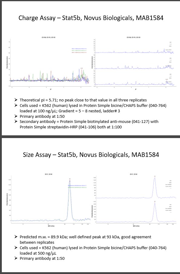
Application: Simple WesternSample Tested: k562 cellsSpecies: HumanVerified Customer | Posted 11/28/2016Charge Assay – Stat5b, Novus Biologicals, MAB1584 Theoretical pI = 5.71; no peak close to that value in all three replicates Cells used = K562 (human) lysed in Protein Simple bicine/CHAPS buffer (040-764) loaded at 100 ng/µL; Gradient = 5 – 8 nested, ladder# 3 Primary antibody at 1:50 Secondary antibody = Protein Simple biotinylated anti-mouse (041-127) with Protein Simple streptavidin-HRP (041-106) both at 1:100 Size Assay – Stat5b, Novus Biologicals, MAB1584 Predicted m.w. = 89.9 kDa; well defined peak at 93 kDa, good agreement between replicates Cells used = K562 (human) lysed in Protein Simple bicine/CHAPS buffer (040-764) loaded at 500 ng/µL Primary antibody at 1:50

There are no reviews that match your criteria.
Protocols
Find general support by application which include: protocols, troubleshooting, illustrated assays, videos and webinars.
- 7-Amino Actinomycin D (7-AAD) Cell Viability Flow Cytometry Protocol
- Cellular Response to Hypoxia Protocols
- Extracellular Membrane Flow Cytometry Protocol
- Flow Cytometry Protocol for Cell Surface Markers
- Flow Cytometry Protocol for Staining Membrane Associated Proteins
- Flow Cytometry Staining Protocols
- Flow Cytometry Troubleshooting Guide
- Immunoprecipitation Protocol
- Intracellular Flow Cytometry Protocol Using Alcohol (Methanol)
- Intracellular Flow Cytometry Protocol Using Detergents
- Intracellular Nuclear Staining Flow Cytometry Protocol Using Detergents
- Intracellular Staining Flow Cytometry Protocol Using Alcohol Permeabilization
- Intracellular Staining Flow Cytometry Protocol Using Detergents to Permeabilize Cells
- Propidium Iodide Cell Viability Flow Cytometry Protocol
- Protocol for Liperfluo
- Protocol for the Characterization of Human Th22 Cells
- Protocol for the Characterization of Human Th9 Cells
- Protocol: Annexin V and PI Staining by Flow Cytometry
- Protocol: Annexin V and PI Staining for Apoptosis by Flow Cytometry
- R&D Systems Quality Control Western Blot Protocol
- Troubleshooting Guide: Fluorokine Flow Cytometry Kits
- Troubleshooting Guide: Western Blot Figures
- Western Blot Conditions
- Western Blot Protocol
- Western Blot Protocol for Cell Lysates
- Western Blot Troubleshooting
- Western Blot Troubleshooting Guide
- View all Protocols, Troubleshooting, Illustrated assays and Webinars
Loading...
Associated Pathways


IL-15 Signaling Pathways
 IL-21 Signaling Pathways
 Jak/STAT Signaling Pathway
 Th1 Differentiation Pathway
 Th17 Differentiation Pathway
