Human Syndecan-1 (also known as CD138) is a variably glycosylated, dimeric, type I transmembrane (TM) protein that belongs to the Syndecan family. It is synthesized as a 310 amino acid (aa) precursor that contains a 17 aa signal sequence, a 234 aa extracellular domain (ECD), a 25 aa TM segment, and a 34 aa cytoplasmic region. The ECD shows various degrees of heparan sulfate and chondroitin sulfate modification, leading to native molecular weights for Syndecan-1 of 120 - 200 kDa. Proteolytic cleavage of the membrane-bound ECD yields soluble forms of approximately the same molecular weight. Syndecan-1 is an epithelial cell Syndecan involved in Wnt and chemokine signaling. Human Syndecan-1 ECD shares 71% aa identity with the ECD of both rat and mouse Syndecan-1.

Key Product Details
Species Reactivity
Validated:
Human
Cited:
Human, Mouse
Applications
Validated:
Immunohistochemistry, Western Blot, ELISA Capture (Matched Antibody Pair)
Cited:
Immunohistochemistry, Immunohistochemistry-Paraffin, Western Blot, Flow Cytometry, Immunocytochemistry, Immunoprecipitation, Immunodepletion
Label
Unconjugated
Antibody Source
Polyclonal Goat IgG
Loading...
Product Specifications
Immunogen
Mouse myeloma cell line NS0-derived recombinant human Syndecan-1/CD138
Gln18-Glu251
Accession # NP_002988
Gln18-Glu251
Accession # NP_002988
Specificity
Detects human Syndecan-1/CD138 in ELISAs and Western blots. In sandwich ELISAs, less than 0.3% cross-reactivity with recombinant human (rh) Syndecan-2, rhSyndecan-3, rhSyndecan-4, and recombinant mouse Syndecan-1 is observed.
Clonality
Polyclonal
Host
Goat
Isotype
IgG
Scientific Data Images for Human Syndecan‑1/CD138 Antibody
Syndecan‑1/CD138 in Human Ileum.
Syndecan‑1/CD138 was detected in immersion fixed paraffin-embedded sections of human ileum using Human Syndecan‑1/CD138 Antigen Affinity-purified Polyclonal Antibody (Catalog # AF2780) at 15 µg/mL overnight at 4 °C. Tissue was stained using the Anti-Goat HRP-DAB Cell & Tissue Staining Kit (brown; Catalog # CTS008) and counterstained with hematoxylin (blue). View our protocol for Chromogenic IHC Staining of Paraffin-embedded Tissue Sections.
Syndecan‑1/CD138 in Human Jejunum.
Syndecan-1/CD138 was detected in immersion fixed paraffin-embedded sections of human jejunum using Human Syndecan-1/CD138 Antigen Affinity-purified Polyclonal Antibody (Catalog # AF2780) at 15 µg/mL overnight at 4 °C. Tissue was stained using the Anti-Goat HRP-DAB Cell & Tissue Staining Kit (brown; Catalog # CTS008) and counterstained with hematoxylin (blue). Lower panel shows a lack of labeling if primary antibodies are omitted and tissue is stained only with secondary antibody followed by incubation with detection reagents. View our protocol for Chromogenic IHC Staining of Paraffin-embedded Tissue Sections.Applications for Human Syndecan‑1/CD138 Antibody
Application
Recommended Usage
Immunohistochemistry
5-15 µg/mL
Sample: Perfusion fixed frozen sections of mouse intestine and immersion fixed paraffin-embedded sections of normal human ileum and jejunum
Sample: Perfusion fixed frozen sections of mouse intestine and immersion fixed paraffin-embedded sections of normal human ileum and jejunum
Western Blot
0.1 µg/mL
Sample: Recombinant Human Syndecan‑1/CD138 (Catalog # 2780-SD)
Sample: Recombinant Human Syndecan‑1/CD138 (Catalog # 2780-SD)
Human Syndecan-1/CD138 Sandwich Immunoassay
Please Note: Optimal dilutions of this antibody should be experimentally determined.
Reviewed Applications
Read 1 review rated 5 using AF2780 in the following applications:
Formulation, Preparation, and Storage
Purification
Antigen Affinity-purified
Reconstitution
Reconstitute at 0.2 mg/mL in sterile PBS. For liquid material, refer to CoA for concentration.
Loading...
Formulation
Lyophilized from a 0.2 μm filtered solution in PBS with Trehalose. *Small pack size (SP) is supplied either lyophilized or as a 0.2 µm filtered solution in PBS.
Shipping
Lyophilized product is shipped at ambient temperature. Liquid small pack size (-SP) is shipped with polar packs. Upon receipt, store immediately at the temperature recommended below.
Stability & Storage
Use a manual defrost freezer and avoid repeated freeze-thaw cycles.
- 12 months from date of receipt, -20 to -70 °C as supplied.
- 1 month, 2 to 8 °C under sterile conditions after reconstitution.
- 6 months, -20 to -70 °C under sterile conditions after reconstitution.
Calculators
Background: Syndecan-1/CD138
References
Alternate Names
CD138, SDC1, Syndecan1
Gene Symbol
SDC1
UniProt
Additional Syndecan-1/CD138 Products
Product Documents for Human Syndecan‑1/CD138 Antibody
Certificate of Analysis
To download a Certificate of Analysis, please enter a lot or batch number in the search box below.
Note: Certificate of Analysis not available for kit components.
Product Specific Notices for Human Syndecan‑1/CD138 Antibody
For research use only
Related Research Areas
Citations for Human Syndecan‑1/CD138 Antibody
 Powered by Bioz
Customer Reviews for Human Syndecan‑1/CD138 Antibody (1)
5 out of 5
1 Customer Rating
Have you used Human Syndecan‑1/CD138 Antibody?
Submit a review and receive an Amazon gift card!
$25/€18/£15/$25CAN/¥2500 Yen for a review with an image
$10/€7/£6/$10CAN/¥1110 Yen for a review without an image
Submit a review
Customer Images
Showing
1
-
1 of
1 review
Showing All
Filter By:
-
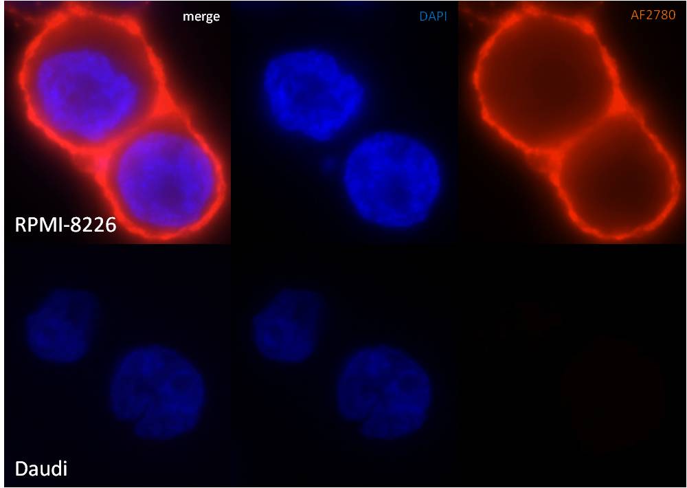
Application: ImmunofluorescenceSample Tested: RPMI-8226 multiple myeloma cell line, and Daudi Burkitt lymphoma cell lineSpecies: HumanVerified Customer | Posted 03/13/2015AF2780 immunofluorescence staining in cell lines

There are no reviews that match your criteria.
Protocols
Find general support by application which include: protocols, troubleshooting, illustrated assays, videos and webinars.
- Antigen Retrieval Protocol (PIER)
- Antigen Retrieval for Frozen Sections Protocol
- Appropriate Fixation of IHC/ICC Samples
- Cellular Response to Hypoxia Protocols
- Chromogenic IHC Staining of Formalin-Fixed Paraffin-Embedded (FFPE) Tissue Protocol
- Chromogenic Immunohistochemistry Staining of Frozen Tissue
- ClariTSA™ Fluorophore Kits
- Detection & Visualization of Antibody Binding
- Fluorescent IHC Staining of Frozen Tissue Protocol
- Graphic Protocol for Heat-induced Epitope Retrieval
- Graphic Protocol for the Preparation and Fluorescent IHC Staining of Frozen Tissue Sections
- Graphic Protocol for the Preparation and Fluorescent IHC Staining of Paraffin-embedded Tissue Sections
- Graphic Protocol for the Preparation of Gelatin-coated Slides for Histological Tissue Sections
- IHC Sample Preparation (Frozen sections vs Paraffin)
- Immunofluorescent IHC Staining of Formalin-Fixed Paraffin-Embedded (FFPE) Tissue Protocol
- Immunohistochemistry (IHC) and Immunocytochemistry (ICC) Protocols
- Immunohistochemistry Frozen Troubleshooting
- Immunohistochemistry Paraffin Troubleshooting
- Preparing Samples for IHC/ICC Experiments
- Preventing Non-Specific Staining (Non-Specific Binding)
- Primary Antibody Selection & Optimization
- Protocol for Heat-Induced Epitope Retrieval (HIER)
- Protocol for Making a 4% Formaldehyde Solution in PBS
- Protocol for VisUCyte™ HRP Polymer Detection Reagent
- Protocol for the Preparation & Fixation of Cells on Coverslips
- Protocol for the Preparation and Chromogenic IHC Staining of Frozen Tissue Sections
- Protocol for the Preparation and Chromogenic IHC Staining of Frozen Tissue Sections - Graphic
- Protocol for the Preparation and Chromogenic IHC Staining of Paraffin-embedded Tissue Sections
- Protocol for the Preparation and Chromogenic IHC Staining of Paraffin-embedded Tissue Sections - Graphic
- Protocol for the Preparation and Fluorescent IHC Staining of Frozen Tissue Sections
- Protocol for the Preparation and Fluorescent IHC Staining of Paraffin-embedded Tissue Sections
- Protocol for the Preparation of Gelatin-coated Slides for Histological Tissue Sections
- R&D Systems Quality Control Western Blot Protocol
- TUNEL and Active Caspase-3 Detection by IHC/ICC Protocol
- The Importance of IHC/ICC Controls
- Troubleshooting Guide: Immunohistochemistry
- Troubleshooting Guide: Western Blot Figures
- Western Blot Conditions
- Western Blot Protocol
- Western Blot Protocol for Cell Lysates
- Western Blot Troubleshooting
- Western Blot Troubleshooting Guide
- View all Protocols, Troubleshooting, Illustrated assays and Webinars


