Tissue inhibitors of metalloproteinases or TIMPs are a family of proteins that regulate the activation and proteolytic activity of the zinc enzymes known as matrix metalloproteinases (MMPs). There are four members of the family, TIMP-1, TIMP-2, TIMP-3, and TIMP-4. TIMP-1 is a glycoprotein with a molecular mass of 28 kDa produced by a wide range of cell types. TIMP-1 inhibits active MMP-mediated proteolysis by forming an N-terminal, non-covalent binary complex with the MMP active site. TIMP-1 also associates C-terminally with Pro-MMP-9 in a complex which may play a role in regulating activation. Independent of MMPs, TIMP-1 has been shown to have a role in tissue homeostasis.

Key Product Details
Species Reactivity
Validated:
Human
Cited:
Human, Primate
Applications
Validated:
Immunohistochemistry, Western Blot, ELISA Capture (Matched Antibody Pair), Intracellular Staining by Flow Cytometry, Immunocytochemistry, CyTOF-ready
Cited:
Western Blot, Immunocytochemistry, Cell-based ELISA, Dot Blot, ELISA Capture, Luminex Development, Mass Spectrometry
Label
Unconjugated
Antibody Source
Monoclonal Mouse IgG2B Clone # 63515
Loading...
Product Specifications
Immunogen
Mouse myeloma cell line NS0-derived recombinant human TIMP-1
Cys24-Ala207
Accession # P01033
Cys24-Ala207
Accession # P01033
Specificity
Detects human TIMP-1 in ELISAs and Western blots. In ELISAs, this antibody does not cross-react with recombinant mouse TIMP‑1 or recombinant human TIMP‑2.
Clonality
Monoclonal
Host
Mouse
Isotype
IgG2B
Scientific Data Images for Human TIMP-1 Antibody (63515)
TIMP‑1 in Wi-38 Human Cell Line.
TIMP-1 was detected in immersion fixed Wi-38 human lung fibroblast cell line using Mouse Anti-Human TIMP-1 Monoclonal Antibody (Catalog # MAB970) at 3 µg/mL for 3 hours at room temperature. Cells were stained using the NorthernLights™ 557-conjugated Anti-Mouse IgG Secondary Antibody (red; Catalog # NL007) and counterstained with DAPI (blue). Specific staining was localized to cytoplasm and endoplasmic reticulum. View our protocol for Fluorescent ICC Staining of Cells on Coverslips.
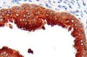
TIMP‑1 in Human Astrocytoma.
TIMP-1 was detected in immersion fixed paraffin-embedded sections of human astrocytoma using Mouse Anti-Human TIMP-1 Monoclonal Antibody (Catalog # MAB970) at 15 µg/mL overnight at 4 °C. Before incubation with the primary antibody tissue was subjected to heat-induced epitope retrieval using Antigen Retrieval Reagent-Basic (Catalog # CTS013). Tissue was stained using the Anti-Mouse HRP-DAB Cell & Tissue Staining Kit (brown; Catalog # CTS002) and counterstained with hematoxylin (blue). View our protocol for Chromogenic IHC Staining of Paraffin-embedded Tissue Sections.
Detection of TIMP-1 by Western Blot
Abolishment of TGF-beta 1-mediated matrix remodeling by p38 and JNK inhibitors in the primary cultured orbital fibroblasts from patients with Graves’ ophthalmopathy (GO). (A) Orbital fibroblasts from GO patients were preincubated with the p38 inhibitor SB202190 (20 μM) and the JNK inhibitor SP600125 (20 μM), respectively, for 1 h, followed by TGF-beta 1 (5 ng/mL) treatment for another 24 h. Then, the expression levels of TIMP-1, TIMP-3, MMP-2, and MMP-9 were analyzed by Western blots; (B) The relative intensities of TIMP-1, TIMP-3, MMP-2, and MMP-9 expression normalized to each GAPDH control and DMSO control without TGF-beta 1 treatment were defined as 1.0. Then, the other relative intensity (folds) was presented. By three independent Western blot experiments, together data from the same patient strain were averaged. Then, the means of different patient (GO1–GO4, n = 4) strains were averaged; (C) The enzyme activities of MMP-2/-9 from the cultured medium were determined. The representative histogram was plotted based on the mean values of relative fluorescence units from the primary cultured orbital fibroblasts from the four GO patients. Data were presented as means ± S.D. of the results from three independent experiments. * p < 0.05 and ** p < 0.01 vs. control without TGF-beta 1 treatment; # p < 0.05 and ## p < 0.01 vs. TGF-beta 1 treatment. Image collected and cropped by CiteAb from the following open publication (https://pubmed.ncbi.nlm.nih.gov/33799469), licensed under a CC-BY license. Not internally tested by R&D Systems.
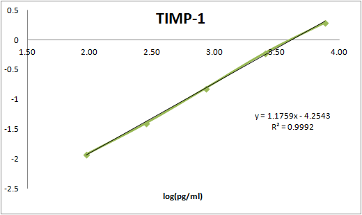
Human TIMP-1 ELISA Standard Curve
Recombinant Human TIMP‑1 (Catalog # 970-TM) was serially diluted and captured by Mouse Anti-Human TIMP‑1 Monoclonal Antibody (Catalog # MAB970) coated on a Clear Polystyrene Microplate (Catalog # DY990). Goat Anti-Human TIMP‑1 Antigen Affinity-purified Polyclonal Antibody (Catalog # AF970) was biotinylated and incubated with the protein captured on the plate. Detection of the standard curve was achieved by incubating Streptavidin-HRP (Catalog # DY998)Applications for Human TIMP-1 Antibody (63515)
Application
Recommended Usage
CyTOF-ready
Ready to be labeled using established conjugation methods. No BSA or other carrier proteins that could interfere with conjugation.
Immunocytochemistry
3-25 µg/mL
Sample: Immersion fixed Wi-38 human lung fibroblast cell line
Sample: Immersion fixed Wi-38 human lung fibroblast cell line
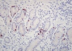
Immunohistochemistry
8-25 µg/mL
Sample:
Sample:
Immersion fixed paraffin-embedded sections of human ovarian cancer tissue and astrocytoma tissue
Intracellular Staining by Flow Cytometry
2.5 µg/106 cells
Sample: Human T cells fixed with paraformaldehyde and permeabilized with saponin
Sample: Human T cells fixed with paraformaldehyde and permeabilized with saponin
Western Blot
1 µg/mL
Sample: Recombinant Human TIMP-1 Western Blot Standard (Catalog # WBC021)
under non-reducing conditions only
Sample: Recombinant Human TIMP-1 Western Blot Standard (Catalog # WBC021)
under non-reducing conditions only
Human TIMP-1 Sandwich Immunoassay
Please Note: Optimal dilutions of this antibody should be experimentally determined.
Reviewed Applications
Read 4 reviews rated 5 using MAB970 in the following applications:
Flow Cytometry Panel Builder
Bio-Techne Knows Flow Cytometry
Save time and reduce costly mistakes by quickly finding compatible reagents using the Panel Builder Tool.
Advanced Features
- Spectra Viewer - Custom analysis of spectra from multiple fluorochromes
- Spillover Popups - Visualize the spectra of individual fluorochromes
- Antigen Density Selector - Match fluorochrome brightness with antigen density

Formulation, Preparation, and Storage
Purification
Protein A or G purified from hybridoma culture supernatant
Reconstitution
Reconstitute at 0.5 mg/mL in sterile PBS. For liquid material, refer to CoA for concentration.
Loading...
Formulation
Lyophilized from a 0.2 μm filtered solution in PBS with Trehalose. *Small pack size (SP) is supplied either lyophilized or as a 0.2 µm filtered solution in PBS.
Shipping
Lyophilized product is shipped at ambient temperature. Liquid small pack size (-SP) is shipped with polar packs. Upon receipt, store immediately at the temperature recommended below.
Stability & Storage
Use a manual defrost freezer and avoid repeated freeze-thaw cycles.
- 12 months from date of receipt, -20 to -70 °C as supplied.
- 1 month, 2 to 8 °C under sterile conditions after reconstitution.
- 6 months, -20 to -70 °C under sterile conditions after reconstitution.
Calculators
Background: TIMP-1
Long Name
Tissue Inhibitors of Metalloproteinases 1
Alternate Names
TIMP1
Gene Symbol
TIMP1
UniProt
Additional TIMP-1 Products
Product Documents for Human TIMP-1 Antibody (63515)
Certificate of Analysis
To download a Certificate of Analysis, please enter a lot or batch number in the search box below.
Note: Certificate of Analysis not available for kit components.
Product Specific Notices for Human TIMP-1 Antibody (63515)
For research use only
Related Research Areas
Citations for Human TIMP-1 Antibody (63515)
 Powered by Bioz
Customer Reviews for Human TIMP-1 Antibody (63515) (4)
5 out of 5
4 Customer Ratings
Have you used Human TIMP-1 Antibody (63515)?
Submit a review and receive an Amazon gift card!
$25/€18/£15/$25CAN/¥2500 Yen for a review with an image
$10/€7/£6/$10CAN/¥1110 Yen for a review without an image
Submit a review
Customer Images
Showing
1
-
4 of
4 reviews
Showing All
Filter By:
-
Application: ImmunohistochemistrySample Tested: Prostate tissueSpecies: HumanVerified Customer | Posted 10/15/2021

-
Application: ImmunohistochemistrySample Tested: gastric cancer tissueSpecies: HumanVerified Customer | Posted 08/27/2021
-
Application: ELISASample Tested: EDTA PlasmaSpecies: HumanVerified Customer | Posted 12/06/2017
-
Application: ELISASample Tested: Serum and PlasmaSpecies: HumanVerified Customer | Posted 11/16/2017This antibody was paired with BAF970 to build an ELISA. The assay was used to measure TIMP-1 in human serum samples.
There are no reviews that match your criteria.
Protocols
Find general support by application which include: protocols, troubleshooting, illustrated assays, videos and webinars.
- 7-Amino Actinomycin D (7-AAD) Cell Viability Flow Cytometry Protocol
- Antigen Retrieval Protocol (PIER)
- Antigen Retrieval for Frozen Sections Protocol
- Appropriate Fixation of IHC/ICC Samples
- Cellular Response to Hypoxia Protocols
- Chromogenic IHC Staining of Formalin-Fixed Paraffin-Embedded (FFPE) Tissue Protocol
- Chromogenic Immunohistochemistry Staining of Frozen Tissue
- ClariTSA™ Fluorophore Kits
- Detection & Visualization of Antibody Binding
- Extracellular Membrane Flow Cytometry Protocol
- Flow Cytometry Protocol for Cell Surface Markers
- Flow Cytometry Protocol for Staining Membrane Associated Proteins
- Flow Cytometry Staining Protocols
- Flow Cytometry Troubleshooting Guide
- Fluorescent IHC Staining of Frozen Tissue Protocol
- Graphic Protocol for Heat-induced Epitope Retrieval
- Graphic Protocol for the Preparation and Fluorescent IHC Staining of Frozen Tissue Sections
- Graphic Protocol for the Preparation and Fluorescent IHC Staining of Paraffin-embedded Tissue Sections
- Graphic Protocol for the Preparation of Gelatin-coated Slides for Histological Tissue Sections
- ICC Cell Smear Protocol for Suspension Cells
- ICC Immunocytochemistry Protocol Videos
- ICC for Adherent Cells
- IHC Sample Preparation (Frozen sections vs Paraffin)
- Immunocytochemistry (ICC) Protocol
- Immunocytochemistry Troubleshooting
- Immunofluorescence of Organoids Embedded in Cultrex Basement Membrane Extract
- Immunofluorescent IHC Staining of Formalin-Fixed Paraffin-Embedded (FFPE) Tissue Protocol
- Immunohistochemistry (IHC) and Immunocytochemistry (ICC) Protocols
- Immunohistochemistry Frozen Troubleshooting
- Immunohistochemistry Paraffin Troubleshooting
- Intracellular Flow Cytometry Protocol Using Alcohol (Methanol)
- Intracellular Flow Cytometry Protocol Using Detergents
- Intracellular Nuclear Staining Flow Cytometry Protocol Using Detergents
- Intracellular Staining Flow Cytometry Protocol Using Alcohol Permeabilization
- Intracellular Staining Flow Cytometry Protocol Using Detergents to Permeabilize Cells
- Preparing Samples for IHC/ICC Experiments
- Preventing Non-Specific Staining (Non-Specific Binding)
- Primary Antibody Selection & Optimization
- Propidium Iodide Cell Viability Flow Cytometry Protocol
- Protocol for Heat-Induced Epitope Retrieval (HIER)
- Protocol for Liperfluo
- Protocol for Making a 4% Formaldehyde Solution in PBS
- Protocol for VisUCyte™ HRP Polymer Detection Reagent
- Protocol for the Characterization of Human Th22 Cells
- Protocol for the Characterization of Human Th9 Cells
- Protocol for the Fluorescent ICC Staining of Cell Smears - Graphic
- Protocol for the Fluorescent ICC Staining of Cultured Cells on Coverslips - Graphic
- Protocol for the Preparation & Fixation of Cells on Coverslips
- Protocol for the Preparation and Chromogenic IHC Staining of Frozen Tissue Sections
- Protocol for the Preparation and Chromogenic IHC Staining of Frozen Tissue Sections - Graphic
- Protocol for the Preparation and Chromogenic IHC Staining of Paraffin-embedded Tissue Sections
- Protocol for the Preparation and Chromogenic IHC Staining of Paraffin-embedded Tissue Sections - Graphic
- Protocol for the Preparation and Fluorescent ICC Staining of Cells on Coverslips
- Protocol for the Preparation and Fluorescent ICC Staining of Non-adherent Cells
- Protocol for the Preparation and Fluorescent ICC Staining of Stem Cells on Coverslips
- Protocol for the Preparation and Fluorescent IHC Staining of Frozen Tissue Sections
- Protocol for the Preparation and Fluorescent IHC Staining of Paraffin-embedded Tissue Sections
- Protocol for the Preparation of Gelatin-coated Slides for Histological Tissue Sections
- Protocol for the Preparation of a Cell Smear for Non-adherent Cell ICC - Graphic
- Protocol: Annexin V and PI Staining by Flow Cytometry
- Protocol: Annexin V and PI Staining for Apoptosis by Flow Cytometry
- R&D Systems Quality Control Western Blot Protocol
- TUNEL and Active Caspase-3 Detection by IHC/ICC Protocol
- The Importance of IHC/ICC Controls
- Troubleshooting Guide: Fluorokine Flow Cytometry Kits
- Troubleshooting Guide: Immunohistochemistry
- Troubleshooting Guide: Western Blot Figures
- Western Blot Conditions
- Western Blot Protocol
- Western Blot Protocol for Cell Lysates
- Western Blot Troubleshooting
- Western Blot Troubleshooting Guide
- View all Protocols, Troubleshooting, Illustrated assays and Webinars
Loading...