Klotho, also called Klotho-alpha, is the founding member of the Klotho family within the glycosidase-1 superfamily. Klotho is expressed in areas concerned with calcium regulation, predominantly in the kidney distal convoluted tubules, but also in the brain choroid plexus (which produces cerebrospinal fluid) and the parathyroid. The 1014 amino acid (aa) type I transmembrane protein contains a 34 aa signal sequence, a 948 aa extracellular domain (ECD) containing two extracellular glycosidase-like domains, a 21 aa transmembrane domain and an 11 aa intracellular domain. Within the ECD, mouse Klotho shares 95%, 87%, and 87% aa identity with rat, human, and equine Klotho, respectively. Although a truncated 554 aa isoform predicts a soluble 70 kDa form, the soluble form found in plasma and cerebrospinal fluid is a 130 kDa form produced by proteolytic cleavage of the glycosylated 135 kDa full-length Klotho. A prominent intracellular 120 kDa form of Klotho is localized to endoplasmic reticulum and Golgi membranes. Klotho is named for the Greek goddess who spins the thread of life. The phenotype of Klotho-deficient mice resembles premature aging, including arteriosclerosis, osteoporosis, skin atrophy, infertility, emphysema, and premature death. Conversely, excess Klotho extends lifespan. Klotho acts as a cofactor for interaction of FGF-23 with FGF R1. This interaction negatively regulates 1 alpha -hydroxylase, the rate-limiting enzyme in the synthesis of 1,25(OH)2D3 (vitamin D). Klotho-deficient mice show severe hyperphosphatemia and ectopic calcification of soft tissues due to excess vitamin D. Both Klotho and beta ‑Klotho are co-factors for FGF-19 binding. Klotho also shows glucuronidase activity which activates the renal ion channel TRPV5 to reabsorb urinary calcium. Klotho has been reported to downregulate insulin or IGF-I signaling in adipocytes, to bind and antagonize Wnt molecules, and to facilitate release of parathyroid hormone.

Key Product Details
Validated by
Species Reactivity
Validated:
Cited:
Applications
Validated:
Cited:
Label
Antibody Source
Product Specifications
Immunogen
Arg31-His550
Accession # BAA25307
Specificity
Clonality
Host
Isotype
Scientific Data Images for Mouse Klotho Antibody
Detection of Mouse Klotho by Western Blot.
Western blot shows lysates of mouse kidney tissue. PVDF membrane was probed with 0.2 µg/mL of Goat Anti-Mouse Klotho Antigen Affinity-purified Polyclonal Antibody (Catalog # AF1819) followed by HRP-conjugated Anti-Goat IgG Secondary Antibody (Catalog # HAF109). A specific band was detected for Klotho at approximately 145 kDa (as indicated). This experiment was conducted under reducing conditions and using Immunoblot Buffer Group 1.
Klotho in Mouse Kidney.
Klotho was detected in perfusion fixed frozen sections of mouse kidney using 1.7 µg/mL Goat Anti-Mouse Klotho Antigen Affinity-purified Polyclonal Antibody (Catalog # AF1819) overnight at 4 °C. Tissue was stained with the Anti-Goat HRP-DAB Cell & Tissue Staining Kit (brown; Catalog # CTS008) and counterstained with hematoxylin (blue). View our protocol for Chromogenic IHC Staining of Frozen Tissue Sections.
Mouse Klotho ELISA Standard Curve.
Recombinant Mouse Klotho protein was serially diluted 2-fold and captured by Rat Anti-Mouse Klotho Monoclonal Antibody (Catalog # MAB1819) coated on a Clear Polystyrene Microplate (Catalog # DY990). Goat Anti-Mouse Klotho Antigen Affinity-purified Polyclonal Antibody (Catalog # AF1819) was biotinylated and incubated with the protein captured on the plate. Detection of the standard curve was achieved by incubating Streptavidin-HRP (Catalog # DY998) followed by Substrate Solution (Catalog # DY999) and stopping the enzymatic reaction with Stop Solution (Catalog # DY994).
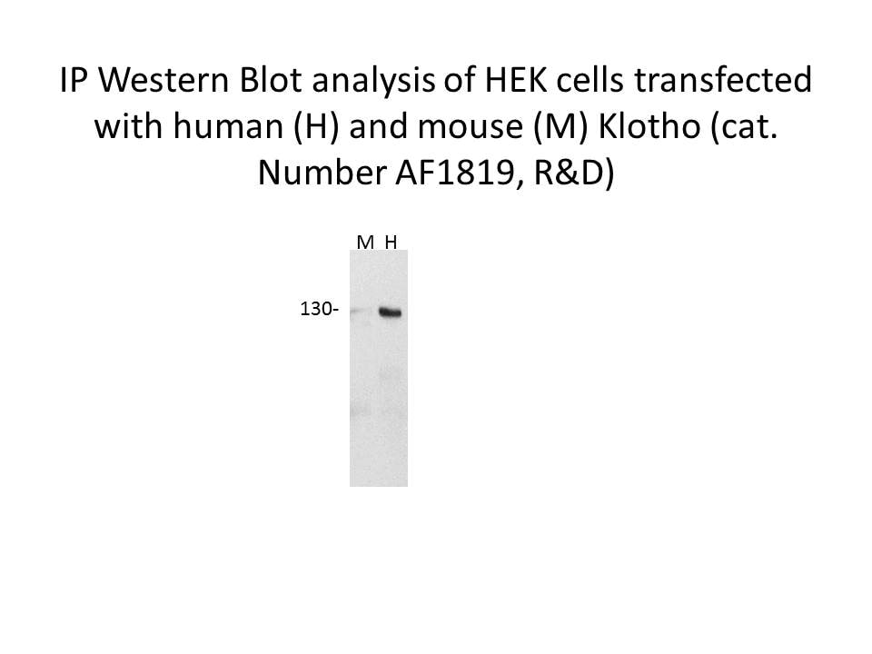
Detection of Human Klotho by Western Blot
Ectopic expression of Klotho protects renal mitochondrial homeostasis and inhibits tubular senescence in an accelerated aging mouse model. (a) Renal expression of PGC‐1 alpha mRNA in different groups was assessed by real‐time PCR. *p < .05 versus control mice; †p < .05 versus d‐gal‐treated mice (n = 5–6). (b) Representative micrographs show renal expression of PGC‐1 alpha. Paraffin sections were immunostained for PGC‐1 alpha. Arrows indicate positive staining. Scale bar, 50 µm. (c–h) Representative (c) Western blots and graphical representations of (d) phospho‐PGC‐1 alpha, (e) TFAM, (f) COX1, (g) Cytb, and (h) TOMM20 protein expression levels in different groups are presented. *p < .05 versus control mice; †p < .05 versus d‐gal‐treated mice (n = 5–6). (i) Quantification of renal mitochondrial mass is shown. Mitochondrial mass was determined by the fluorescence intensity of MitoTracker deep red staining normalized to DAPI. *p < .05 versus control mice; †p < .05 versus d‐gal‐treated mice (n = 5–6). (j) Representative micrographs show mitochondrial ROS production and ultrastructure. They were assessed by mitoSOX staining and electron microscopy analyses, respectively. The administration of d‐gal induced mitochondrial ROS production, mitochondrial swelling, and cristae disorganization in renal tubular cells. Arrows indicate positive staining. Ectopic expression of Klotho inhibited mitochondrial ROS production and preserved normal structure of mitochondria. Scale bar in mitoSOX staining, 50 μm. Scale bar in TEM, 1 μm. TEM, transmission electron microscopy. (k) Representative staining micrographs show renal expression of p16INK4A, SA‐ beta ‐gal activity, and gamma H2AX. Paraffin kidney sections were stained with antibodies against p16INK4A and gamma H2AX. Frozen kidney sections were stained for SA‐ beta ‐gal activity. Arrows indicate positive staining. Scale bar, 25 μm. (l–n) Representative (l) Western blots and graphical representations of (m) p16INK4A and (n) p19ARF protein expression levels in kidneys are pr
Detection of Mouse Klotho by Immunocytochemistry/ Immunofluorescence
Klotho-EGFP N614Q is not transported to the cell surface. (A,B) HEK293 cells stably expressing Klotho-EGFP variants were subjected to surface immunofluorescence using an anti-Klotho antibody (red) and imaged via immunofluorescence. (B) Cells processed as in (A) were imaged using higher exposure times to visualize the small amounts of Klotho-EGFP N614Q at the PM. Nuclei were stained with Hoechst 33342 in blue. Single Apotome sections are shown. Arrows indicated PM localization. Scale bar 10 μm. (C,D) Lysates and supernatants of HEK293T cells stably expressing Klotho-EGFP variants were separated by SDS-PAGE followed by Western blotting using Klotho antibody. (D) Quantification of the ratio of mature/total Klotho-EGFP. sKl indicates shed Klotho. n = 4 independent experiments. (E,F) HEK293 cells stably expressing Klotho-EGFP variants were subjected to surface biotinylation, lysed, precipitated with streptavidin beads, separated on SDS-PAGE, and blotted for Klotho and EEA1 as a cytosolic control protein. * indicates Klotho remnant staining from incomplete stripping. (F) Quantification of n = 4 independent experiments from (E). The ratio of surface/total of Klotho-EGFP WT was set to 100, and the ratio of surface/total of Klotho-EGFP N614Q related to that. A Student’s t-test was used. Data are shown as mean ± standard deviation (SD). * denote levels of significance, **** indicating p < 0.0001, ns: non-significant. For full-size blots, see Supplementary Figure S1. Image collected and cropped by CiteAb from the following open publication (https://pubmed.ncbi.nlm.nih.gov/39451260), licensed under a CC-BY license. Not internally tested by R&D Systems.Applications for Mouse Klotho Antibody
ELISA
This antibody functions as an ELISA detection antibody when paired with Rat Anti-Mouse Klotho Monoclonal Antibody (Catalog # MAB1819).
This product is intended for assay development on various assay platforms requiring antibody pairs.
Immunohistochemistry
Sample: Perfusion fixed frozen sections of mouse kidney
Western Blot
Sample: Mouse Kidney Tissue
Reviewed Applications
Read 3 reviews rated 4 using AF1819 in the following applications:
Formulation, Preparation, and Storage
Purification
Reconstitution
Reconstitute at 0.2 mg/mL in sterile PBS. For liquid material, refer to CoA for concentration.
Formulation
Shipping
Stability & Storage
- 12 months from date of receipt, -20 to -70 °C as supplied.
- 1 month, 2 to 8 °C under sterile conditions after reconstitution.
- 6 months, -20 to -70 °C under sterile conditions after reconstitution.
Calculators
Background: Klotho
Additional Klotho Products
Product Documents for Mouse Klotho Antibody
Certificate of Analysis
To download a Certificate of Analysis, please enter a lot or batch number in the search box below.
Note: Certificate of Analysis not available for kit components.
Product Specific Notices for Mouse Klotho Antibody
For research use only
Related Research Areas
Citations for Mouse Klotho Antibody
 Powered by Bioz
Customer Reviews for Mouse Klotho Antibody (3)
Have you used Mouse Klotho Antibody?
Submit a review and receive an Amazon gift card!
$25/€18/£15/$25CAN/¥2500 Yen for a review with an image
$10/€7/£6/$10CAN/¥1110 Yen for a review without an image
Submit a review
Customer Images
-
Application: Western BlotSample Tested: HEK293 human embryonic kidney cell lineSpecies: HEK cells and HEK293 cells transfected with mouse klothoVerified Customer | Posted 12/21/2017

-
Application: Western BlotSample Tested: Kidney tissueSpecies: MouseVerified Customer | Posted 12/02/2016
-
Application: Western BlotSample Tested: Adult kidneySpecies: RatVerified Customer | Posted 06/10/2016
There are no reviews that match your criteria.
Protocols
Find general support by application which include: protocols, troubleshooting, illustrated assays, videos and webinars.
- Antigen Retrieval Protocol (PIER)
- Antigen Retrieval for Frozen Sections Protocol
- Appropriate Fixation of IHC/ICC Samples
- Cellular Response to Hypoxia Protocols
- Chromogenic IHC Staining of Formalin-Fixed Paraffin-Embedded (FFPE) Tissue Protocol
- Chromogenic Immunohistochemistry Staining of Frozen Tissue
- ClariTSA™ Fluorophore Kits
- Detection & Visualization of Antibody Binding
- ELISA Sample Preparation & Collection Guide
- ELISA Troubleshooting Guide
- Fluorescent IHC Staining of Frozen Tissue Protocol
- Graphic Protocol for Heat-induced Epitope Retrieval
- Graphic Protocol for the Preparation and Fluorescent IHC Staining of Frozen Tissue Sections
- Graphic Protocol for the Preparation and Fluorescent IHC Staining of Paraffin-embedded Tissue Sections
- Graphic Protocol for the Preparation of Gelatin-coated Slides for Histological Tissue Sections
- How to Run an R&D Systems DuoSet ELISA
- How to Run an R&D Systems Quantikine ELISA
- How to Run an R&D Systems Quantikine™ QuicKit™ ELISA
- IHC Sample Preparation (Frozen sections vs Paraffin)
- Immunofluorescent IHC Staining of Formalin-Fixed Paraffin-Embedded (FFPE) Tissue Protocol
- Immunohistochemistry (IHC) and Immunocytochemistry (ICC) Protocols
- Immunohistochemistry Frozen Troubleshooting
- Immunohistochemistry Paraffin Troubleshooting
- Preparing Samples for IHC/ICC Experiments
- Preventing Non-Specific Staining (Non-Specific Binding)
- Primary Antibody Selection & Optimization
- Protocol for Heat-Induced Epitope Retrieval (HIER)
- Protocol for Making a 4% Formaldehyde Solution in PBS
- Protocol for VisUCyte™ HRP Polymer Detection Reagent
- Protocol for the Preparation & Fixation of Cells on Coverslips
- Protocol for the Preparation and Chromogenic IHC Staining of Frozen Tissue Sections
- Protocol for the Preparation and Chromogenic IHC Staining of Frozen Tissue Sections - Graphic
- Protocol for the Preparation and Chromogenic IHC Staining of Paraffin-embedded Tissue Sections
- Protocol for the Preparation and Chromogenic IHC Staining of Paraffin-embedded Tissue Sections - Graphic
- Protocol for the Preparation and Fluorescent IHC Staining of Frozen Tissue Sections
- Protocol for the Preparation and Fluorescent IHC Staining of Paraffin-embedded Tissue Sections
- Protocol for the Preparation of Gelatin-coated Slides for Histological Tissue Sections
- Quantikine HS ELISA Kit Assay Principle, Alkaline Phosphatase
- Quantikine HS ELISA Kit Principle, Streptavidin-HRP Polymer
- R&D Systems Quality Control Western Blot Protocol
- Sandwich ELISA (Colorimetric) – Biotin/Streptavidin Detection Protocol
- Sandwich ELISA (Colorimetric) – Direct Detection Protocol
- TUNEL and Active Caspase-3 Detection by IHC/ICC Protocol
- The Importance of IHC/ICC Controls
- Troubleshooting Guide: ELISA
- Troubleshooting Guide: Immunohistochemistry
- Troubleshooting Guide: Western Blot Figures
- Western Blot Conditions
- Western Blot Protocol
- Western Blot Protocol for Cell Lysates
- Western Blot Troubleshooting
- Western Blot Troubleshooting Guide
- View all Protocols, Troubleshooting, Illustrated assays and Webinars