TIM-1 (T cell-immunoglobulin-mucin; also KIM-1 and HAVcr-1) is a 100 kDa, type I transmembrane glycoprotein member of the TIM family of immunoglobulin superfamily molecules (1-3). This gene family is involved in the regulation of Th1 and Th2-cell-mediated immunity. Human TIM-1 is synthesized as a 359 amino acid (aa) precursor that contains a 20 aa signal sequence, a 270 aa extracellular domain (ECD), a 21 aa transmembrane segment and a 48 aa cytoplasmic domain (4-6). The ECD contains oneV-type Ig-like domain and a mucin region characterized by multiple PTTTTL motifs. The mucin region undergoes extensive O-linked glycosylation. The TIM-1 gene is highly polymorphic and undergoes alternate splicing (1). For instance, the presence of a six aa sequence (MTTTVP) at position #137 of the mature molecule is associated with protection from atopy in people with a history of hepatitis A (7, 8). There are two cytoplasmic alternate splice forms of
TIM‑1. One is a long (359 aa) kidney form termed TIM-1b, and one is a short (334 aa) liver form termed TIM-1a. Both are identical through the first 323 aa of their precursors. TIM-1b contains a tyrosine phosphorylation motif that is not present in 1a (6). TIM-1 is also known to circulate as a soluble form. Constitutive cleavage by an undefined MMP (possibly ADAM33) releases an 85 - 90 kDa soluble molecule (6). The ECD of human TIM-1 is 50% and 43% aa identical to mouse and canine TIM-1 ECD, respectively. The only two reported ligands for TIM-1 are TIM-4 and the hepatitis A virus (4, 9). However, others are believed to exist, and based on the ligand for TIM-3, one may well be an S-type lectin (10). TIM-1 ligation induces T cell proliferation and promotes cytokine production (1, 10).
Human TIM-1/KIM-1/HAVCR Antibody (219211)
R&D Systems | Catalog # MAB1750

Key Product Details
Validated by
Species Reactivity
Validated:
Cited:
Applications
Validated:
Cited:
Label
Antibody Source
Product Specifications
Immunogen
Ser21-Thr288
Accession # AAC39862
Specificity
Clonality
Host
Isotype
Scientific Data Images for Human TIM-1/KIM-1/HAVCR Antibody (219211)
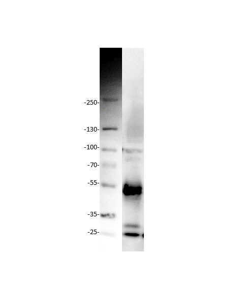
Detection of Human TIM‑1/KIM‑1/HAVCR by Western Blot.
Western blot shows lysates of human CD4+cells treated (+) with 5 µg/mL of Hamster Anti-Mouse CD3e Monoclonal Antibody (MAB484) and 1 µg/mL of Rat Anti-Mouse CD28 Monoclonal Antibody (MAB4831) for 24 hours. PVDF membrane was probed with 1 µg/mL of Mouse Anti-Human TIM-1/KIM-1/HAVCR Monoclonal Antibody (Catalog # MAB1750) followed by HRP-conjugated Anti-Mouse IgG Secondary Antibody (HAF007). A specific band was detected for TIM-1/KIM-1/HAVCR at approximately 80 kDa (as indicated). This experiment was conducted under reducing conditions and using Immunoblot Buffer Group 1.
Detection of TIM‑1/KIM‑1/HAVCR in Th2-stimulated Human PBMCs by Flow Cytometry.
(A) Unstimulated and (B) Th2-stimulated human PBMCs were stained with Mouse Anti-Human TIM-1/KIM-1/HAVCR Monoclonal Antibody (Catalog # MAB1750) followed by Allophycocyanin-conjugated Anti-Mouse IgG Secondary Antibody (F0101B) and Human CD4 PerCP-conjugated Monoclonal Antibody (FAB3791C). Quadrant markers were set based on control antibody staining (Catalog # MAB0041).
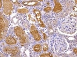
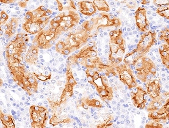
TIM‑1/KIM‑1/HAVCR in Human Kidney.
TIM-1/KIM-1/HAVCR was detected in immersion fixed paraffin-embedded sections of human kidney using 25 µg/mL Mouse Anti-Human TIM-1/ KIM-1/HAVCR Monoclonal Antibody (Catalog # MAB1750) overnight at 4 °C. Tissue was stained with the Anti-Mouse HRP-DAB Cell & Tissue Staining Kit (brown; CTS002) and counterstained with hematoxylin (blue). View our protocol for Chromogenic IHC Staining of Paraffin-embedded Tissue Sections.
Detection of TIM‑1/KIM‑1/HAVCR in Huh‑7 human hepatoma cell line by Flow Cytometry
Huh‑7 human hepatoma cell line were stained with Mouse Anti-Human TIM‑1/KIM‑1/HAVCR Monoclonal Antibody (Catalog # MAB1750, filled histogram) or isotype control antibody (Catalog # MAB0041, open histogram) followed by Allophycocyanin-conjugated Anti-Mouse IgG Secondary Antibody (Catalog # F0101B). View our protocol for Staining Membrane-associated Proteins.Applications for Human TIM-1/KIM-1/HAVCR Antibody (219211)
CyTOF-ready
Flow Cytometry
Sample: stimulated Human CD4+ cells
Immunohistochemistry
Sample: Immersion fixed paraffin-embedded sections of human kidney
Western Blot
Sample: Human CD4+ cells treated with Hamster Anti-Mouse CD3 epsilon Monoclonal Antibody (Catalog # MAB484) and Rat Anti-Mouse CD28 Monoclonal Antibody (Catalog # MAB4831)
Reviewed Applications
Read 4 reviews rated 4.8 using MAB1750 in the following applications:
Flow Cytometry Panel Builder
Bio-Techne Knows Flow Cytometry
Save time and reduce costly mistakes by quickly finding compatible reagents using the Panel Builder Tool.
Advanced Features
- Spectra Viewer - Custom analysis of spectra from multiple fluorochromes
- Spillover Popups - Visualize the spectra of individual fluorochromes
- Antigen Density Selector - Match fluorochrome brightness with antigen density

Formulation, Preparation, and Storage
Purification
Reconstitution
Reconstitute at 0.5 mg/mL in sterile PBS. For liquid material, refer to CoA for concentration.
Formulation
*Small pack size (-SP) is supplied either lyophilized or as a 0.2 µm filtered solution in PBS.
Shipping
Stability & Storage
- 12 months from date of receipt, -20 to -70 °C as supplied.
- 1 month, 2 to 8 °C under sterile conditions after reconstitution.
- 6 months, -20 to -70 °C under sterile conditions after reconstitution.
Calculators
Background: TIM-1/KIM-1/HAVCR
References
- Meyers, J.H. et al. (2005) Trends Mol. Med. 11:1471.
- Kuchroo, V.K. et al. (2003) Nat. Rev. Immunol. 3:454.
- Mariat, C. et al. (2005) Phil. Trans. R. Soc. B 360:1681.
- Feigelstock, D. et al. (1998) J. Virol. 72:6621.
- Ichimura, T. et al. (1998) J. Biol. Chem. 273:4135.
- Bailly, V. et al. (2002) J. Biol. Chem. 277:39739.
- Umetsu, D.T. et al. (2005) J. Pediatr. Gastroenterol. Nutr. 40:S43.
- Gao, P-S. et al. (2005) J. Allergy Clin. Immunol. 115:982.
- Zhu, C. et al. (2005) Nat. Immunol. 6:1245.
- Meyers, J.H. et al. (2005) Nat. Immunol. 6:455.
Long Name
Alternate Names
Gene Symbol
UniProt
Additional TIM-1/KIM-1/HAVCR Products
Product Documents for Human TIM-1/KIM-1/HAVCR Antibody (219211)
Certificate of Analysis
To download a Certificate of Analysis, please enter a lot or batch number in the search box below.
Note: Certificate of Analysis not available for kit components.
Product Specific Notices for Human TIM-1/KIM-1/HAVCR Antibody (219211)
This product is covered by one or more of the following US Patents 7,300,652; 7,041,290; 6,664,385 and other US and foreign patents pending or issued.
For research use only
Related Research Areas
Citations for Human TIM-1/KIM-1/HAVCR Antibody (219211)
 Powered by Bioz
Customer Reviews for Human TIM-1/KIM-1/HAVCR Antibody (219211) (4)
Have you used Human TIM-1/KIM-1/HAVCR Antibody (219211)?
Submit a review and receive an Amazon gift card!
$25/€18/£15/$25CAN/¥2500 Yen for a review with an image
$10/€7/£6/$10CAN/¥1110 Yen for a review without an image
Submit a review
Customer Images

-
Application: Western BlotSample Tested: A549 human lung carcinoma cell line and HEK293T human embryonic kidney cell lineSpecies: HumanVerified Customer | Posted 04/08/2022

-
Application: ImmunohistochemistrySample Tested: Kidney tissueSpecies: HumanVerified Customer | Posted 11/22/2021
-
Application: Western BlotSample Tested: B cellsSpecies: HumanVerified Customer | Posted 11/08/2021
-
Application: ImmunohistochemistrySample Tested: Kidney tissueSpecies: HumanVerified Customer | Posted 08/27/2021
There are no reviews that match your criteria.
Protocols
Find general support by application which include: protocols, troubleshooting, illustrated assays, videos and webinars.
- 7-Amino Actinomycin D (7-AAD) Cell Viability Flow Cytometry Protocol
- Antigen Retrieval Protocol (PIER)
- Antigen Retrieval for Frozen Sections Protocol
- Appropriate Fixation of IHC/ICC Samples
- Cellular Response to Hypoxia Protocols
- Chromogenic IHC Staining of Formalin-Fixed Paraffin-Embedded (FFPE) Tissue Protocol
- Chromogenic Immunohistochemistry Staining of Frozen Tissue
- ClariTSA™ Fluorophore Kits
- Detection & Visualization of Antibody Binding
- Extracellular Membrane Flow Cytometry Protocol
- Flow Cytometry Protocol for Cell Surface Markers
- Flow Cytometry Protocol for Staining Membrane Associated Proteins
- Flow Cytometry Staining Protocols
- Flow Cytometry Troubleshooting Guide
- Fluorescent IHC Staining of Frozen Tissue Protocol
- Graphic Protocol for Heat-induced Epitope Retrieval
- Graphic Protocol for the Preparation and Fluorescent IHC Staining of Frozen Tissue Sections
- Graphic Protocol for the Preparation and Fluorescent IHC Staining of Paraffin-embedded Tissue Sections
- Graphic Protocol for the Preparation of Gelatin-coated Slides for Histological Tissue Sections
- IHC Sample Preparation (Frozen sections vs Paraffin)
- Immunofluorescent IHC Staining of Formalin-Fixed Paraffin-Embedded (FFPE) Tissue Protocol
- Immunohistochemistry (IHC) and Immunocytochemistry (ICC) Protocols
- Immunohistochemistry Frozen Troubleshooting
- Immunohistochemistry Paraffin Troubleshooting
- Intracellular Flow Cytometry Protocol Using Alcohol (Methanol)
- Intracellular Flow Cytometry Protocol Using Detergents
- Intracellular Nuclear Staining Flow Cytometry Protocol Using Detergents
- Intracellular Staining Flow Cytometry Protocol Using Alcohol Permeabilization
- Intracellular Staining Flow Cytometry Protocol Using Detergents to Permeabilize Cells
- Preparing Samples for IHC/ICC Experiments
- Preventing Non-Specific Staining (Non-Specific Binding)
- Primary Antibody Selection & Optimization
- Propidium Iodide Cell Viability Flow Cytometry Protocol
- Protocol for Heat-Induced Epitope Retrieval (HIER)
- Protocol for Liperfluo
- Protocol for Making a 4% Formaldehyde Solution in PBS
- Protocol for VisUCyte™ HRP Polymer Detection Reagent
- Protocol for the Characterization of Human Th22 Cells
- Protocol for the Characterization of Human Th9 Cells
- Protocol for the Preparation & Fixation of Cells on Coverslips
- Protocol for the Preparation and Chromogenic IHC Staining of Frozen Tissue Sections
- Protocol for the Preparation and Chromogenic IHC Staining of Frozen Tissue Sections - Graphic
- Protocol for the Preparation and Chromogenic IHC Staining of Paraffin-embedded Tissue Sections
- Protocol for the Preparation and Chromogenic IHC Staining of Paraffin-embedded Tissue Sections - Graphic
- Protocol for the Preparation and Fluorescent IHC Staining of Frozen Tissue Sections
- Protocol for the Preparation and Fluorescent IHC Staining of Paraffin-embedded Tissue Sections
- Protocol for the Preparation of Gelatin-coated Slides for Histological Tissue Sections
- Protocol: Annexin V and PI Staining by Flow Cytometry
- Protocol: Annexin V and PI Staining for Apoptosis by Flow Cytometry
- R&D Systems Quality Control Western Blot Protocol
- TUNEL and Active Caspase-3 Detection by IHC/ICC Protocol
- The Importance of IHC/ICC Controls
- Troubleshooting Guide: Fluorokine Flow Cytometry Kits
- Troubleshooting Guide: Immunohistochemistry
- Troubleshooting Guide: Western Blot Figures
- Western Blot Conditions
- Western Blot Protocol
- Western Blot Protocol for Cell Lysates
- Western Blot Troubleshooting
- Western Blot Troubleshooting Guide
- View all Protocols, Troubleshooting, Illustrated assays and Webinars