Insulin is a peptide hormone that facilitates the cellular uptake of glucose by regulating the appearance of membrane glucose transporters. The single chain insulin propeptide consists of a 30 amino acid B chain (aa 25-54), a C-Peptide (aa 55-89), and a 21 aa A chain (aa 90-110). Removal of the C-Peptide by proteolysis enables the formation of mature Insulin, a disulfide-linked heterodimer of the A and B chains. Circulating C-peptide levels are elevated in hyperinsulinism, obesity, and type II diabetes. The human C-Peptide shares 61% and 68% aa sequence identity with mouse and rat C-Peptide, respectively.
Human C-Peptide Antibody (790904)
R&D Systems | Catalog # MAB14171

Key Product Details
Species Reactivity
Human
Applications
Immunohistochemistry, Immunocytochemistry
Label
Unconjugated
Antibody Source
Monoclonal Mouse IgG2B Clone # 790904
Loading...
Product Specifications
Immunogen
Human C-Peptide synthetic peptide
EAEDLQVGQVELGGGPGAGSLQPLALEGSLQ
Accession # P01308
EAEDLQVGQVELGGGPGAGSLQPLALEGSLQ
Accession # P01308
Specificity
Detects human C-Peptide in direct ELISAs.
Clonality
Monoclonal
Host
Mouse
Isotype
IgG2B
Scientific Data Images for Human C-Peptide Antibody (790904)
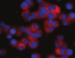
C-Peptide in BG01V Human Embryonic Stem Cells.
C-Peptide was detected in immersion fixed BG01V human embryonic stem cells differentiated into pancreatic beta cells using Mouse Anti-Human C-Peptide Monoclonal Antibody (Catalog # MAB14171) at 10 µg/mL for 3 hours at room temperature. Cells were stained using the NorthernLights™ 557-conjugated Anti-Mouse IgG Secondary Antibody (red; Catalog # NL007) and counterstained with DAPI (blue). Specific staining was localized to cytoplasm. View our protocol for Fluorescent ICC Staining of Stem Cells on Coverslips.
C-Peptide in Human Pancreas.
C-Peptide was detected in immersion fixed paraffin-embedded sections of human pancreas using Mouse Anti-Human C-Peptide Monoclonal Antibody (Catalog # MAB14171) at 15 µg/mL overnight at 4 °C. Before incubation with the primary antibody, tissue was subjected to heat-induced epitope retrieval using Antigen Retrieval Reagent-Basic (Catalog # CTS013). Tissue was stained using the Anti-Mouse HRP-DAB Cell & Tissue Staining Kit (brown; Catalog # CTS002) and counter-stained with hematoxylin (blue). Specific staining was localized to the cytoplasm of islet cells. View our protocol for Chromogenic IHC Staining of Paraffin-embedded Tissue Sections.
Immunofluorescent Staining of iPSC-derived Beta Islets.
iPSC-derived beta islets were fixed, frozen, and sectioned. Sections were stained with a Mouse Anti-Human C-peptide Monoclonal Antibody (Catalog # MAB14171), and a Rat Anti-Human/Mouse Somatostatin Monoclonal Antibody (Catalog # MAB2358), followed by secondary antibody staining with the NorthernLights NL493-conjugated Donkey Anti-Mouse IgG Antigen Affinity-purified Polyclonal Antibody (Catalog # NL009; green) and NorthernLights NL557-conjugated Goat Anti-Rat IgG Antigen Affinity-purified Polyclonal Antibody (Catalog # NL013; red). Cell nuclei were stained with DAPI (Catalog # 5748; blue) and the images were overlaid.Applications for Human C-Peptide Antibody (790904)
Application
Recommended Usage
Immunocytochemistry
8-25 µg/mL
Sample: Immersion fixed BG01V human embryonic stem cells differentiated into pancreatic beta cells
Sample: Immersion fixed BG01V human embryonic stem cells differentiated into pancreatic beta cells
Immunohistochemistry
8-25 µg/mL
Sample: Immersion fixed paraffin-embedded sections of human pancreas
Sample: Immersion fixed paraffin-embedded sections of human pancreas
Reviewed Applications
Read 1 review rated 5 using MAB14171 in the following applications:
Formulation, Preparation, and Storage
Purification
Protein A or G purified from hybridoma culture supernatant
Reconstitution
Sterile PBS to a final concentration of 0.5 mg/mL. For liquid material, refer to CoA for concentration.
Loading...
Formulation
Lyophilized from a 0.2 μm filtered solution in PBS with Trehalose. *Small pack size (SP) is supplied either lyophilized or as a 0.2 µm filtered solution in PBS.
Shipping
Lyophilized product is shipped at ambient temperature. Liquid small pack size (-SP) is shipped with polar packs. Upon receipt, store immediately at the temperature recommended below.
Stability & Storage
Use a manual defrost freezer and avoid repeated freeze-thaw cycles.
- 12 months from date of receipt, -20 to -70 °C as supplied.
- 1 month, 2 to 8 °C under sterile conditions after reconstitution.
- 6 months, -20 to -70 °C under sterile conditions after reconstitution.
Calculators
Background: C-Peptide
Long Name
Proinsulin C-Peptide
Alternate Names
C-P, Insulin C-Peptide, Proinsulin C-Peptide
Gene Symbol
INS
UniProt
Additional C-Peptide Products
Product Documents for Human C-Peptide Antibody (790904)
Certificate of Analysis
To download a Certificate of Analysis, please enter a lot or batch number in the search box below.
Note: Certificate of Analysis not available for kit components.
Product Specific Notices for Human C-Peptide Antibody (790904)
For research use only
Citations for Human C-Peptide Antibody (790904)
 Powered by Bioz
Customer Reviews for Human C-Peptide Antibody (790904) (1)
5 out of 5
1 Customer Rating
Have you used Human C-Peptide Antibody (790904)?
Submit a review and receive an Amazon gift card!
$25/€18/£15/$25CAN/¥2500 Yen for a review with an image
$10/€7/£6/$10CAN/¥1110 Yen for a review without an image
Submit a review
Customer Images
Showing
1
-
1 of
1 review
Showing All
Filter By:
-
Application: Immunocytochemistry/ImmunofluorescenceSample Tested: Human bone marrow-derived stem cellsSpecies: HumanVerified Customer | Posted 11/19/2021

There are no reviews that match your criteria.
Protocols
Find general support by application which include: protocols, troubleshooting, illustrated assays, videos and webinars.
- Antigen Retrieval Protocol (PIER)
- Antigen Retrieval for Frozen Sections Protocol
- Appropriate Fixation of IHC/ICC Samples
- Cellular Response to Hypoxia Protocols
- Chromogenic IHC Staining of Formalin-Fixed Paraffin-Embedded (FFPE) Tissue Protocol
- Chromogenic Immunohistochemistry Staining of Frozen Tissue
- ClariTSA™ Fluorophore Kits
- Detection & Visualization of Antibody Binding
- Fluorescent IHC Staining of Frozen Tissue Protocol
- Graphic Protocol for Heat-induced Epitope Retrieval
- Graphic Protocol for the Preparation and Fluorescent IHC Staining of Frozen Tissue Sections
- Graphic Protocol for the Preparation and Fluorescent IHC Staining of Paraffin-embedded Tissue Sections
- Graphic Protocol for the Preparation of Gelatin-coated Slides for Histological Tissue Sections
- ICC Cell Smear Protocol for Suspension Cells
- ICC Immunocytochemistry Protocol Videos
- ICC for Adherent Cells
- IHC Sample Preparation (Frozen sections vs Paraffin)
- Immunocytochemistry (ICC) Protocol
- Immunocytochemistry Troubleshooting
- Immunofluorescence of Organoids Embedded in Cultrex Basement Membrane Extract
- Immunofluorescent IHC Staining of Formalin-Fixed Paraffin-Embedded (FFPE) Tissue Protocol
- Immunohistochemistry (IHC) and Immunocytochemistry (ICC) Protocols
- Immunohistochemistry Frozen Troubleshooting
- Immunohistochemistry Paraffin Troubleshooting
- Preparing Samples for IHC/ICC Experiments
- Preventing Non-Specific Staining (Non-Specific Binding)
- Primary Antibody Selection & Optimization
- Protocol for Heat-Induced Epitope Retrieval (HIER)
- Protocol for Making a 4% Formaldehyde Solution in PBS
- Protocol for VisUCyte™ HRP Polymer Detection Reagent
- Protocol for the Fluorescent ICC Staining of Cell Smears - Graphic
- Protocol for the Fluorescent ICC Staining of Cultured Cells on Coverslips - Graphic
- Protocol for the Preparation & Fixation of Cells on Coverslips
- Protocol for the Preparation and Chromogenic IHC Staining of Frozen Tissue Sections
- Protocol for the Preparation and Chromogenic IHC Staining of Frozen Tissue Sections - Graphic
- Protocol for the Preparation and Chromogenic IHC Staining of Paraffin-embedded Tissue Sections
- Protocol for the Preparation and Chromogenic IHC Staining of Paraffin-embedded Tissue Sections - Graphic
- Protocol for the Preparation and Fluorescent ICC Staining of Cells on Coverslips
- Protocol for the Preparation and Fluorescent ICC Staining of Non-adherent Cells
- Protocol for the Preparation and Fluorescent ICC Staining of Stem Cells on Coverslips
- Protocol for the Preparation and Fluorescent IHC Staining of Frozen Tissue Sections
- Protocol for the Preparation and Fluorescent IHC Staining of Paraffin-embedded Tissue Sections
- Protocol for the Preparation of Gelatin-coated Slides for Histological Tissue Sections
- Protocol for the Preparation of a Cell Smear for Non-adherent Cell ICC - Graphic
- TUNEL and Active Caspase-3 Detection by IHC/ICC Protocol
- The Importance of IHC/ICC Controls
- Troubleshooting Guide: Immunohistochemistry
- View all Protocols, Troubleshooting, Illustrated assays and Webinars
Loading...