RANTES (Regulated upon Activation, Normal T cell Expressed and presumably Secreted) is a member of the beta (C-C) chemokine subfamily and is now designated CCL5. It binds and activates the chemokine receptors CCR1, 3 and 5.

Key Product Details
Validated by
Biological Validation
Species Reactivity
Validated:
Human
Cited:
Human, Mouse, Primate - Macaca mulatta (Rhesus Macaque)
Applications
Validated:
Immunohistochemistry, Western Blot, Neutralization, Immunocytochemistry, Simple Western
Cited:
Immunohistochemistry, Immunohistochemistry-Paraffin, Immunohistochemistry-Frozen, Western Blot, Neutralization, Flow Cytometry, Immunocytochemistry, Immunoprecipitation, Bioassay
Label
Unconjugated
Antibody Source
Polyclonal Goat IgG
Loading...
Product Specifications
Immunogen
E. coli-derived recombinant human CCL5/RANTES
Ser24-Ser91
Accession # P13501
Ser24-Ser91
Accession # P13501
Specificity
Detects human CCL5/RANTES in direct ELISAs. Detects human CCL5/RANTES and mouse CCL5/RANTES Western blots. In direct ELISAs, approximately 40% cross-reactivity with recombinant canine CCL5/RANTES is observed and approximately 20% cross-reactivity with recombinant cotton rat CCL5/RANTES is observed. In Western blots, no cross-reactivity with recombinant human (rh) CCL3 and rhCCL4 is observed.
Clonality
Polyclonal
Host
Goat
Isotype
IgG
Endotoxin Level
<0.10 EU per 1 μg of the antibody by the LAL method.
Scientific Data Images for Human CCL5/RANTES Antibody
Detection of Recombinant Human and Mouse CCL5/RANTES by Western Blot.
Western blot shows 25 ng of Recombinant Human CCL5/RANTES (278-RN), Recombinant Mouse CCL5/RANTES (478-MR), Recombinant Human CCL3/MIP-1a Isoform LD78a (270-LD), and Recombinant Human CCL4/MIP-1 beta (271-BME). PVDF Membrane was probed with 0.1 µg/mL of Goat Anti-Human CCL5/RANTES Antigen Affinity-purified Polyclonal Antibody (Catalog # AF-278-NA) followed by HRP-conjugated Anti-Goat IgG Secondary Antibody (HAF109). A specific band was detected for CCL5/RANTES at approximately 10 kDa (as indicated). This experiment was conducted under reducing conditions and using Immunoblot Buffer Group 3.
CCL5/RANTES in Human PBMCs.
CCL5/RANTES was detected in immersion fixed human peripheral blood mononuclear cells (PBMCs) using Goat Anti-Human CCL5/RANTES Antigen Affinity-purified Polyclonal Antibody (Catalog # AF-278-NA) at 15 µg/mL for 3 hours at room temperature. Cells were stained using the NorthernLights™ 557-conjugated Anti-Goat IgG Secondary Antibody (orange; NL001) and counterstained with DAPI (blue). Specific staining was localized to cytoplasm. View our protocol for Fluorescent ICC Staining of Non-adherent Cells.
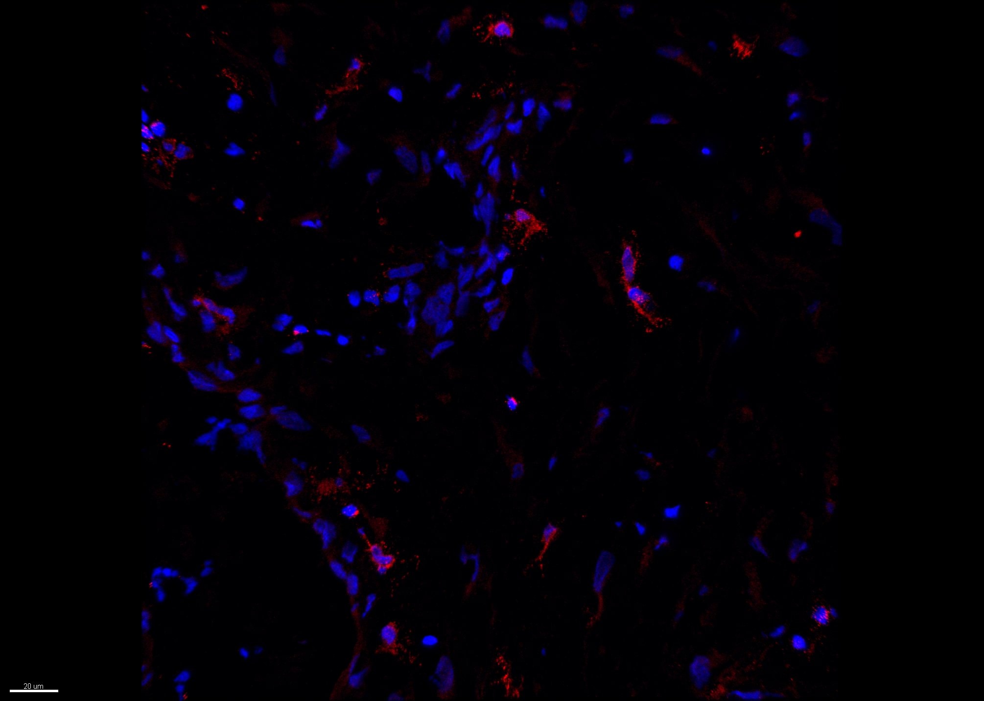
CCL5/RANTES in Human Tonsil.
CCL5/RANTES was detected in immersion fixed paraffin-embedded sections of human tonsil using Goat Anti-Human CCL5/RANTES Antigen Affinity-purified Polyclonal Antibody (Catalog # AF-278-NA) at 10 µg/mL overnight at 4 °C. Tissue was stained using the NorthernLights™ 557-conjugated Anti-Goat IgG Secondary Antibody (red; NL001) and counterstained with DAPI (blue). View our protocol for Chromogenic IHC Staining of Paraffin-embedded Tissue Sections.
Detection of Human CCL5/RANTES by Simple WesternTM.
Simple Western lane view shows lysates of THP-1 human acute monocytic leukemia cell line untreated (-) or treated (+) with 200nM PMA for 24 hrs and 10ug/mL LPS for 3 hrs, loaded at 0.2 mg/mL. A specific band was detected for CCL5/RANTES at approximately 7 kDa (as indicated) using 10 µg/mL of Goat Anti-Human CCL5/RANTES Antigen Affinity-purified Polyclonal Antibody (Catalog # AF-278-NA) followed by 1:50 dilution of HRP-conjugated Anti-Goat IgG Secondary Antibody (HAF109). This experiment was conducted under reducing conditions and using the 2-40 kDa separation system.
Chemotaxis Induced by CCL5/RANTES and Neutralization by Human CCL5/RANTES Antibody.
Recombinant Human CCL5/RANTES (Catalog # 278-RN) chemoattracts the BaF3 mouse pro-B cell line transfected with human CCR5 in a dose-dependent manner (orange line). The amount of cells that migrated through to the lower chemotaxis chamber was measured by Resazurin (AR002). Chemotaxis elicited by Recombinant Human CCL5/RANTES (0.01 µg/mL) is neutralized (green line) by increasing concentrations of Goat Anti-Human CCL5/RANTES Antigen Affinity-purified Polyclonal Antibody (Catalog # AF-278-NA). The ND50 is typically 0.1‑0.4 µg/mL.
Detection of CCL5/RANTES in Human Tonsil.
CCL5/RANTES was detected in immersion fixed paraffin-embedded sections of Human Tonsil using Goat Anti-Human CCL5/RANTES Antigen Affinity-purified Polyclonal Antibody (Catalog # AF-278-NA) at 15 µg/mL for 1 hour at room temperature followed by incubation with the Anti-Goat IgG VisUCyte™ HRP Polymer Antibody (Catalog # VC004). Before incubation with the primary antibody, tissue was subjected to heat-induced epitope retrieval using VisUCyte Antigen Retrieval Reagent-Basic (Catalog # VCTS021). Tissue was stained using DAB (brown) and counterstained with hematoxylin (blue). Specific staining was localized to cytoplasm in lymphocytes. View our protocol for IHC Staining with VisUCyte HRP Polymer Detection Reagents.
Detection of Human CCL5/RANTES by Immunohistochemistry
CCL5-CCR5 axis induced aerobic glycolysis by regulation of AMPK signaling(A) Western blot for AMPK, c-Myc, HIF-1 alpha and Akt in breast cancer cells co-cultured with 15mM lactic acid-activated THP-1 macrophages (ratio 1:1) for 72 h. Results presented were representatives of at least three independent experiments. (B) The expression of AMPK downstream signaling target ACC in breast cancer cells co-cultured as in (A). (C) MDA-MB-231 and MCF-7 cells were transfected with 50 nM AMPK alpha 1 siRNA, or pretreated with 10μM compound C for 4 h, and then incubated with 15mM lactic acid-activated THP-1 macrophages (ratio 1:1) for 48 h. The glucose uptake, lactic acid production and ATP levels were detected. (D) The inhibition of AMPK abrogated macrophage-induced EMT in MCF-7 cells. Cells were treated as described in (C). After co-culture, the expression of EMT markers, E-cadherin and vimentin, was measured by western blot. (E) Recombinant human CCL5 induced the phosphorylation of AMPK in MDA-MB-231 and MCF-7/CCR5 cells. 106 cells were treated with 50ng/ml CCL5 for defferent time points as indicated, and phosphorylated AMPK and total AMPK were investigated by western blot. (F) Inhibition of CCR5 in MDA-MB-231 cells significantly attenuated macrophage-induced AMPK phosphorylation. MDA-MB-231 cells were transfected with shRNAs designed against CCR5, or pre-treated with 5μM Maraviroc for 2 h, then co-cultured with 15 mM lactate-activated macrophages as described in (A). After co-culture, the phosphorylation of AMPK was detected by western blot. (G) Expressions of CCL5, CCR5 and p-AMPK in samples obtained from breast cancer patients (n =28). Scale bars represent 50 μm. *, P<0.05; **, P<0.01. Image collected and cropped by CiteAb from the following open publication (https://www.oncotarget.com/lookup/doi/10.18632/oncotarget.22786), licensed under a CC-BY license. Not internally tested by R&D Systems.
Detection of Human CCL5/RANTES by Western Blot
Lactate-activated macrophages induced glycolysis through CCL5-CCR5 axis(A) Glucose uptake, lactic acid production and ATP levels in breast cancer cells co-cultured with lactate-activated THP-1 macrophages, with or without 5μg/ml anti-CCL5 neutralizing antibody. The co-culture system was described in Figure 5C. (B) Western blots for glycolytic enzymes in breast cancer cells treated as in (A). (C) MDA-MB-231 cells were transfected with shRNAs designed against CCR5, or pre-treated with 5μM Maraviroc for 2 h, and then subjected to cell co-culture. Glucose uptake, lactic acid production and ATP levels were measured after co-culture. The co-culture system was described in Figure 5C. (D) The protein levels of HK2, PKM2 and LDHA in MDA-MB-231 cells cultured as in (C). (E) Recombinant human CCL5 induced aerobic glycolysis in breast cancer cells. MDA-MB-231 and MCF-7/CCR5 cells were treated with increasing concentrations of CCL5 for 12 h, and glucose uptake, lactic acid production and ATP levels were detected. (F) Western blots for glycolytic enzymes in MDA-MB-231 and MCF-7/CCR5 cells after stimulation with CCL5. *, P<0.05; **, P<0.01. Image collected and cropped by CiteAb from the following open publication (https://www.oncotarget.com/lookup/doi/10.18632/oncotarget.22786), licensed under a CC-BY license. Not internally tested by R&D Systems.
Detection of Human Human CCL5/RANTES Antibody by Immunohistochemistry
Lactic acid induced the secretion of CCL5 in human macrophages(A) 3×10⁵ THP-1 macrophages were treated with 15 mM lactate for 24 h, and the mRNA levels of chemokines were measured by quantitative PCR. The growth medium of control macrophages was titrated to pH6.1 using sterile HCl. (B) 3×10⁵ THP-1 macrophages were incubated with different concentrations of lactate for 24 h, and CCL5 gene expression was determined with quantitative PCR. (C) 106 THP-1 macrophages were exposed to increasing concentrations of lactate for 48 h, and the secretion of CCL5 was measured by ELISA. (D) 106 human primary macrophages from breast cancer patients (n=9) were cultured with different concentrations of lactate for 48 h, and CCL5 production was detected. (E) 106 MDA-MB-231 cells were pre-treated with 15μM GSK 2837808A for 2 h, then the media were changed, and cells were cultured for another 24 h. The conditional media (MD-231 CM) were collected and applied to 106 THP-1 macrophages. CCL5 concentrations were detected with ELISA. (F) Immunohistochemical staining of CD68 and CCL5 in tumor adjacent tissues (control) and breast tumors (n=28). Scale bars represent 50 μm. *, P<0.05; **, P<0.01. Image collected and cropped by CiteAb from the following publication (https://pubmed.ncbi.nlm.nih.gov/29299159), licensed under a CC-BY license. Not internally tested by R&D Systems.
Human CCL5 / RANTES ELISA Standard Curve
Recombinant Human CCL5/RANTES (Catalog # 278-RN) was serially diluted and captured by Mouse Anti-Human/Primate CCL5/RANTES Monoclonal Antibody (Catalog # MAB678) coated on a Clear Polystyrene Microplate (Catalog # DY990). Goat Anti-Human CCL5/RANTES Antigen Affinity-purified Polyclonal Antibody (Catalog # AF-278-NA) was biotinylated and incubated with the protein captured on the plate. Detection of the standard curve was achieved by incubating Streptavidin-HRP (Catalog # DY998)Applications for Human CCL5/RANTES Antibody
Application
Recommended Usage
Immunocytochemistry
5-15 µg/mL
Sample: Immersion fixed human peripheral blood mononuclear cells (PBMC)
Sample: Immersion fixed human peripheral blood mononuclear cells (PBMC)
Immunohistochemistry
5-15 µg/mL
Sample: Immersion fixed paraffin-embedded sections of human lymphoma and human tonsil
Sample: Immersion fixed paraffin-embedded sections of human lymphoma and human tonsil
Simple Western
10 µg/mL
Sample: THP‑1 human acute monocytic leukemia cell line treated with PMA and LPS
Sample: THP‑1 human acute monocytic leukemia cell line treated with PMA and LPS
Western Blot
0.1 µg/mL
Sample: Recombinant Human CCL5/RANTES (Catalog # 278-RN)
Sample: Recombinant Human CCL5/RANTES (Catalog # 278-RN)
Neutralization
Measured by its ability to neutralize CCL5/RANTES-induced chemotaxis in the BaF3 mouse pro‑B cell line transfected with human CCR5. The Neutralization Dose (ND50) is typically 0.1-0.4 µg/mL in the presence of 0.01 µg/mL Recombinant Human CCL5/RANTES.
Reviewed Applications
Read 1 review rated 4 using AF-278-NA in the following applications:
Formulation, Preparation, and Storage
Purification
Antigen Affinity-purified
Reconstitution
Reconstitute at 0.2 mg/mL in sterile PBS. For liquid material, refer to CoA for concentration.
Loading...
Formulation
Lyophilized from a 0.2 μm filtered solution in PBS with Trehalose. *Small pack size (SP) is supplied either lyophilized or as a 0.2 µm filtered solution in PBS.
Shipping
Lyophilized product is shipped at ambient temperature. Liquid small pack size (-SP) is shipped with polar packs. Upon receipt, store immediately at the temperature recommended below.
Stability & Storage
Use a manual defrost freezer and avoid repeated freeze-thaw cycles.
- 12 months from date of receipt, -20 to -70 °C as supplied.
- 1 month, 2 to 8 °C under sterile conditions after reconstitution.
- 6 months, -20 to -70 °C under sterile conditions after reconstitution.
Calculators
Background: CCL5/RANTES
Alternate Names
RANTES, SISd
Gene Symbol
CCL5
UniProt
Additional CCL5/RANTES Products
Product Documents for Human CCL5/RANTES Antibody
Certificate of Analysis
To download a Certificate of Analysis, please enter a lot or batch number in the search box below.
Note: Certificate of Analysis not available for kit components.
Product Specific Notices for Human CCL5/RANTES Antibody
For research use only
Citations for Human CCL5/RANTES Antibody
 Powered by Bioz
Customer Reviews for Human CCL5/RANTES Antibody (1)
4 out of 5
1 Customer Rating
Have you used Human CCL5/RANTES Antibody?
Submit a review and receive an Amazon gift card!
$25/€18/£15/$25CAN/¥2500 Yen for a review with an image
$10/€7/£6/$10CAN/¥1110 Yen for a review without an image
Submit a review
Customer Images
Showing
1
-
1 of
1 review
Showing All
Filter By:
-
Application: Immunofluorescence-FrozenSample Tested: human melanomaSpecies: HumanVerified Customer | Posted 11/15/2018Human melanoma stained for CCL5 (red) counterstained with DAPI (blue)Acetone fixation

There are no reviews that match your criteria.
Protocols
Find general support by application which include: protocols, troubleshooting, illustrated assays, videos and webinars.
- Antigen Retrieval Protocol (PIER)
- Antigen Retrieval for Frozen Sections Protocol
- Appropriate Fixation of IHC/ICC Samples
- Cellular Response to Hypoxia Protocols
- Chromogenic IHC Staining of Formalin-Fixed Paraffin-Embedded (FFPE) Tissue Protocol
- Chromogenic Immunohistochemistry Staining of Frozen Tissue
- ClariTSA™ Fluorophore Kits
- Detection & Visualization of Antibody Binding
- Fluorescent IHC Staining of Frozen Tissue Protocol
- Graphic Protocol for Heat-induced Epitope Retrieval
- Graphic Protocol for the Preparation and Fluorescent IHC Staining of Frozen Tissue Sections
- Graphic Protocol for the Preparation and Fluorescent IHC Staining of Paraffin-embedded Tissue Sections
- Graphic Protocol for the Preparation of Gelatin-coated Slides for Histological Tissue Sections
- ICC Cell Smear Protocol for Suspension Cells
- ICC Immunocytochemistry Protocol Videos
- ICC for Adherent Cells
- IHC Sample Preparation (Frozen sections vs Paraffin)
- Immunocytochemistry (ICC) Protocol
- Immunocytochemistry Troubleshooting
- Immunofluorescence of Organoids Embedded in Cultrex Basement Membrane Extract
- Immunofluorescent IHC Staining of Formalin-Fixed Paraffin-Embedded (FFPE) Tissue Protocol
- Immunohistochemistry (IHC) and Immunocytochemistry (ICC) Protocols
- Immunohistochemistry Frozen Troubleshooting
- Immunohistochemistry Paraffin Troubleshooting
- Preparing Samples for IHC/ICC Experiments
- Preventing Non-Specific Staining (Non-Specific Binding)
- Primary Antibody Selection & Optimization
- Protocol for Heat-Induced Epitope Retrieval (HIER)
- Protocol for Making a 4% Formaldehyde Solution in PBS
- Protocol for VisUCyte™ HRP Polymer Detection Reagent
- Protocol for the Fluorescent ICC Staining of Cell Smears - Graphic
- Protocol for the Fluorescent ICC Staining of Cultured Cells on Coverslips - Graphic
- Protocol for the Preparation & Fixation of Cells on Coverslips
- Protocol for the Preparation and Chromogenic IHC Staining of Frozen Tissue Sections
- Protocol for the Preparation and Chromogenic IHC Staining of Frozen Tissue Sections - Graphic
- Protocol for the Preparation and Chromogenic IHC Staining of Paraffin-embedded Tissue Sections
- Protocol for the Preparation and Chromogenic IHC Staining of Paraffin-embedded Tissue Sections - Graphic
- Protocol for the Preparation and Fluorescent ICC Staining of Cells on Coverslips
- Protocol for the Preparation and Fluorescent ICC Staining of Non-adherent Cells
- Protocol for the Preparation and Fluorescent ICC Staining of Stem Cells on Coverslips
- Protocol for the Preparation and Fluorescent IHC Staining of Frozen Tissue Sections
- Protocol for the Preparation and Fluorescent IHC Staining of Paraffin-embedded Tissue Sections
- Protocol for the Preparation of Gelatin-coated Slides for Histological Tissue Sections
- Protocol for the Preparation of a Cell Smear for Non-adherent Cell ICC - Graphic
- R&D Systems Quality Control Western Blot Protocol
- TUNEL and Active Caspase-3 Detection by IHC/ICC Protocol
- The Importance of IHC/ICC Controls
- Troubleshooting Guide: Immunohistochemistry
- Troubleshooting Guide: Western Blot Figures
- Western Blot Conditions
- Western Blot Protocol
- Western Blot Protocol for Cell Lysates
- Western Blot Troubleshooting
- Western Blot Troubleshooting Guide
- View all Protocols, Troubleshooting, Illustrated assays and Webinars
Loading...
Associated Pathways
