CXCL10 was originally identified as an IFN-gamma -inducible gene in monocytes, fibroblasts, and endothelial cells. It has since been shown that CXCL10 mRNA is also induced by LPS, IL-1 beta, TNF-alpha, IL-12, and viruses. Additional cell types that have been shown to express CXCL10 include activated T-lymphocytes, splenocytes, keratinocytes, osteoblasts, astrocytes, and smooth muscle cells. CXCL10 is also expressed in psoriatic and lepromatous lesions of skin. The mouse homologue of human CXCL10, CRG-2, has been cloned and shown to share approximately 67% amino acid sequence identity with human CXCL10. Human CXCL10 cDNA encodes a 98 amino acid (aa) residue precursor protein with a 21 aa residue signal peptide that is cleaved to form the 77 aa residue secreted protein. The amino acid sequence of CXCL10 identified the protein as a member of the chemokine alpha subfamily that lacks the ELR domain. CXCL10 has been shown to be a chemoattractant for activated T-lymphocytes. CXCL10 has been reported to be a potent inhibitor of angiogenesis and to display a potent thymus-dependent antitumor effect. A chemokine receptor specific for CXCL10 and MIG has been cloned and shown to be highly expressed in IL-2-activated T-lymphocytes.
Human CXCL10/IP-10/CRG-2 Antibody
R&D Systems | Catalog # AF-266-NA

Key Product Details
Validated by
Species Reactivity
Validated:
Cited:
Applications
Validated:
Cited:
Label
Antibody Source
Product Specifications
Immunogen
Val22-Pro98
Accession # P02778
Specificity
Clonality
Host
Isotype
Endotoxin Level
Scientific Data Images for Human CXCL10/IP-10/CRG-2 Antibody
CXCL10/IP-10 in Human PBMCs.
CXCL10/IP-10 was detected in immersion fixed PHA-treated human peripheral blood mononuclear cells (PBMCs) using 10 µg/mL Goat Anti-Human CXCL10/IP-10 Antigen Affinity-purified Polyclonal Antibody (Catalog # AF-266-NA) for 3 hours at room temperature. Cells were stained with the NorthernLights™ 557-conjugated Anti-Goat IgG Secondary Antibody (red; Catalog # NL001) and counterstained with DAPI (blue). View our protocol for Fluorescent ICC Staining of Non-adherent Cells.
CXCL10/IP‑10/CRG‑2 in Human Tonsil.
CXCL10/IP-10/CRG-2 was detected in immersion fixed paraffin-embedded sections of human tonsil using Goat Anti-Human CXCL10/IP-10/CRG-2 Antigen Affinity-purified Polyclonal Antibody (Catalog # AF-266-NA) at 1 µg/mL for 1 hour at room temperature followed by incubation with the Anti-Goat IgG VisUCyte™ HRP Polymer Antibody (Catalog # VC004). Tissue was stained using DAB (brown) and counterstained with hematoxylin (blue). Specific staining was localized to cytoplasm. View our protocol for IHC Staining with VisUCyte HRP Polymer Detection Reagents.
Chemotaxis Induced by CXCL10/IP‑10 and Neutralization by Human CXCL10/IP‑10 Antibody.
Recombinant Human CXCL10/IP-10 (Catalog # 266-IP) chemoattracts the BaF3 mouse pro-B cell line transfected with human CXCR3 in a dose-dependent manner (orange line). The amount of cells that migrated through to the lower chemotaxis chamber was measured by Resazurin (Catalog # AR002). Chemotaxis elicited by Recombinant Human CXCL10/IP-10 (0.2 µg/mL) is neutralized (green line) by increasing concentrations of Goat Anti-Human CXCL10/IP-10 Antigen Affinity-purified Polyclonal Antibody (Catalog # AF-266-NA). The ND50 is typically 1-4 µg/mL.
Human CXCL10/IP‑10/CRG‑2 ELISA Standard Curve.
Recombinant Human CXCL10/IP-10/CRG-2 protein was serially diluted 2-fold and captured by Mouse Anti-Human CXCL10/IP-10/CRG-2 Monoclonal Antibody (Catalog # MAB2661) coated on a Clear Polystyrene Microplate (Catalog # DY990). Goat Anti-Human CXCL10/IP-10/CRG-2 Antigen Affinity-purified Polyclonal Antibody (Catalog # AF-266-NA) was biotinylated and incubated with the protein captured on the plate. Detection of the standard curve was achieved by incubating Streptavidin-HRP (Catalog # DY998) followed by Substrate Solution (Catalog # DY999) and stopping the enzymatic reaction with Stop Solution (Catalog # DY994).
Detection of CXCL10/IP-10/CRG-2 by Immunohistochemistry
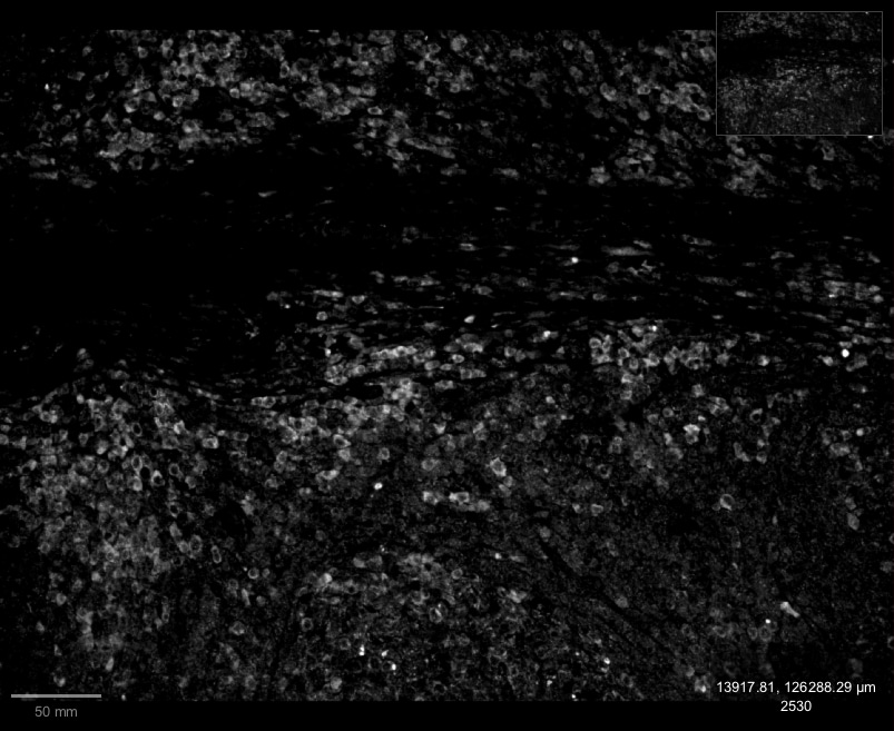
The expression of CXCR3 and CXCL10 in AIH. (A) Multiplex immunofluorescence staining of CXCR3 (red), CD19 (yellow), and TSPAN1 (green) in the liver of AIH patients. (B) Representative confocal staining of TSPAN1 and CXCL10 in the livers of AIH. (C) Immunohistochemical staining of CXCL10 in AIH and HC (×400). Correlation analysis of hepatic CXCL10 expression degree with the numbers of TSPAN1. (D) CD19+ B cells were placed in the upper chambers and complete medium was added in the lower chambers with rhCXCL10 (3 μg/mL) or not for 6 hours. The percentage of TSPAN1+ cells was measured by flow cytometry. Boxes represent the 25th–75th percentile of the distribution; the median is shown as a thick line in the middle of the box; whiskers extend to values with 1.5 times the difference between the 25th and 75th percentiles. *P < 0.05, **P < 0.01. Image collected and cropped by CiteAb from the following open publication (https://pubmed.ncbi.nlm.nih.gov/36591302), licensed under a CC-BY license. Not internally tested by R&D Systems.
Detection of CXCL10/IP-10/CRG-2 by Immunohistochemistry
The expression of CXCR3 and CXCL10 in AIH. (A) Multiplex immunofluorescence staining of CXCR3 (red), CD19 (yellow), and TSPAN1 (green) in the liver of AIH patients. (B) Representative confocal staining of TSPAN1 and CXCL10 in the livers of AIH. (C) Immunohistochemical staining of CXCL10 in AIH and HC (×400). Correlation analysis of hepatic CXCL10 expression degree with the numbers of TSPAN1. (D) CD19+ B cells were placed in the upper chambers and complete medium was added in the lower chambers with rhCXCL10 (3 μg/mL) or not for 6 hours. The percentage of TSPAN1+ cells was measured by flow cytometry. Boxes represent the 25th–75th percentile of the distribution; the median is shown as a thick line in the middle of the box; whiskers extend to values with 1.5 times the difference between the 25th and 75th percentiles. *P < 0.05, **P < 0.01. Image collected and cropped by CiteAb from the following open publication (https://pubmed.ncbi.nlm.nih.gov/36591302), licensed under a CC-BY license. Not internally tested by R&D Systems.
Human CXCL10 / IP-10 / CRG-2 ELISA Standard Curve
Recombinant Human CXCL10/IP‑10/CRG‑2 (Catalog # 266-IP) was serially diluted and captured by Mouse Anti-Human CXCL10/IP‑10/CRG‑2 Monoclonal Antibody (Catalog # MAB266) coated on a Clear Polystyrene Microplate (Catalog # DY990). Goat Anti-Human CXCL10/IP‑10/CRG‑2 Antigen Affinity-purified Polyclonal Antibody (Catalog # AF-266-NA) was biotinylated and incubated with the protein captured on the plate. Detection of the standard curve was achieved by incubating Streptavidin-HRP (Catalog # DY998)Applications for Human CXCL10/IP-10/CRG-2 Antibody
CyTOF-ready
ELISA
This antibody functions as an ELISA detection antibody when paired with Mouse Anti-Human CXCL10/IP‑10/CRG‑2 Monoclonal Antibody (Catalog # MAB2661).
This product is intended for assay development on various assay platforms requiring antibody pairs. We recommend the Human CXCL10/IP-10 DuoSet ELISA Kit (Catalog # DY266) for convenient development of a sandwich ELISA or the Human CXCL10/IP-10 Quantikine ELISA Kit (Catalog # DIP100) for a complete optimized ELISA.
Immunocytochemistry
Sample: Immersion fixed human peripheral blood mononuclear cells treated with PHA
Immunohistochemistry
Sample: Immersion fixed paraffin-embedded sections of human tonsil
Intracellular Staining by Flow Cytometry
Sample: Human peripheral blood monocytes treated with Recombinant Human IFN‑ gamma (Catalog # 285‑IF), fixed with paraformaldehyde, and permeabilized with saponin
Neutralization
Reviewed Applications
Read 3 reviews rated 4.3 using AF-266-NA in the following applications:
Flow Cytometry Panel Builder
Bio-Techne Knows Flow Cytometry
Save time and reduce costly mistakes by quickly finding compatible reagents using the Panel Builder Tool.
Advanced Features
- Spectra Viewer - Custom analysis of spectra from multiple fluorochromes
- Spillover Popups - Visualize the spectra of individual fluorochromes
- Antigen Density Selector - Match fluorochrome brightness with antigen density

Formulation, Preparation, and Storage
Purification
Reconstitution
Reconstitute at 0.2 mg/mL in sterile PBS. For liquid material, refer to CoA for concentration.
Formulation
Shipping
Stability & Storage
- 12 months from date of receipt, -20 to -70 °C as supplied.
- 1 month, 2 to 8 °C under sterile conditions after reconstitution.
- 6 months, -20 to -70 °C under sterile conditions after reconstitution.
Calculators
Background: CXCL10/IP-10/CRG-2
References
- Loetscher, M. et al. (1996) J. Exp. Med. 184:963.
- Wang, X. et al.(1996) J. Biol. Chem. 271:24286.
Alternate Names
Gene Symbol
UniProt
Additional CXCL10/IP-10/CRG-2 Products
Product Documents for Human CXCL10/IP-10/CRG-2 Antibody
Certificate of Analysis
To download a Certificate of Analysis, please enter a lot or batch number in the search box below.
Note: Certificate of Analysis not available for kit components.
Product Specific Notices for Human CXCL10/IP-10/CRG-2 Antibody
For research use only
Citations for Human CXCL10/IP-10/CRG-2 Antibody
 Powered by Bioz
Customer Reviews for Human CXCL10/IP-10/CRG-2 Antibody (3)
Have you used Human CXCL10/IP-10/CRG-2 Antibody?
Submit a review and receive an Amazon gift card!
$25/€18/£15/$25CAN/¥2500 Yen for a review with an image
$10/€7/£6/$10CAN/¥1110 Yen for a review without an image
Submit a review
Customer Images
-
Application: Immunohistochemistry-ParaffinSample Tested: Human Tonsil tissueSpecies: HumanVerified Customer | Posted 01/18/2023CXCL10 stained human tonsil tissue

-
Application: ImmunofluorescenceSample Tested: See PMID 23423575Species: HumanVerified Customer | Posted 01/07/2015
-
Application: Immunohistochemistry-ParaffinSample Tested: See PMID 24080276Species: HumanVerified Customer | Posted 01/07/2015
There are no reviews that match your criteria.
Protocols
Find general support by application which include: protocols, troubleshooting, illustrated assays, videos and webinars.
- 7-Amino Actinomycin D (7-AAD) Cell Viability Flow Cytometry Protocol
- Antigen Retrieval Protocol (PIER)
- Antigen Retrieval for Frozen Sections Protocol
- Appropriate Fixation of IHC/ICC Samples
- Cellular Response to Hypoxia Protocols
- Chromogenic IHC Staining of Formalin-Fixed Paraffin-Embedded (FFPE) Tissue Protocol
- Chromogenic Immunohistochemistry Staining of Frozen Tissue
- ClariTSA™ Fluorophore Kits
- Detection & Visualization of Antibody Binding
- ELISA Sample Preparation & Collection Guide
- ELISA Troubleshooting Guide
- Extracellular Membrane Flow Cytometry Protocol
- Flow Cytometry Protocol for Cell Surface Markers
- Flow Cytometry Protocol for Staining Membrane Associated Proteins
- Flow Cytometry Staining Protocols
- Flow Cytometry Troubleshooting Guide
- Fluorescent IHC Staining of Frozen Tissue Protocol
- Graphic Protocol for Heat-induced Epitope Retrieval
- Graphic Protocol for the Preparation and Fluorescent IHC Staining of Frozen Tissue Sections
- Graphic Protocol for the Preparation and Fluorescent IHC Staining of Paraffin-embedded Tissue Sections
- Graphic Protocol for the Preparation of Gelatin-coated Slides for Histological Tissue Sections
- How to Run an R&D Systems DuoSet ELISA
- How to Run an R&D Systems Quantikine ELISA
- How to Run an R&D Systems Quantikine™ QuicKit™ ELISA
- ICC Cell Smear Protocol for Suspension Cells
- ICC Immunocytochemistry Protocol Videos
- ICC for Adherent Cells
- IHC Sample Preparation (Frozen sections vs Paraffin)
- Immunocytochemistry (ICC) Protocol
- Immunocytochemistry Troubleshooting
- Immunofluorescence of Organoids Embedded in Cultrex Basement Membrane Extract
- Immunofluorescent IHC Staining of Formalin-Fixed Paraffin-Embedded (FFPE) Tissue Protocol
- Immunohistochemistry (IHC) and Immunocytochemistry (ICC) Protocols
- Immunohistochemistry Frozen Troubleshooting
- Immunohistochemistry Paraffin Troubleshooting
- Intracellular Flow Cytometry Protocol Using Alcohol (Methanol)
- Intracellular Flow Cytometry Protocol Using Detergents
- Intracellular Nuclear Staining Flow Cytometry Protocol Using Detergents
- Intracellular Staining Flow Cytometry Protocol Using Alcohol Permeabilization
- Intracellular Staining Flow Cytometry Protocol Using Detergents to Permeabilize Cells
- Preparing Samples for IHC/ICC Experiments
- Preventing Non-Specific Staining (Non-Specific Binding)
- Primary Antibody Selection & Optimization
- Propidium Iodide Cell Viability Flow Cytometry Protocol
- Protocol for Heat-Induced Epitope Retrieval (HIER)
- Protocol for Liperfluo
- Protocol for Making a 4% Formaldehyde Solution in PBS
- Protocol for VisUCyte™ HRP Polymer Detection Reagent
- Protocol for the Characterization of Human Th22 Cells
- Protocol for the Characterization of Human Th9 Cells
- Protocol for the Fluorescent ICC Staining of Cell Smears - Graphic
- Protocol for the Fluorescent ICC Staining of Cultured Cells on Coverslips - Graphic
- Protocol for the Preparation & Fixation of Cells on Coverslips
- Protocol for the Preparation and Chromogenic IHC Staining of Frozen Tissue Sections
- Protocol for the Preparation and Chromogenic IHC Staining of Frozen Tissue Sections - Graphic
- Protocol for the Preparation and Chromogenic IHC Staining of Paraffin-embedded Tissue Sections
- Protocol for the Preparation and Chromogenic IHC Staining of Paraffin-embedded Tissue Sections - Graphic
- Protocol for the Preparation and Fluorescent ICC Staining of Cells on Coverslips
- Protocol for the Preparation and Fluorescent ICC Staining of Non-adherent Cells
- Protocol for the Preparation and Fluorescent ICC Staining of Stem Cells on Coverslips
- Protocol for the Preparation and Fluorescent IHC Staining of Frozen Tissue Sections
- Protocol for the Preparation and Fluorescent IHC Staining of Paraffin-embedded Tissue Sections
- Protocol for the Preparation of Gelatin-coated Slides for Histological Tissue Sections
- Protocol for the Preparation of a Cell Smear for Non-adherent Cell ICC - Graphic
- Protocol: Annexin V and PI Staining by Flow Cytometry
- Protocol: Annexin V and PI Staining for Apoptosis by Flow Cytometry
- Quantikine HS ELISA Kit Assay Principle, Alkaline Phosphatase
- Quantikine HS ELISA Kit Principle, Streptavidin-HRP Polymer
- Sandwich ELISA (Colorimetric) – Biotin/Streptavidin Detection Protocol
- Sandwich ELISA (Colorimetric) – Direct Detection Protocol
- TUNEL and Active Caspase-3 Detection by IHC/ICC Protocol
- The Importance of IHC/ICC Controls
- Troubleshooting Guide: ELISA
- Troubleshooting Guide: Fluorokine Flow Cytometry Kits
- Troubleshooting Guide: Immunohistochemistry
- View all Protocols, Troubleshooting, Illustrated assays and Webinars
Associated Pathways
