Cytokeratin 19 (Keratin, type I cytoskeletal 19; also KRT-19, CK19 and Keratin-19) is a 40-45 kDa, acidic Class I keratin member of the intermediate filament family of proteins. Individual keratins are always expressed in tandem with a second keratin, and these are found in all epithelial cells. The class I KRT-19 heterodimerizes/polymerizes with 50-52 kDa class II KRT-8 (plus KRT-5 and -7) to form 8-10 nm filaments in epidermal stem cells, secretory gland (sweat; mammary; bile duct) simple epithelium, and neuroendocrine epidermal Merkel cells. It may represent a viable marker for skin stem cells. In skin, Cytokeratin 19 forms filaments in the fetal epithelium, and then progressively decreases with age, being virtually absent by age 17. Human Cytokeratin 19 is 400 amino acids (aa) in length. It contains an N-terminal "head" region (aa 1-79) and a subsequent "rod" region (aa 80-387), but is absent a typical C-terminal tail region. Cytokeratin 19 possesses at least 5 utilized phosphorylation sites plus one acetylated Lys residue. Based on other keratins, and the presence of an Asp at position 238, there may be caspase cleavage-generated isoforms. Full length human Cytokeratin 19 (aa 2-400) shares 82% aa sequence identity with mouse Cytokeratin 19.

Key Product Details
Validated by
Orthogonal Validation
Species Reactivity
Human
Applications
Immunohistochemistry, Western Blot, Dual RNAscope ISH-IHC Compatible, Immunocytochemistry, Simple Western
Label
Unconjugated
Antibody Source
Monoclonal Mouse IgG2A Clone # 963420
Loading...
Product Specifications
Immunogen
E.coli-derived recombinant human Cytokeratin 19
Gln311-Lys370
Accession # P08727
Gln311-Lys370
Accession # P08727
Specificity
Detects human Cytokeratin 19 in direct ELISAs and Western blots.
Clonality
Monoclonal
Host
Mouse
Isotype
IgG2A
Scientific Data Images for Human Cytokeratin 19 Antibody (963420)
Cytokeratin 19 in Human Breast Cancer Tissue Using Dual RNAscope®ISH and IHC.
Cytokeratin 19 mRNA (red) and protein (green) was detected in formalin-fixed paraffin-embedded tissue sections of human breast cancer tissue probed with ACD RNAScope®Probe (Catalog # 310221) followed by immunohistochemistry using R&D Systems Mouse Anti-Human Cytokeratin 19 Monoclonal Antibody (Catalog # MAB35061) at 15ug/mL for 1 hour at room temperature followed by incubation with the Anti-Mouse IgG VisUCyte HRP Polymer Antibody (R&D Systems, Catalog # VC001). Tissue was stained using ACD RNAscope®2.5 HD Duplex Detection Reagents (Catalog # 322500).
Detection of Human Cytokeratin 19 by Western Blot.
Western blot shows lysates of HT-29 human colon adenocarcinoma cell line, ZR-75 human breast cancer cell line, DU145 human prostate carcinoma cell line, MCF-7 human breast cancer cell line, and A431 human epithelial carcinoma cell line. PVDF membrane was probed with 0.25 µg/mL of Mouse Anti-Human Cytokeratin 19 Monoclonal Antibody (Catalog # MAB35061) followed by HRP-conjugated Anti-Mouse IgG Secondary Antibody (Catalog # HAF018). A specific band was detected for Cytokeratin 19 at approximately 40 kDa (as indicated). This experiment was conducted under reducing conditions and using Immunoblot Buffer Group 1.
Cytokeratin 19 in MCF‑7 Human Cell Line.
Cytokeratin 19 was detected in immersion fixed MCF-7 human breast cancer cell line using Mouse Anti-Human Cytokeratin 19 Monoclonal Antibody (Catalog # MAB35061) at 8 µg/mL for 3 hours at room temperature. Cells were stained using the NorthernLights™ 557-conjugated Anti-Mouse IgG Secondary Antibody (red; Catalog # NL007) and counterstained with DAPI (blue). Specific staining was localized to cytoplasm. View our protocol for Fluorescent ICC Staining of Cells on Coverslips.
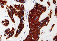
Cytokeratin 19 in Human Breast Cancer Tissue.
Cytokeratin 19 was detected in immersion fixed paraffin-embedded sections of human breast cancer tissue using Mouse Anti-Human Cytokeratin 19 Monoclonal Antibody (Catalog # MAB35061) at 5 µg/mL for 1 hour at room temperature followed by incubation with the Anti-Mouse IgG VisUCyte™ HRP Polymer Antibody (Catalog # VC001). Tissue was stained using DAB (brown) and counterstained with hematoxylin (blue). Specific staining was localized to cytoplasm. View our protocol for IHC Staining with VisUCyte HRP Polymer Detection Reagents.
Cytokeratin 19 in Human Gastric Cancer Tissue.
Cytokeratin 19 was detected in immersion fixed paraffin-embedded sections of human gastric cancer tissue using Mouse Anti-Human Cytokeratin 19 Monoclonal Antibody (Catalog # MAB35061) at 0.5 µg/mL for 1 hour at room temperature followed by incubation with the Anti-Mouse IgG VisUCyte™ HRP Polymer Antibody (Catalog # VC001). Tissue was stained using DAB (brown) and counterstained with hematoxylin (blue). Specific staining was localized to cytoplasm in gastric glands. View our protocol for IHC Staining with VisUCyte HRP Polymer Detection Reagents.
Cytokeratin 19 in Human Papillary Thyroid Cancer Tissue.
Cytokeratin 19 was detected in immersion fixed paraffin-embedded sections of human papillary thyroid cancer tissue using Mouse Anti-Human Cytokeratin 19 Monoclonal Antibody (Catalog # MAB35061) at 0.5 µg/mL for 1 hour at room temperature followed by incubation with the Anti-Mouse IgG VisUCyte™ HRP Polymer Antibody (Catalog # VC001). Tissue was stained using DAB (brown) and counterstained with hematoxylin (blue). Specific staining was localized to cytoplasm in epithelial cells. View our protocol for IHC Staining with VisUCyte HRP Polymer Detection Reagents.
Cytokeratin 19 in Human Lymphoma Tissue.
Cytokeratin 19 was detected in immersion fixed paraffin-embedded sections of human lymphoma tissue using Mouse Anti-Human Cytokeratin 19 Monoclonal Antibody (Catalog # MAB35061) at 0.5 µg/mL for 1 hour at room temperature followed by incubation with the Anti-Mouse IgG VisUCyte™ HRP Polymer Antibody (Catalog # VC001). Tissue was stained using DAB (brown) and counterstained with hematoxylin (blue). Specific staining was localized to cell surfaces. View our protocol for IHC Staining with VisUCyte HRP Polymer Detection Reagents.
Detection of Human Cytokeratin 19 by Simple WesternTM.
Simple Western lane view shows lysates of MCF‑7 human breast cancer cell line and HT‑29 human colon adenocarcinoma cell line, loaded at 0.2 mg/mL. A specific band was detected for Cytokeratin 19 at approximately 50 kDa (as indicated) using 10 µg/mL of Mouse Anti-Human Cytokeratin 19 Monoclonal Antibody (Catalog # MAB35061). This experiment was conducted under reducing conditions and using the 12-230 kDa separation system.
Immunofluorescent Staining of iPSC-derived Pancreatic Duct-like Organoids
iPSC-derived pancreatic duct-like organoids were fixed and stained with Mouse Anti-Human KRT19 Monoclonal Antibody (Catalog # MAB35061; green) and a Rat Anti-Human Claudin-1 Monoclonal Antibody (Catalog # MAB4618; red), followed by secondary antibody staining with the NorthernLights™ 493-conjugated Donkey Anti-Mouse IgG Antigen Affinity-purified Polyclonal Antibody (Catalog # NL009) and NorthernLights 557-conjugated Goat Anti-Rat IgG Antigen Affinity-purified Polyclonal Antibody (Catalog # NL013). Cell nuclei were stained with DAPI (Catalog # 5748; blue) and the images were overlaid. Images were taken using a Leica THUNDER Imaging System (scale bar: 20 mm).Applications for Human Cytokeratin 19 Antibody (963420)
Application
Recommended Usage
Dual RNAscope ISH-IHC Compatible
Optimal dilutions of this antibody should be experimentally determined.
Immunocytochemistry
5-25 µg/mL
Sample: Immersion fixed MCF-7 human breast cancer cell line
Sample: Immersion fixed MCF-7 human breast cancer cell line
Immunohistochemistry
5-25 µg/mL
Sample: Immersion fixed paraffin-embedded sections of human breast cancer tissue, human gastric cancer tissue, human papillary thyroid cancer tissue and human lymphoma tissue
Sample: Immersion fixed paraffin-embedded sections of human breast cancer tissue, human gastric cancer tissue, human papillary thyroid cancer tissue and human lymphoma tissue
Simple Western
50 µg/mL
Sample: MCF‑7 human breast cancer cell line and HT‑29 human colon adenocarcinoma cell line
Sample: MCF‑7 human breast cancer cell line and HT‑29 human colon adenocarcinoma cell line
Western Blot
0.25 µg/mL
Sample: HT‑29 human colon adenocarcinoma cell line, ZR‑75 human breast cancer cell line, DU145 human prostate carcinoma cell line, MCF‑7 human breast cancer cell line, and A431 human epithelial carcinoma cell line
Sample: HT‑29 human colon adenocarcinoma cell line, ZR‑75 human breast cancer cell line, DU145 human prostate carcinoma cell line, MCF‑7 human breast cancer cell line, and A431 human epithelial carcinoma cell line
Reviewed Applications
Read 1 review rated 5 using MAB35061 in the following applications:
Formulation, Preparation, and Storage
Purification
Protein A or G purified from hybridoma culture supernatant
Reconstitution
Reconstitute at 0.5 mg/mL in sterile PBS. For liquid material, refer to CoA for concentration.
Loading...
Formulation
Lyophilized from a 0.2 μm filtered solution in PBS with Trehalose. *Small pack size (SP) is supplied either lyophilized or as a 0.2 µm filtered solution in PBS.
Shipping
Lyophilized product is shipped at ambient temperature. Liquid small pack size (-SP) is shipped with polar packs. Upon receipt, store immediately at the temperature recommended below.
Stability & Storage
Use a manual defrost freezer and avoid repeated freeze-thaw cycles.
- 12 months from date of receipt, -20 to -70 °C as supplied.
- 1 month, 2 to 8 °C under sterile conditions after reconstitution.
- 6 months, -20 to -70 °C under sterile conditions after reconstitution.
Calculators
Background: Cytokeratin 19
Alternate Names
CK19, EndoC, K19, Krt19
Gene Symbol
KRT19
UniProt
Additional Cytokeratin 19 Products
Product Documents for Human Cytokeratin 19 Antibody (963420)
Certificate of Analysis
To download a Certificate of Analysis, please enter a lot or batch number in the search box below.
Note: Certificate of Analysis not available for kit components.
Product Specific Notices for Human Cytokeratin 19 Antibody (963420)
For research use only
Related Research Areas
Customer Reviews for Human Cytokeratin 19 Antibody (963420) (1)
5 out of 5
1 Customer Rating
Have you used Human Cytokeratin 19 Antibody (963420)?
Submit a review and receive an Amazon gift card!
$25/€18/£15/$25CAN/¥2500 Yen for a review with an image
$10/€7/£6/$10CAN/¥1110 Yen for a review without an image
Submit a review
Customer Images
Showing
1
-
1 of
1 review
Showing All
Filter By:
-
Application: ImmunohistochemistrySample Tested: breast carcinomaSpecies: HumanVerified Customer | Posted 11/23/2021

There are no reviews that match your criteria.
Protocols
Find general support by application which include: protocols, troubleshooting, illustrated assays, videos and webinars.
- Antigen Retrieval Protocol (PIER)
- Antigen Retrieval for Frozen Sections Protocol
- Appropriate Fixation of IHC/ICC Samples
- Cellular Response to Hypoxia Protocols
- Chromogenic IHC Staining of Formalin-Fixed Paraffin-Embedded (FFPE) Tissue Protocol
- Chromogenic Immunohistochemistry Staining of Frozen Tissue
- ClariTSA™ Fluorophore Kits
- Detection & Visualization of Antibody Binding
- Fluorescent IHC Staining of Frozen Tissue Protocol
- Graphic Protocol for Heat-induced Epitope Retrieval
- Graphic Protocol for the Preparation and Fluorescent IHC Staining of Frozen Tissue Sections
- Graphic Protocol for the Preparation and Fluorescent IHC Staining of Paraffin-embedded Tissue Sections
- Graphic Protocol for the Preparation of Gelatin-coated Slides for Histological Tissue Sections
- ICC Cell Smear Protocol for Suspension Cells
- ICC Immunocytochemistry Protocol Videos
- ICC for Adherent Cells
- IHC Sample Preparation (Frozen sections vs Paraffin)
- ISH-IHC Protocol for Chromogenic Detection on Formalin Fixed Paraffin Embedded (FFPE) Tissue
- Immunocytochemistry (ICC) Protocol
- Immunocytochemistry Troubleshooting
- Immunofluorescence of Organoids Embedded in Cultrex Basement Membrane Extract
- Immunofluorescent IHC Staining of Formalin-Fixed Paraffin-Embedded (FFPE) Tissue Protocol
- Immunohistochemistry (IHC) and Immunocytochemistry (ICC) Protocols
- Immunohistochemistry Frozen Troubleshooting
- Immunohistochemistry Paraffin Troubleshooting
- Preparing Samples for IHC/ICC Experiments
- Preventing Non-Specific Staining (Non-Specific Binding)
- Primary Antibody Selection & Optimization
- Protocol for Heat-Induced Epitope Retrieval (HIER)
- Protocol for Making a 4% Formaldehyde Solution in PBS
- Protocol for VisUCyte™ HRP Polymer Detection Reagent
- Protocol for the Fluorescent ICC Staining of Cell Smears - Graphic
- Protocol for the Fluorescent ICC Staining of Cultured Cells on Coverslips - Graphic
- Protocol for the Preparation & Fixation of Cells on Coverslips
- Protocol for the Preparation and Chromogenic IHC Staining of Frozen Tissue Sections
- Protocol for the Preparation and Chromogenic IHC Staining of Frozen Tissue Sections - Graphic
- Protocol for the Preparation and Chromogenic IHC Staining of Paraffin-embedded Tissue Sections
- Protocol for the Preparation and Chromogenic IHC Staining of Paraffin-embedded Tissue Sections - Graphic
- Protocol for the Preparation and Fluorescent ICC Staining of Cells on Coverslips
- Protocol for the Preparation and Fluorescent ICC Staining of Non-adherent Cells
- Protocol for the Preparation and Fluorescent ICC Staining of Stem Cells on Coverslips
- Protocol for the Preparation and Fluorescent IHC Staining of Frozen Tissue Sections
- Protocol for the Preparation and Fluorescent IHC Staining of Paraffin-embedded Tissue Sections
- Protocol for the Preparation of Gelatin-coated Slides for Histological Tissue Sections
- Protocol for the Preparation of a Cell Smear for Non-adherent Cell ICC - Graphic
- R&D Systems Quality Control Western Blot Protocol
- TUNEL and Active Caspase-3 Detection by IHC/ICC Protocol
- The Importance of IHC/ICC Controls
- Troubleshooting Guide: Immunohistochemistry
- Troubleshooting Guide: Western Blot Figures
- Western Blot Conditions
- Western Blot Protocol
- Western Blot Protocol for Cell Lysates
- Western Blot Troubleshooting
- Western Blot Troubleshooting Guide
- View all Protocols, Troubleshooting, Illustrated assays and Webinars
Loading...
Associated Pathways
