Human IL‑1 beta /IL‑1F2 Antibody
R&D Systems | Catalog # MAB201

Key Product Details
Validated by
Biological Validation
Species Reactivity
Validated:
Human
Cited:
Human, Mouse, Transgenic Mouse
Applications
Validated:
Western Blot, Neutralization, Intracellular Staining by Flow Cytometry, Immunocytochemistry, Simple Western, CyTOF-ready
Cited:
Immunohistochemistry-Paraffin, Western Blot, Neutralization, Flow Cytometry, Immunocytochemistry, ELISA Development, Mass Cytometry
Label
Unconjugated
Antibody Source
Monoclonal Mouse IgG1 Clone # 8516
Loading...
Product Specifications
Immunogen
E. coli-derived recombinant human IL-1 beta /IL-1F2
aa 117-269
Accession # P01584
aa 117-269
Accession # P01584
Specificity
Detects human IL-1 beta /IL-1F2 in Western blots. Shows less than 5% cross-reactivity with recombinant mouse (rm) IL‑1 beta and rpIL‑1 beta and no cross-reactivity with rrIL‑1 beta, rmIL‑1 alpha, rhIL‑1ra, rmIL‑1ra, or rrIL‑1 alpha.
Clonality
Monoclonal
Host
Mouse
Isotype
IgG1
Endotoxin Level
<0.10 EU per 1 μg of the antibody by the LAL method.
Scientific Data Images for Human IL‑1 beta /IL‑1F2 Antibody
Detection of Human IL‑1 beta /IL‑1F2 by Western Blot.
Western blot shows lysates of THP-1 human acute monocytic leukemia cell line untreated (-) or treated (+) with 200 nM PMA for 24 hours and 10 µg/mL LPS and 3 hours. PVDF membrane was probed with 1 µg/mL of Mouse Anti-Human IL-1 beta /IL-1F2 Monoclonal Antibody (Catalog # MAB201) followed by HRP-conjugated Anti-Mouse IgG Secondary Antibody (Catalog # HAF018). A specific band was detected for IL-1 beta /IL-1F2 at approximately 36 kDa (as indicated). This experiment was conducted under reducing conditions and using Immunoblot Buffer Group 1.
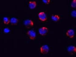
IL‑1 beta /IL‑1F2 in Human PBMCs.
IL‑1 beta /IL‑1F2 was detected in immersion fixed human peripheral blood mononuclear cells (PBMCs) using Human IL‑1 beta /IL‑1F2 Monoclonal Antibody (Catalog # MAB201) at 10 µg/mL for 3 hours at room temperature. Cells were stained using the NorthernLights™ 557-conjugated Anti-Mouse IgG Secondary Antibody (red; Catalog # NL007) and counterstained with DAPI (blue). View our protocol for Fluorescent ICC Staining of Non-adherent Cells.
IL‑1 beta /IL‑1F2 in Human PBMCs.
IL-1 beta /IL-1F2 was detected in immersion fixed human peripheral blood mononuclear cells (PBMCs) treated with LPS and monensin using Human IL-1 beta /IL-1F2 Monoclonal Antibody (Catalog # MAB201) at 10 µg/mL for 3 hours at room temperature. Cells were stained using the NorthernLights™ 557-conjugated Anti-Mouse IgG Secondary Antibody (yellow; Catalog # NL007) and counterstained with DAPI (blue). View our protocol for Fluorescent ICC Staining of Non-adherent Cells.
Detection of Human IL‑1 beta /IL‑1F2 by Simple WesternTM.
Simple Western lane view shows lysates of THP‑1 human acute monocytic leukemia cell line untreated (-) or treated (+) with 200 nm PMA and 10 ug/ml LPS for 24 hrs and 3 hrs, respectively, loaded at 0.2 mg/mL. A specific band was detected for IL‑1 beta /IL‑1F2 at approximately 38 kDa (as indicated) using 10 µg/mL of Mouse Anti-Human IL‑1 beta /IL‑1F2 Monoclonal Antibody (Catalog # MAB201). This experiment was conducted under reducing conditions and using the 12-230 kDa separation system.
Cell Proliferation Induced by IL‑1 beta /IL‑1F2 and Neutralization by Human IL‑1 beta /IL‑1F2 Antibody.
Recombinant Human IL-1 beta /IL-1F2 (Catalog # 201-LB) stimulates proliferation in the the D10.G4.1 mouse helper T cell line in a dose-dependent manner (orange line). Proliferation elicited by Recombinant Human IL-1 beta /IL-1F2 (50 pg/mL) is neutralized (green line) by increasing concentrations of Mouse Anti-Human IL-1 beta /IL-1F2 Monoclonal Antibody (Catalog # MAB201). The ND50 is typically 0.001-0.003 µg/mL.
Detection of Human IL-1 beta/IL-1F2 by Western Blot
Hsp27 modulates IL-1 beta production in monocytes.(a) Immunoblot analysis of Hsp27 protein expression in classical (CL) and non-classical (NC) monocytes (left), and relative band densities from 2 combined experiments (right), with GAPDH as loading control. (b) Representative immunoblot of Hsp27 and pro-IL-1 beta (p31) in lysates from siRNA-transfected total monocytes for si-control (scrambled control siRNA) or si-Hsp27 (siRNA to Hsp27) either untreated or stimulated with LPS for 3 h. Data in (a) and (b) are cropped images and the corresponding full-length images are presentated in Supplementary Fig. 3. (c) Expression of IL-1 beta mRNA in siRNA-transfected total monocytes (n = 3). (d) Mature IL-1 beta release measured by ELISA of supernatants from siRNA-transfected total monocytes upon LPS stimulation in the presence or absence of BzATP (n = 4). (e) Fold change in IL-1 beta production by LPS and BzATP-treated total monocytes transfected with si-Hsp27, with respect to si-control. Data plotted are mean ± SEM, student t-test (a and b) and two-way ANOVA with Tukey’s multiple comparison test (c), *p < 0.05, **p < 0.01. Image collected and cropped by CiteAb from the following publication (https://pubmed.ncbi.nlm.nih.gov/27976724), licensed under a CC-BY license. Not internally tested by R&D Systems.
Detection of Human IL-1 beta/IL-1F2 by Western Blot
Non-classical monocytes secrete less mature IL-1 beta compared to other monocyte subsets following LPS and BzATP stimulation.(a) IL-1 beta ELISA on supernatants from monocyte subsets either untreated or stimulated with LPS for 24 h in the presence or absence of the P2X7 antagonist, A438079, followed by a secondary stimulation by BzATP in some conditions as indicated. Data plotted are mean ± SEM; n = 8. ****p < 0.0001 ***p < 0.001. ##p < 0.01 and #p < 0.05 is comparison to the respective classical and intermediate subsets treated with LPS + BzATP. Statistics are calculated based on Two-way ANOVA with Tukey’s multiple comparison test (*p < 0.05). (b) Representative cropped immunoblot images for mature 17 kDa (p17) IL-1 beta in culture supernatants of monocyte subsets stimulated as described in (a). The samples were derived from the same experiment and were ran on the same gel. Full-length images are shown in Supplementary Fig. 3. (c) YoPro-1 dye uptake was measured by flow cytometry to reflect level of P2X7R activity, and is shown as the fold change of BzATP-induced YoPro-1 uptake normalized to the respective untreated subset uptake. Data plotted are mean ± SEM; n = 6 based on one-way ANOVA with Tukey’s multiple comparison test. CL: classical, INT: intermediate, NC: non-classical. Image collected and cropped by CiteAb from the following publication (https://pubmed.ncbi.nlm.nih.gov/27976724), licensed under a CC-BY license. Not internally tested by R&D Systems.
Detection of Rabbit IL-1 beta/IL-1F2 by Immunocytochemistry/Immunofluorescence
Immunohistochemistry for CD3, IL-1 beta and CD79 expression.Some expression for these markers were found in the CS and CSVT groups at 21 days (positive signal by brown staining). The same overall expression to the materials was seen at the other time points. Image collected and cropped by CiteAb from the following publication (https://dx.plos.org/10.1371/journal.pone.0136514), licensed under a CC-BY license. Not internally tested by R&D Systems.
Detection of IL-1 beta /IL-1F2 by Western Blot
Yptb effectors enable evasion of T3SS-dependent inflammasome activation in human cells. BMDMs (A), Caco-2 cells (B, D, and E), or primary hMDMs from three healthy human donors (C, F, and G) were infected with PBS (mock), WT Yptb, or delta 6 Yptb. (A–C) Cell death was measured as percentage of cytotoxicity normalized to cells treated with 2% triton at 6 hpi. (D) Release of IL-18 into the supernatant was measured by ELISA at 6 hpi. (E) Lysates and supernatants collected 6 hpi were immunoblotted for IL-18 and beta -actin. (F) Release of IL-1 beta into the supernatant was measured by ELISA at 6 hpi. (G) Lysates and supernatants collected 6 hpi were immunoblotted for IL-1 beta and beta -actin. ns: not significant; *P < 0.05; **P < 0.01 by t-test (A–C) or one-way ANOVA (D and F). Shown are averages and error bars representing the standard deviation from at least three pooled experiments. ANOVA, analysis of variance; BMDMs, bone marrow-derived macrophages; ELISA, enzyme-linked immunosorbent assay; PBS, phosphate-buffered saline. Image collected and cropped by CiteAb from the following open publication (https://pubmed.ncbi.nlm.nih.gov/37615436), licensed under a CC-BY license. Not internally tested by R&D Systems.
Detection of IL-1 beta /IL-1F2 by Western Blot
Yptb effectors enable evasion of T3SS-dependent inflammasome activation in human cells. BMDMs (A), Caco-2 cells (B, D, and E), or primary hMDMs from three healthy human donors (C, F, and G) were infected with PBS (mock), WT Yptb, or delta 6 Yptb. (A–C) Cell death was measured as percentage of cytotoxicity normalized to cells treated with 2% triton at 6 hpi. (D) Release of IL-18 into the supernatant was measured by ELISA at 6 hpi. (E) Lysates and supernatants collected 6 hpi were immunoblotted for IL-18 and beta -actin. (F) Release of IL-1 beta into the supernatant was measured by ELISA at 6 hpi. (G) Lysates and supernatants collected 6 hpi were immunoblotted for IL-1 beta and beta -actin. ns: not significant; *P < 0.05; **P < 0.01 by t-test (A–C) or one-way ANOVA (D and F). Shown are averages and error bars representing the standard deviation from at least three pooled experiments. ANOVA, analysis of variance; BMDMs, bone marrow-derived macrophages; ELISA, enzyme-linked immunosorbent assay; PBS, phosphate-buffered saline. Image collected and cropped by CiteAb from the following open publication (https://pubmed.ncbi.nlm.nih.gov/37615436), licensed under a CC-BY license. Not internally tested by R&D Systems.Applications for Human IL‑1 beta /IL‑1F2 Antibody
Application
Recommended Usage
CyTOF-ready
Ready to be labeled using established conjugation methods. No BSA or other carrier proteins that could interfere with conjugation.
Immunocytochemistry
8-25 µg/mL
Sample: Immersion fixed human peripheral blood mononuclear cells (PBMCs) stimulated with LPS, or with LPS and monensin
Sample: Immersion fixed human peripheral blood mononuclear cells (PBMCs) stimulated with LPS, or with LPS and monensin
Intracellular Staining by Flow Cytometry
2.5 µg/106 cells
Sample: Human peripheral blood mononuclear cells treated with LPS, fixed with paraformaldehyde, and permeabilized with saponin
Sample: Human peripheral blood mononuclear cells treated with LPS, fixed with paraformaldehyde, and permeabilized with saponin
Simple Western
10 µg/mL
Sample: TF‑1 human erythroleukemic cell line
Sample: TF‑1 human erythroleukemic cell line
Western Blot
1 µg/mL
Sample: THP‑1 human acute monocytic leukemia cell line treated with PMA and LPS
Sample: THP‑1 human acute monocytic leukemia cell line treated with PMA and LPS
Neutralization
Measured by its ability to neutralize IL‑1 beta /IL‑1F2-induced proliferation in the D10.G4.1 mouse helper T cell line. Symons, J.A. et al. (1987) in Lymphokines and Interferons, a Practical Approach. Clemens, M.J. et al. (eds): IRL Press. 272. The Neutralization Dose (ND50) is typically 0.001-0.003 µg/mL in the presence of 50 pg/mL Recombinant Human IL‑1 beta /IL‑1F2.
Reviewed Applications
Read 8 reviews rated 4.4 using MAB201 in the following applications:
Flow Cytometry Panel Builder
Bio-Techne Knows Flow Cytometry
Save time and reduce costly mistakes by quickly finding compatible reagents using the Panel Builder Tool.
Advanced Features
- Spectra Viewer - Custom analysis of spectra from multiple fluorochromes
- Spillover Popups - Visualize the spectra of individual fluorochromes
- Antigen Density Selector - Match fluorochrome brightness with antigen density

Formulation, Preparation, and Storage
Purification
Protein A or G purified from hybridoma culture supernatant
Reconstitution
Reconstitute at 0.5 mg/mL in sterile PBS. For liquid material, refer to CoA for concentration.
Loading...
Formulation
Lyophilized from a 0.2 μm filtered solution in PBS with Trehalose. *Small pack size (SP) is supplied either lyophilized or as a 0.2 µm filtered solution in PBS.
Shipping
Lyophilized product is shipped at ambient temperature. Liquid small pack size (-SP) is shipped with polar packs. Upon receipt, store immediately at the temperature recommended below.
Stability & Storage
Use a manual defrost freezer and avoid repeated freeze-thaw cycles.
- 12 months from date of receipt, -20 to -70 °C as supplied.
- 1 month, 2 to 8 °C under sterile conditions after reconstitution.
- 6 months, -20 to -70 °C under sterile conditions after reconstitution.
Calculators
Background: IL-1 beta/IL-1F2
Long Name
Interleukin 1 beta
Alternate Names
IL-1b, IL-1F2, IL1 beta, IL1B
Entrez Gene IDs
Gene Symbol
IL1B
UniProt
Additional IL-1 beta/IL-1F2 Products
Product Documents for Human IL‑1 beta /IL‑1F2 Antibody
Certificate of Analysis
To download a Certificate of Analysis, please enter a lot or batch number in the search box below.
Note: Certificate of Analysis not available for kit components.
Product Specific Notices for Human IL‑1 beta /IL‑1F2 Antibody
For research use only
Related Research Areas
Citations for Human IL‑1 beta /IL‑1F2 Antibody
 Powered by Bioz
Customer Reviews for Human IL‑1 beta /IL‑1F2 Antibody (8)
4.4 out of 5
8 Customer Ratings
Have you used Human IL‑1 beta /IL‑1F2 Antibody?
Submit a review and receive an Amazon gift card!
$25/€18/£15/$25CAN/¥2500 Yen for a review with an image
$10/€7/£6/$10CAN/¥1110 Yen for a review without an image
Submit a review
Customer Images
Showing
1
-
5 of
8 reviews
Showing All
Filter By:
-
Application: Immunocytochemistry/ImmunofluorescenceSample Tested: Peripheral blood mononuclear cells (PBMCs)Species: HumanVerified Customer | Posted 06/18/2022

-
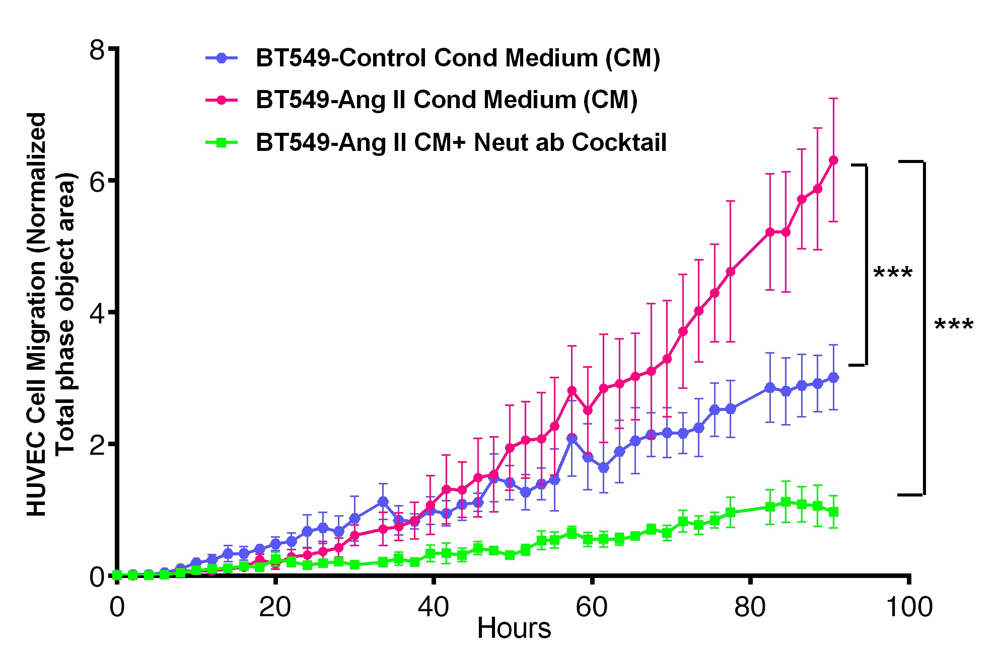
Application: Block/NeutralizeSample Tested: Serum-free Cell Culture SupernatesSpecies: HumanVerified Customer | Posted 07/07/2020We used this IL1B (5ng/mL) neutralizing antibody as part of an antibody cocktail containing several other antibodies to neutralize IL1B present in the Conditioned Medium (CM) from BT549 Cells treated with Angiotensin II. The effect of cytokines in the CM (+/-Neutralizing ab Cocktail) on Endothelial (HUVEC) Cell migration was measured using Incucyte Zoom Chemotaxis Assay.
-
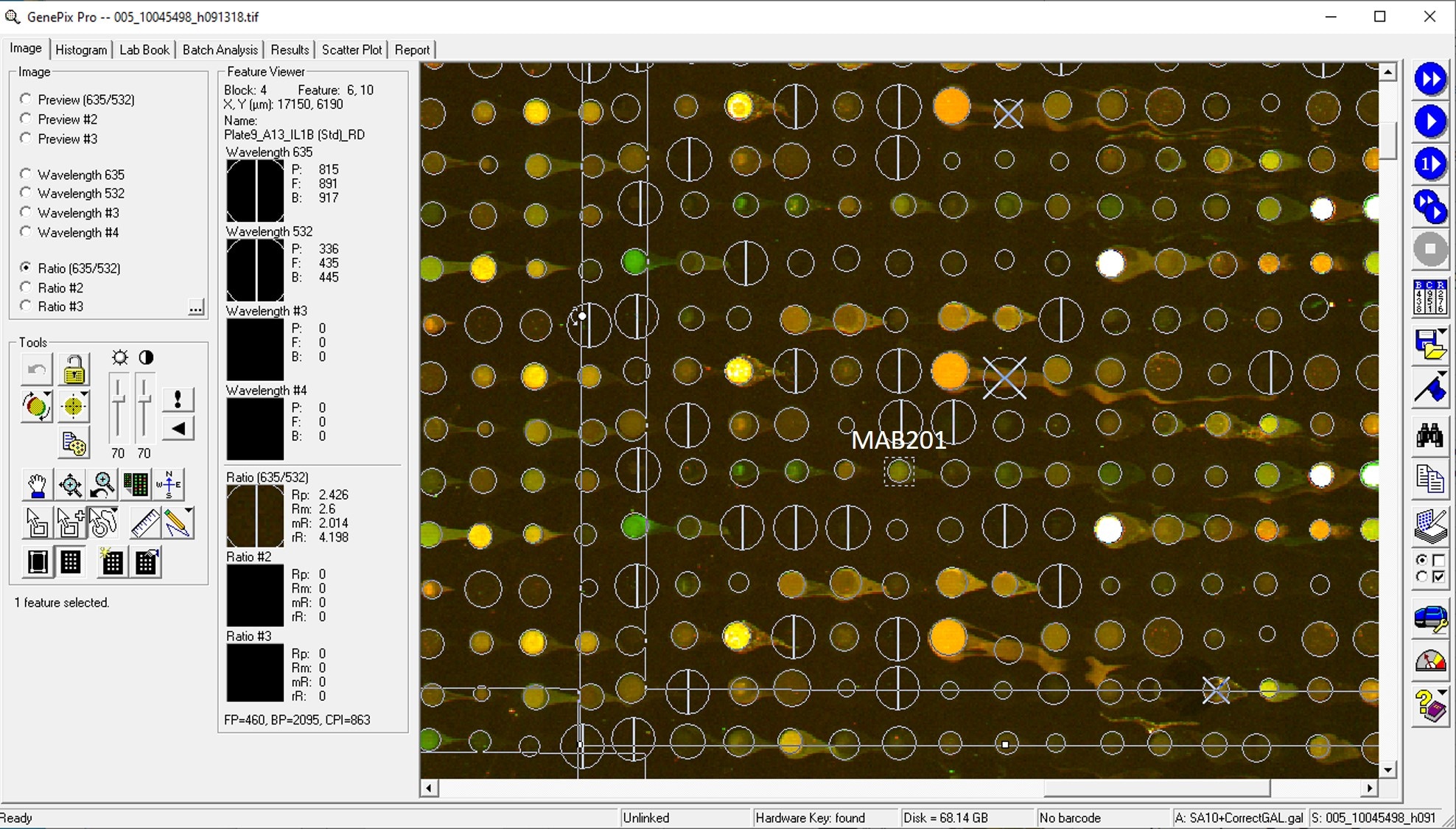
Application: MicroarraysSample Tested: EDTA PlasmaSpecies: HumanVerified Customer | Posted 06/10/2020
-
Application: Block/NeutralizeSample Tested: Serum-free Cell Culture Media and Breast cancer cellsSpecies: HumanVerified Customer | Posted 05/27/2019
-
Application: ELISASample Tested: Serum and PlasmaSpecies: HumanVerified Customer | Posted 11/11/2018
-
Application: MicroarraysSample Tested: EDTA PlasmaSpecies: HumanVerified Customer | Posted 11/07/2018
-
Application: MicroarraySample Tested: EDTA PlasmaSpecies: HumanVerified Customer | Posted 11/02/2018
-
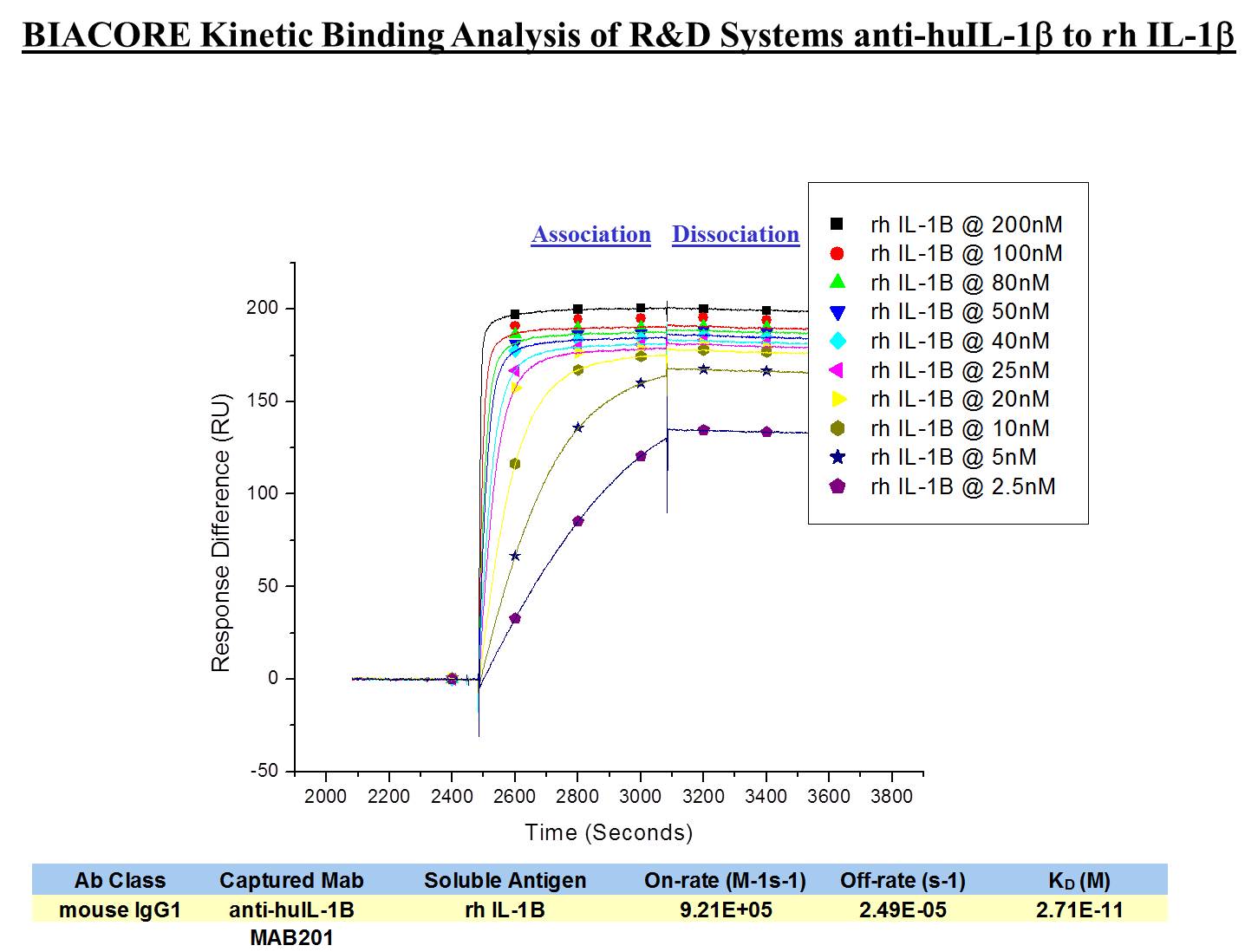
Application: Affinity measurementSample Tested: Recombinant proteinSpecies: HumanVerified Customer | Posted 02/07/2018
There are no reviews that match your criteria.
Protocols
Find general support by application which include: protocols, troubleshooting, illustrated assays, videos and webinars.
- 7-Amino Actinomycin D (7-AAD) Cell Viability Flow Cytometry Protocol
- Appropriate Fixation of IHC/ICC Samples
- Cellular Response to Hypoxia Protocols
- ClariTSA™ Fluorophore Kits
- Detection & Visualization of Antibody Binding
- Extracellular Membrane Flow Cytometry Protocol
- Flow Cytometry Protocol for Cell Surface Markers
- Flow Cytometry Protocol for Staining Membrane Associated Proteins
- Flow Cytometry Staining Protocols
- Flow Cytometry Troubleshooting Guide
- ICC Cell Smear Protocol for Suspension Cells
- ICC Immunocytochemistry Protocol Videos
- ICC for Adherent Cells
- Immunocytochemistry (ICC) Protocol
- Immunocytochemistry Troubleshooting
- Immunofluorescence of Organoids Embedded in Cultrex Basement Membrane Extract
- Immunohistochemistry (IHC) and Immunocytochemistry (ICC) Protocols
- Intracellular Flow Cytometry Protocol Using Alcohol (Methanol)
- Intracellular Flow Cytometry Protocol Using Detergents
- Intracellular Nuclear Staining Flow Cytometry Protocol Using Detergents
- Intracellular Staining Flow Cytometry Protocol Using Alcohol Permeabilization
- Intracellular Staining Flow Cytometry Protocol Using Detergents to Permeabilize Cells
- Preparing Samples for IHC/ICC Experiments
- Preventing Non-Specific Staining (Non-Specific Binding)
- Primary Antibody Selection & Optimization
- Propidium Iodide Cell Viability Flow Cytometry Protocol
- Protocol for Liperfluo
- Protocol for VisUCyte™ HRP Polymer Detection Reagent
- Protocol for the Characterization of Human Th22 Cells
- Protocol for the Characterization of Human Th9 Cells
- Protocol for the Fluorescent ICC Staining of Cell Smears - Graphic
- Protocol for the Fluorescent ICC Staining of Cultured Cells on Coverslips - Graphic
- Protocol for the Preparation and Fluorescent ICC Staining of Cells on Coverslips
- Protocol for the Preparation and Fluorescent ICC Staining of Non-adherent Cells
- Protocol for the Preparation and Fluorescent ICC Staining of Stem Cells on Coverslips
- Protocol for the Preparation of a Cell Smear for Non-adherent Cell ICC - Graphic
- Protocol: Annexin V and PI Staining by Flow Cytometry
- Protocol: Annexin V and PI Staining for Apoptosis by Flow Cytometry
- R&D Systems Quality Control Western Blot Protocol
- TUNEL and Active Caspase-3 Detection by IHC/ICC Protocol
- The Importance of IHC/ICC Controls
- Troubleshooting Guide: Fluorokine Flow Cytometry Kits
- Troubleshooting Guide: Western Blot Figures
- Western Blot Conditions
- Western Blot Protocol
- Western Blot Protocol for Cell Lysates
- Western Blot Troubleshooting
- Western Blot Troubleshooting Guide
- View all Protocols, Troubleshooting, Illustrated assays and Webinars
Loading...
Associated Pathways


Innate Lymphoid Cell Differentiation Pathways
 NOD-like Receptor Signaling Pathways
 Th17 Differentiation Pathway
 Toll-Like Receptor Signaling Pathways
