Human IL-6 Antibody (1936R)
R&D Systems | Catalog # MAB2061R
Recombinant Monoclonal Antibody.

Key Product Details
Validated by
Biological Validation
Species Reactivity
Validated:
Human
Cited:
Human
Applications
Validated:
Neutralization, Intracellular Staining by Flow Cytometry
Cited:
Immunohistochemistry
Label
Unconjugated
Antibody Source
Recombinant Monoclonal Mouse IgG2B Clone # 1936R
Loading...
Product Specifications
Immunogen
E. coli-derived recombinant human IL-6
Val30-Met212
Accession # P05231
Val30-Met212
Accession # P05231
Specificity
Detects human IL-6 in direct ELISAs. Does not cross-react with recombinant IL-6 from mouse, rat, or pig.
Clonality
Monoclonal
Host
Mouse
Isotype
IgG2B
Endotoxin Level
<0.10 EU per 1 μg of the antibody by the LAL method.
Scientific Data Images for Human IL-6 Antibody (1936R)
Detection of IL‑6 in Human PBMCs by Flow Cytometry.
Human peripheral blood mononuclear cells (PBMCs) treated with 100 ng/mL LPS for 24 hours were stained with Mouse Anti-Human IL-6 Monoclonal Antibody (Catalog # MAB2061R, filled histogram) or isotype control antibody (MAB004, open histogram), followed by Phycoerythrin-conjugated Anti-Mouse IgG Secondary Antibody (F0102B). To facilitate intracellular staining, cells were fixed with Flow Cytometry Fixation Buffer (FC004) and permeabilized with Flow Cytometry Permeabilization/Wash Buffer I (FC005). View our protocol for Staining Intracellular Molecules.
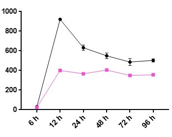
Cell Proliferation Induced by IL-6 and Neutralization by Human IL-6 Antibody.
Recombinant Human IL-6 (206-IL) stimulates proliferation in the T1165.85.2.1 mouse plasmacytoma cell line in a dose-dependent manner (orange line), as measured by Resazurin (AR002). Proliferation elicited by Recombinant Human IL-6 (2.5 ng/mL) is neutralized (green line) by increasing concentrations of Mouse Anti-Human IL-6 Monoclonal Antibody (Catalog # MAB2061R). The ND50 is typically 8.00-80.0 ng/mL.
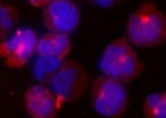
Detection of IL‑6 in PBMC cells by Flow Cytometry.
PBMC stimulated with 100 ng/ml of LPS and Brefeldin A for 24 hrs (A) or unstimulated (B) were stained with Mouse Anti-Human IL‑6 Monoclonal Antibody (Catalog # MAB2061R) and Mouse Anti-Human CD14 PE‑conjugated Monoclonal Antibody (Catalog # FAB3832P). To facilitate intracellular staining, cells were fixed with Flow Cytometry Fixation Buffer (1X) (FC004) and permeabilized with Saponin. View our protocol for Staining Intracellular Molecules.Applications for Human IL-6 Antibody (1936R)
Application
Recommended Usage
Intracellular Staining by Flow Cytometry
0.25 µg/106 cells
Sample: PBMCs treated with LPS were fixed with Flow Cytometry Fixation Buffer (Catalog # FC004) and permeabilized with Flow Cytometry Permeabilization/Wash Buffer I (Catalog # FC005) and PBMC stimulated with 100 ng/ml of LPS and Brefeldin A for 24 hrs or unstimulated
Sample: PBMCs treated with LPS were fixed with Flow Cytometry Fixation Buffer (Catalog # FC004) and permeabilized with Flow Cytometry Permeabilization/Wash Buffer I (Catalog # FC005) and PBMC stimulated with 100 ng/ml of LPS and Brefeldin A for 24 hrs or unstimulated
Neutralization
Measured by its ability to neutralize IL‑6-induced proliferation in the
T1165.85.2.1 mouse plasmacytoma cell line. The Neutralization Dose (ND50) is typically 8.00-80.0 ng/mL in the presence of 2.5 ng/mL Recombinant Human IL‑6.
Reviewed Applications
Read 2 reviews rated 5 using MAB2061R in the following applications:
Flow Cytometry Panel Builder
Bio-Techne Knows Flow Cytometry
Save time and reduce costly mistakes by quickly finding compatible reagents using the Panel Builder Tool.
Advanced Features
- Spectra Viewer - Custom analysis of spectra from multiple fluorochromes
- Spillover Popups - Visualize the spectra of individual fluorochromes
- Antigen Density Selector - Match fluorochrome brightness with antigen density

Formulation, Preparation, and Storage
Purification
Protein A or G purified from cell culture supernatant
Reconstitution
Reconstitute at 0.5 mg/mL in sterile PBS. For liquid material, refer to CoA for concentration.
Loading...
Formulation
Lyophilized from a 0.2 μm filtered solution in PBS with Trehalose. *Small pack size (SP) is supplied either lyophilized or as a 0.2 µm filtered solution in PBS.
Shipping
Lyophilized product is shipped at ambient temperature. Liquid small pack size (-SP) is shipped with polar packs. Upon receipt, store immediately at the temperature recommended below.
Stability & Storage
Use a manual defrost freezer and avoid repeated freeze-thaw cycles.
- 12 months from date of receipt, -20 to -70 °C as supplied.
- 1 month, 2 to 8 °C under sterile conditions after reconstitution.
- 6 months, -20 to -70 °C under sterile conditions after reconstitution.
Calculators
Background: IL-6
Long Name
Interleukin 6
Alternate Names
BSF-2, BSF2, IFNB2, IL6, MGI-2A
Entrez Gene IDs
Gene Symbol
IL6
UniProt
Additional IL-6 Products
Product Documents for Human IL-6 Antibody (1936R)
Certificate of Analysis
To download a Certificate of Analysis, please enter a lot or batch number in the search box below.
Note: Certificate of Analysis not available for kit components.
Product Specific Notices for Human IL-6 Antibody (1936R)
For research use only
Related Research Areas
Citations for Human IL-6 Antibody (1936R)
 Powered by Bioz
Customer Reviews for Human IL-6 Antibody (1936R) (2)
5 out of 5
2 Customer Ratings
Have you used Human IL-6 Antibody (1936R)?
Submit a review and receive an Amazon gift card!
$25/€18/£15/$25CAN/¥2500 Yen for a review with an image
$10/€7/£6/$10CAN/¥1110 Yen for a review without an image
Submit a review
Customer Images
Showing
1
-
2 of
2 reviews
Showing All
Filter By:
-
Application: Immunocytochemistry/ImmunofluorescenceSample Tested: Peripheral blood mononuclear cells (PBMCs)Species: HumanVerified Customer | Posted 12/13/2021

-
Application: Block/NeutralizeSample Tested: macrophageSpecies: HumanVerified Customer | Posted 07/30/2021
There are no reviews that match your criteria.
Protocols
Find general support by application which include: protocols, troubleshooting, illustrated assays, videos and webinars.
- 7-Amino Actinomycin D (7-AAD) Cell Viability Flow Cytometry Protocol
- Extracellular Membrane Flow Cytometry Protocol
- Flow Cytometry Protocol for Cell Surface Markers
- Flow Cytometry Protocol for Staining Membrane Associated Proteins
- Flow Cytometry Staining Protocols
- Flow Cytometry Troubleshooting Guide
- Intracellular Flow Cytometry Protocol Using Alcohol (Methanol)
- Intracellular Flow Cytometry Protocol Using Detergents
- Intracellular Nuclear Staining Flow Cytometry Protocol Using Detergents
- Intracellular Staining Flow Cytometry Protocol Using Alcohol Permeabilization
- Intracellular Staining Flow Cytometry Protocol Using Detergents to Permeabilize Cells
- Propidium Iodide Cell Viability Flow Cytometry Protocol
- Protocol for Liperfluo
- Protocol for the Characterization of Human Th22 Cells
- Protocol for the Characterization of Human Th9 Cells
- Protocol: Annexin V and PI Staining by Flow Cytometry
- Protocol: Annexin V and PI Staining for Apoptosis by Flow Cytometry
- Troubleshooting Guide: Fluorokine Flow Cytometry Kits
- View all Protocols, Troubleshooting, Illustrated assays and Webinars
Loading...
Associated Pathways


IL-21 Signaling Pathways and their Primary Biological Effects in Different Immune Cell Types
 Jak/STAT Signaling Pathway
 Mesenchymal Stem Cell Differentiation Pathways & Lineage-specific Markers
 NOD-like Receptor Signaling Pathways
 Th17 Differentiation Pathway
 Toll-Like Receptor Signaling Pathways
