Neural cell adhesion molecule 1 (NCAM-1) is a multifunctional member of the Ig superfamily. It belongs to a family of membrane-bound glycoproteins that are involved in Ca++ independent cell matrix and homophilic or heterophilic cell-cell interactions. NCAM-1 specifically binds to heparan sulfate proteoglycans (1), the extracellular matrix protein agrin (2), and several chondroitin sulfate proteoglycans that include neurocan and phosphocan (3). There are three main forms of human NCAM-1 that arise by alternate splicing. These are designated NCAM-120/NCAM-1 (761 amino acids [aa]), NCAM-140 (848 aa), and NCAM-180 (1120 aa). NCAM-120 is GPI-linked, while NCAM-140 and NCAM-180 are type I transmembrane glycoproteins (4‑6). Additional alternate splicing adds considerable diversity to all three forms, and extracellular proteolytic processing is possible for NCAM-180 (7‑8). NCAM-1 is synthesized as a 761 aa preproprecursor that contains a 19 aa signal sequence, a 722 aa GPI-linked mature region, and a 20 aa C-terminal prosegment (4). The molecule contains five C-2 type Ig-like domains and two fibronectin type-III domains. Human to mouse, NCAM-1 is 93% aa identical. NCAM-1 appears to be highly sialylated. The polysialyation of NCAM-1 reduces its adhesive property and increases its neurite outgrowth promoting features (9). NCAM-1 in the adult brain shows a decline of sialylation relative to earlier developmental periods. In regions that retain a high degree of neuronal plasticity, however, the adult brain continues to express polysialylation-NCAM-1, suggesting sialylation of NCAM-1 is involved in regenerative processes and synaptic plasticity (10‑13).

Key Product Details
Validated by
Knockout/Knockdown
Species Reactivity
Validated:
Human, Mouse
Cited:
Human, Mouse, Xenograft
Applications
Validated:
Knockout Validated, Western Blot, Flow Cytometry, Immunocytochemistry, Simple Western, CyTOF-ready
Cited:
Immunohistochemistry, Immunohistochemistry-Paraffin, Western Blot, Flow Cytometry, Immunocytochemistry
Label
Unconjugated
Antibody Source
Polyclonal Goat IgG
Loading...
Product Specifications
Immunogen
Mouse myeloma cell line NS0-derived recombinant human NCAM1/CD56
Leu20-Pro603
Accession # NP_001070150
Leu20-Pro603
Accession # NP_001070150
Specificity
Detects human NCAM-1/CD56 in direct ELISAs and Western blots. In direct ELISAs and Western blots, less than 1% cross‑reactivity with recombinant human (rh) ALCAM, rhBCAM and rhEpCAM is observed.
Clonality
Polyclonal
Host
Goat
Isotype
IgG
Scientific Data Images for NCAM-1/CD56 Antibody
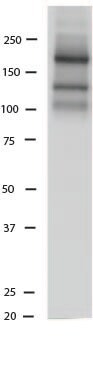
Detection of Human and Mouse NCAM‑1/CD56 by Western Blot.
Western blot shows lysates of human brain (cerebellum and motor cortex) tissue and mouse brain (cerebellum) tissue. PVDF membrane was probed with 0.5 µg/mL of Goat Anti-Human/Mouse NCAM-1/CD56 Antigen Affinity-purified Polyclonal Antibody (Catalog # AF2408) followed by HRP-conjugated Anti-Goat IgG Secondary Antibody (Catalog # HAF017). Specific bands were detected for NCAM-1/CD56 at approximately 100-150 kDa (as indicated). This experiment was conducted under reducing conditions and using Immunoblot Buffer Group 1.
NCAM-1/CD56 in SH-SY5Y Human Neuroblastoma Cells.
SH-SY5Y human neuroblastoma cells were cultured overnight in the presence of 1 μM Retinoic Acid (0695/50) prior to immersion fixation. Neural Cell Adhesion Molecule 1 (NCAM-1)/CD56 was detected using a Goat Anti-Human/Mouse NCAM-1/CD56 Antigen Affinity-purified Polyclonal Antibody (Catalog # AF2408). The cells were stained with the NorthernLights 557-conjugated Donkey Anti-Goat IgG Affinity-purified Secondary Antibody (red; Catalog # NL001). Actin filaments were stained with FITC-conjugated Phalloidin (green) and cell nuclei were counter-stained with DAPI (blue). NCAM-1/CD56 immuno-reactivity was localized to the plasma membrane. View our protocol for Fluorescent ICC Staining of Cells on Coverslips.
NCAM‑1/CD56 in BG01V Human Embryonic Stem Cells.
NCAM-1/CD56 was detected in immersion fixed BG01V human embryonic stem cells differentiated into neural progenitor cells using Goat Anti-Human/Mouse NCAM-1/CD56 Antigen Affinity-purified Polyclonal Antibody (Catalog # AF2408) at 10 µg/mL for 3 hours at room temperature. Cells were stained using the NorthernLights™ 557-conjugated Anti-Goat IgG Secondary Antibody (red; Catalog # NL001) and counterstained with DAPI (blue). Specific staining was localized to cytoplasm. View our protocol for Fluorescent ICC Staining of Stem Cells on Coverslips.
Detection of Human and Mouse NCAM‑1/CD56 by Simple WesternTM.
Simple Western lane view shows lysates of human and mouse brain (cerebellum) tissue, loaded at 0.2 mg/mL. A specific band was detected for NCAM-1/CD56 at approximately 143 kDa (as indicated) using 5 µg/mL for human lysates and 25 µg/mL for mouse lysates of Goat Anti-Human/Mouse NCAM-1/ CD56 Antigen Affinity-purified Polyclonal Antibody (Catalog # AF2408) followed by 1:50 dilution of HRP-conjugated Anti-Goat IgG Secondary Antibody (Catalog # HAF109). This experiment was conducted under reducing conditions and using the 12-230 kDa separation system.
Western Blot Shows Human NCAM‑1/CD56 Specificity by Using Knockout Cell Line.
Western blot shows lysates of U937 human histiocytic lymphoma cell line and human NCAM-1 knockout U937 human histiocytic lymphoma cell line (KO). PVDF membrane was probed with 0.25 µg/mL of Goat Anti-Human/Mouse NCAM‑1/CD56 Antigen Affinity-purified Polyclonal Antibody (Catalog # AF2408) followed by HRP-conjugated Anti-Goat IgG Secondary Antibody (HAF017). A specific band was detected for NCAM‑1/CD56 at approximately 160 kDa (as indicated) in the parental U937 human histiocytic lymphoma cell line, but is not detectable in knockout U937 human histiocytic lymphoma cell line. GAPDH (AF5718) is shown as a loading control. This experiment was conducted under reducing conditions and using Western Blot Buffer Group 1.
Human NCAM-1 / CD56 ELISA Standard Curve
Recombinant Human NCAM‑1/CD56 120 isoform (Catalog # 2408-NC) was serially diluted and captured by Mouse Anti-Human/Primate NCAM‑1/CD56 Monoclonal Antibody (Catalog # MAB2408) coated on a Clear Polystyrene Microplate (Catalog # DY990). Goat Anti-Human/Mouse NCAM‑1/CD56 Antigen Affinity-purified Polyclonal Antibody (Catalog # AF2408) was biotinylated and incubated with the protein captured on the plate. Detection of the standard curve was achieved by incubating Streptavidin-HRP (Catalog # DY998)
Human NCAM-1/CD56 DuoSet ELISA
Recombinant Human NCAM‑1/CD56 120 isoform (Catalog # 2408-NC) was serially diluted and captured by Mouse Anti-Human/Primate NCAM‑1/CD56 Monoclonal Antibody (Catalog # MAB2408) coated on a Clear Polystyrene Microplate (Catalog # DY990). Goat Anti-Human/Mouse NCAM‑1/CD56 Antigen Affinity-purified Polyclonal Antibody (Catalog # AF2408) was biotinylated and incubated with the protein captured on the plate. Detection of the standard curve was achieved by incubating Streptavidin-HRP (Catalog # DY998)Applications for NCAM-1/CD56 Antibody
Application
Recommended Usage
CyTOF-ready
Ready to be labeled using established conjugation methods. No BSA or other carrier proteins that could interfere with conjugation.
Flow Cytometry
0.25 µg/106 cells
Sample: Human peripheral blood mononuclear cells
Sample: Human peripheral blood mononuclear cells
Immunocytochemistry
5-15 µg/mL
Sample: Immersion fixed SH-SY5Y human neuroblastoma cell line and BG01V human embryonic stem cells differentiated into neural progenitor cells
Sample: Immersion fixed SH-SY5Y human neuroblastoma cell line and BG01V human embryonic stem cells differentiated into neural progenitor cells
Knockout Validated
NCAM‑1/CD56
is specifically detected in U937 human histiocytic lymphoma parental cell
line but is not detectable in NCAM‑1/CD56 knockout U937 human
histiocytic lymphoma cell line.
Simple Western
5-25 µg/mL
Sample: Human and mouse brain (cerebellum) tissue
Sample: Human and mouse brain (cerebellum) tissue
Western Blot
0.5 µg/mL
Sample: Human brain (cerebellum and motor cortex) tissue and mouse brain (cerebellum) tissue
Sample: Human brain (cerebellum and motor cortex) tissue and mouse brain (cerebellum) tissue
Reviewed Applications
Read 2 reviews rated 5 using AF2408 in the following applications:
Flow Cytometry Panel Builder
Bio-Techne Knows Flow Cytometry
Save time and reduce costly mistakes by quickly finding compatible reagents using the Panel Builder Tool.
Advanced Features
- Spectra Viewer - Custom analysis of spectra from multiple fluorochromes
- Spillover Popups - Visualize the spectra of individual fluorochromes
- Antigen Density Selector - Match fluorochrome brightness with antigen density

Formulation, Preparation, and Storage
Purification
Antigen Affinity-purified
Reconstitution
Reconstitute at 0.2 mg/mL in sterile PBS. For liquid material, refer to CoA for concentration.
Loading...
Formulation
Lyophilized from a 0.2 μm filtered solution in PBS with Trehalose. *Small pack size (SP) is supplied either lyophilized or as a 0.2 µm filtered solution in PBS.
Shipping
Lyophilized product is shipped at ambient temperature. Liquid small pack size (-SP) is shipped with polar packs. Upon receipt, store immediately at the temperature recommended below.
Stability & Storage
Use a manual defrost freezer and avoid repeated freeze-thaw cycles.
- 12 months from date of receipt, -20 to -70 °C as supplied.
- 1 month, 2 to 8 °C under sterile conditions after reconstitution.
- 6 months, -20 to -70 °C under sterile conditions after reconstitution.
Calculators
Background: NCAM-1/CD56
References
- Burg, M.A. et al. (1995) J. Neurosci. Res. 41:49.
- Storms, S.D. and U. Rutishauser (1998) J Biol. Chem. 273:27124.
- Margolis, R.K. et al. (1996) Perspect. Dev. Neurobiol. 3:273.
- Dickson, G. et al. (1987) Cell 50:1119.
- Lanier, L.L. et al. (1991) J. Immunol. 146:4421.
- Hemperly, J.J. et al. (1990) J. Mol. Neurosci. 2:71.
- Rutishauser, U.and C. Goridis (1986) Trends Genet. 2:72.
- Vawter, M.P. et al. (2001) Exp. Neurol. 172:29.
- Rutihauser, U. (1990) Adv. Exp. Med. Biol. 265:179.
- Becker, C.G. et al. (1996) J. Neurosci. Res. 45:143.
- Doherty, P. et al. (1995) J. Neurobiol. 26:437.
- Eckardt, M. et al. (2000) J. Neurosci. 20:5234.
- Muller, D. et al. (1996) Neuron 17:413.
Long Name
Neural Cell Adhesion Molecule
Alternate Names
CD56, NCAM1
Gene Symbol
NCAM1
UniProt
Additional NCAM-1/CD56 Products
Product Documents for NCAM-1/CD56 Antibody
Certificate of Analysis
To download a Certificate of Analysis, please enter a lot or batch number in the search box below.
Note: Certificate of Analysis not available for kit components.
Product Specific Notices for NCAM-1/CD56 Antibody
For research use only
Related Research Areas
Citations for NCAM-1/CD56 Antibody
 Powered by Bioz
Customer Reviews for NCAM-1/CD56 Antibody (2)
5 out of 5
2 Customer Ratings
Have you used NCAM-1/CD56 Antibody?
Submit a review and receive an Amazon gift card!
$25/€18/£15/$25CAN/¥2500 Yen for a review with an image
$10/€7/£6/$10CAN/¥1110 Yen for a review without an image
Submit a review
Customer Images
Showing
1
-
2 of
2 reviews
Showing All
Filter By:
-
Application: Western BlotSample Tested: Adult brain (hippocampus), Brain olfactory membrane fraction and mouse brainSpecies: MouseVerified Customer | Posted 04/15/2021This antibody is working great without non-specific bands.

-
Application: ImmunohistochemistrySample Tested: Mouse brain cancer sample and Mouse brain cancer tissueSpecies: MouseVerified Customer | Posted 01/19/2017Immunohistochemistry-Floating section
There are no reviews that match your criteria.
Protocols
Find general support by application which include: protocols, troubleshooting, illustrated assays, videos and webinars.
- 7-Amino Actinomycin D (7-AAD) Cell Viability Flow Cytometry Protocol
- Appropriate Fixation of IHC/ICC Samples
- Cellular Response to Hypoxia Protocols
- ClariTSA™ Fluorophore Kits
- Detection & Visualization of Antibody Binding
- Extracellular Membrane Flow Cytometry Protocol
- Flow Cytometry Protocol for Cell Surface Markers
- Flow Cytometry Protocol for Staining Membrane Associated Proteins
- Flow Cytometry Staining Protocols
- Flow Cytometry Troubleshooting Guide
- ICC Cell Smear Protocol for Suspension Cells
- ICC Immunocytochemistry Protocol Videos
- ICC for Adherent Cells
- Immunocytochemistry (ICC) Protocol
- Immunocytochemistry Troubleshooting
- Immunofluorescence of Organoids Embedded in Cultrex Basement Membrane Extract
- Immunohistochemistry (IHC) and Immunocytochemistry (ICC) Protocols
- Intracellular Flow Cytometry Protocol Using Alcohol (Methanol)
- Intracellular Flow Cytometry Protocol Using Detergents
- Intracellular Nuclear Staining Flow Cytometry Protocol Using Detergents
- Intracellular Staining Flow Cytometry Protocol Using Alcohol Permeabilization
- Intracellular Staining Flow Cytometry Protocol Using Detergents to Permeabilize Cells
- Preparing Samples for IHC/ICC Experiments
- Preventing Non-Specific Staining (Non-Specific Binding)
- Primary Antibody Selection & Optimization
- Propidium Iodide Cell Viability Flow Cytometry Protocol
- Protocol for Liperfluo
- Protocol for VisUCyte™ HRP Polymer Detection Reagent
- Protocol for the Characterization of Human Th22 Cells
- Protocol for the Characterization of Human Th9 Cells
- Protocol for the Fluorescent ICC Staining of Cell Smears - Graphic
- Protocol for the Fluorescent ICC Staining of Cultured Cells on Coverslips - Graphic
- Protocol for the Preparation and Fluorescent ICC Staining of Cells on Coverslips
- Protocol for the Preparation and Fluorescent ICC Staining of Non-adherent Cells
- Protocol for the Preparation and Fluorescent ICC Staining of Stem Cells on Coverslips
- Protocol for the Preparation of a Cell Smear for Non-adherent Cell ICC - Graphic
- Protocol: Annexin V and PI Staining by Flow Cytometry
- Protocol: Annexin V and PI Staining for Apoptosis by Flow Cytometry
- R&D Systems Quality Control Western Blot Protocol
- TUNEL and Active Caspase-3 Detection by IHC/ICC Protocol
- The Importance of IHC/ICC Controls
- Troubleshooting Guide: Fluorokine Flow Cytometry Kits
- Troubleshooting Guide: Western Blot Figures
- Western Blot Conditions
- Western Blot Protocol
- Western Blot Protocol for Cell Lysates
- Western Blot Troubleshooting
- Western Blot Troubleshooting Guide
- View all Protocols, Troubleshooting, Illustrated assays and Webinars
Loading...
Associated Pathways


