Tissue inhibitors of metalloproteinases or TIMPs are a family of proteins that regulate the activation and proteolytic activity of the zinc enzymes known as matrix metalloproteinases (MMPs). There are four members of the family, TIMP-1, TIMP-2, TIMP-3, and TIMP-4. TIMP-2 is a non N-glycosylated protein with a molecular mass of 22 kDa produced by a wide range of cell types, which inhibits MMPs non-covalently by the formation of binary complexes. TIMP-2 also has erythroid-potentiating and cell growth promoting activities.

Key Product Details
Validated by
Knockout/Knockdown
Species Reactivity
Validated:
Human, Mouse
Cited:
Human, Mouse
Applications
Validated:
Knockout Validated, Immunohistochemistry, Western Blot, Neutralization, Dual RNAscope ISH-IHC Compatible
Cited:
Immunohistochemistry, Immunohistochemistry-Frozen, Western Blot, Neutralization, Immunocytochemistry, Immunoprecipitation, Functional Assay
Label
Unconjugated
Antibody Source
Polyclonal Goat IgG
Loading...
Product Specifications
Immunogen
Chinese hamster ovary cell line CHO-derived recombinant human TIMP‑2
Cys27-Pro220
Accession # P16035
Cys27-Pro220
Accession # P16035
Specificity
Detects human and mouse TIMP‑2 in direct ELISAs and Western blots. In direct ELISAs, less than 2% cross‑reactivity with recombinant human TIMP-4 is observed.
Clonality
Polyclonal
Host
Goat
Isotype
IgG
Endotoxin Level
<0.10 EU per 1 μg of the antibody by the LAL method.
Scientific Data Images for TIMP-2 Antibody
Detection of Human TIMP‑2 by Western Blot.
Western blot shows Recombinant Human TIMP-2 Western Blot Standard Protein (Catalog # WBC023) and lysates of HeLa human cervical epithelial carcinoma cell line and human placenta tissue. PVDF membrane was probed with 1 µg/mL of Goat Anti-Human/Mouse TIMP-2 Antigen Affinity-purified Polyclonal Antibody (Catalog # AF971) followed by HRP-conjugated Anti-Goat IgG Secondary Antibody (Catalog # HAF017). A specific band was detected for TIMP-2 at approximately 22 kDa (as indicated). This experiment was conducted under reducing conditions and using Immunoblot Buffer Group 1.
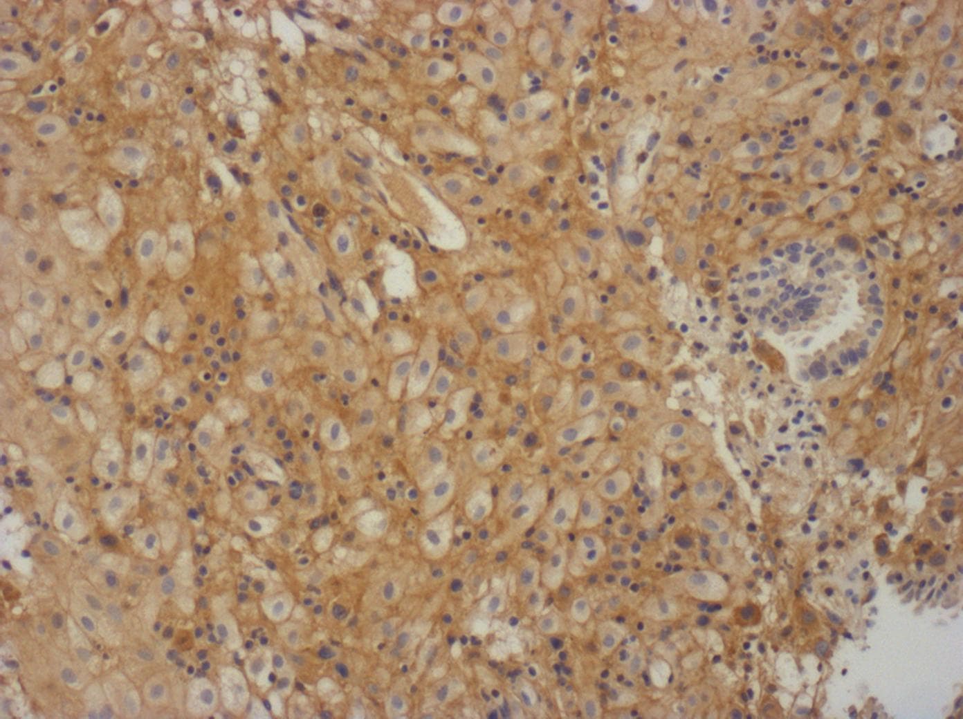
TIMP‑2 in Human Ovarian Cancer Tissue.
TIMP-2 was detected in immersion fixed paraffin-embedded sections of human ovarian cancer tissue using 15 µg/mL Goat Anti-Human/Mouse TIMP-2 Antigen Affinity-purified Polyclonal Antibody (Catalog # AF971) overnight at 4 °C. Tissue was stained with the Anti-Goat HRP-DAB Cell & Tissue Staining Kit (brown; Catalog # CTS008) and counter-stained with hematoxylin (blue). View our protocol for Chromogenic IHC Staining of Paraffin-embedded Tissue Sections.
Western Blot Shows Human TIMP‑2 Specificity by Using Knockout Cell Line.
Western blot shows lysates of HeLa human cervical epithelial carcinoma cell line and human TIMP-2 knockout HeLa human cervical epithelial carcinoma cell line (KO). PVDF membrane was probed with 1 µg/mL of Goat Anti-Human/Mouse TIMP‑2 Antigen Affinity-purified Polyclonal Antibody (Catalog # AF971) followed by HRP-conjugated Anti-Goat IgG Secondary Antibody (HAF017). A specific band was detected for TIMP‑2 at approximately 22 kDa (as indicated) in the parental HeLa human cervical epithelial carcinoma cell line, but is not detectable in knockout HeLa human cervical epithelial carcinoma cell line. GAPDH (AF5718) is shown as a loading control. This experiment was conducted under reducing conditions and using Western Blot Buffer Group 1.
Neutralization of TIMP‑2 Activity by Human TIMP‑2 Antibody.
Recombinant Human MMP-2 (0.2 µg/mL, Catalog # 902-MPNor 902-MP) activity is measured in the presence of Recombinant Human/Mouse TIMP-2 (0.143 µg/mL, Catalog # 971-TM) that has been preincubated with increasing concentrations of Goat Anti-Human TIMP-2 Antigen Affinity-purified Polyclonal Antibody (Catalog # AF971). The ND50 is typically 2.6 µg/mL.
Detection of TIMP‑2 in Human Placenta.
Formalin-fixed paraffin-embedded tissue sections of human placenta were probed for TIMP2 mRNA (ACD RNAScope Probe, catalog #470358; Fast Red chromogen, ACD catalog # 322750). Adjacent tissue section was processed for immunohistochemistry using goat anti-human TIMP2 polyclonal antibody (R&D Systems catalog # AF971) at 5ug/mL with overnight incubation at 4 degrees Celsius followed by incubation with anti-goat IgG VisUCyte HRP Polymer Antibody (Catalog # VC004) and DAB chromogen (yellow-brown). Tissue was counterstained with hematoxylin (blue). Specific staining was localized to decidual cells.
Human TIMP-2 ELISA Standard Curve
Recombinant Human TIMP‑2 (Catalog # 971-TM) was serially diluted and captured by Mouse Anti-Human TIMP‑2 Monoclonal Antibody (Catalog # MAB9711) coated on a Clear Polystyrene Microplate (Catalog # DY990). Goat Anti-Human/Mouse TIMP‑2 Antigen Affinity-purified Polyclonal Antibody (Catalog # AF971) was biotinylated and incubated with the protein captured on the plate. Detection of the standard curve was achieved by incubating Streptavidin-HRP (Catalog # DY998)Applications for TIMP-2 Antibody
Application
Recommended Usage
Dual RNAscope ISH-IHC Compatible
5-15 µg/mL
Sample: Immersion fixed paraffin-embedded sections of human placenta
Sample: Immersion fixed paraffin-embedded sections of human placenta
Immunohistochemistry
5-15 µg/mL
Sample: Immersion fixed paraffin-embedded sections of human ovarian cancer tissue and normal human ovarian array
Sample: Immersion fixed paraffin-embedded sections of human ovarian cancer tissue and normal human ovarian array
Knockout Validated
TIMP‑2
is specifically detected in HeLa human cervical epithelial carcinoma parental
cell line but is not detectable in TIMP‑2 knockout HeLa human
cervical epithelial carcinoma cell line.
Western Blot
1 µg/mL
Sample: Recombinant Human TIMP-2 Western Blot Standard Protein (Catalog # WBC023), HeLa human cervical epithelial carcinoma cell line and human placenta tissue
Sample: Recombinant Human TIMP-2 Western Blot Standard Protein (Catalog # WBC023), HeLa human cervical epithelial carcinoma cell line and human placenta tissue
Neutralization
Measured by its ability to neutralize Recombinant Human TIMP-2 (0.143 µg/mL, Catalog # 971-TM) inhibition of Recombinant Human MMP‑2 (0.2 µg/mL, Catalog # 902-MPN or 902-MP) cleavage of the fluorogenic peptide substrate Mca‑PLGL‑Dpa‑AR‑NH2 (10 µM, Catalog # ES001). The Neutralization Dose (ND50) is typically 2.6 µg/mL.
Reviewed Applications
Read 2 reviews rated 4 using AF971 in the following applications:
Formulation, Preparation, and Storage
Purification
Antigen Affinity-purified
Reconstitution
Reconstitute at 0.2 mg/mL in sterile PBS. For liquid material, refer to CoA for concentration.
Loading...
Formulation
Lyophilized from a 0.2 μm filtered solution in PBS with Trehalose. See Certificate of Analysis for details.
*Small pack size (-SP) is supplied either lyophilized or as a 0.2 µm filtered solution in PBS.
*Small pack size (-SP) is supplied either lyophilized or as a 0.2 µm filtered solution in PBS.
Shipping
Lyophilized product is shipped at ambient temperature. Liquid small pack size (-SP) is shipped with polar packs. Upon receipt, store immediately at the temperature recommended below.
Stability & Storage
Use a manual defrost freezer and avoid repeated freeze-thaw cycles.
- 12 months from date of receipt, -20 to -70 °C as supplied.
- 1 month, 2 to 8 °C under sterile conditions after reconstitution.
- 6 months, -20 to -70 °C under sterile conditions after reconstitution.
Calculators
Background: TIMP-2
Long Name
Tissue Inhibitors of Metalloproteinases 2
Alternate Names
TIMP2
Gene Symbol
TIMP2
UniProt
Additional TIMP-2 Products
Product Documents for TIMP-2 Antibody
Certificate of Analysis
To download a Certificate of Analysis, please enter a lot or batch number in the search box below.
Note: Certificate of Analysis not available for kit components.
Product Specific Notices for TIMP-2 Antibody
For research use only
Related Research Areas
Citations for TIMP-2 Antibody
 Powered by Bioz
Customer Reviews for TIMP-2 Antibody (2)
4 out of 5
2 Customer Ratings
Have you used TIMP-2 Antibody?
Submit a review and receive an Amazon gift card!
$25/€18/£15/$25CAN/¥2500 Yen for a review with an image
$10/€7/£6/$10CAN/¥1110 Yen for a review without an image
Submit a review
Customer Images
Showing
1
-
2 of
2 reviews
Showing All
Filter By:
-
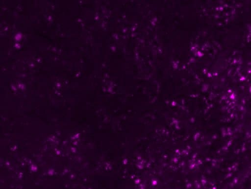
Application: Immunocytochemistry/ImmunofluorescenceSample Tested: Adult brainSpecies: MouseVerified Customer | Posted 02/16/2022Dilution 1:300

-
Application: ImmunohistochemistrySample Tested: Human first trimester deciduaSpecies: GoatVerified Customer | Posted 01/23/2017sodium citrate antigen retrieval
There are no reviews that match your criteria.
Protocols
Find general support by application which include: protocols, troubleshooting, illustrated assays, videos and webinars.
- Antigen Retrieval Protocol (PIER)
- Antigen Retrieval for Frozen Sections Protocol
- Appropriate Fixation of IHC/ICC Samples
- Cellular Response to Hypoxia Protocols
- Chromogenic IHC Staining of Formalin-Fixed Paraffin-Embedded (FFPE) Tissue Protocol
- Chromogenic Immunohistochemistry Staining of Frozen Tissue
- ClariTSA™ Fluorophore Kits
- Detection & Visualization of Antibody Binding
- Fluorescent IHC Staining of Frozen Tissue Protocol
- Graphic Protocol for Heat-induced Epitope Retrieval
- Graphic Protocol for the Preparation and Fluorescent IHC Staining of Frozen Tissue Sections
- Graphic Protocol for the Preparation and Fluorescent IHC Staining of Paraffin-embedded Tissue Sections
- Graphic Protocol for the Preparation of Gelatin-coated Slides for Histological Tissue Sections
- IHC Sample Preparation (Frozen sections vs Paraffin)
- ISH-IHC Protocol for Chromogenic Detection on Formalin Fixed Paraffin Embedded (FFPE) Tissue
- Immunofluorescent IHC Staining of Formalin-Fixed Paraffin-Embedded (FFPE) Tissue Protocol
- Immunohistochemistry (IHC) and Immunocytochemistry (ICC) Protocols
- Immunohistochemistry Frozen Troubleshooting
- Immunohistochemistry Paraffin Troubleshooting
- Preparing Samples for IHC/ICC Experiments
- Preventing Non-Specific Staining (Non-Specific Binding)
- Primary Antibody Selection & Optimization
- Protocol for Heat-Induced Epitope Retrieval (HIER)
- Protocol for Making a 4% Formaldehyde Solution in PBS
- Protocol for VisUCyte™ HRP Polymer Detection Reagent
- Protocol for the Preparation & Fixation of Cells on Coverslips
- Protocol for the Preparation and Chromogenic IHC Staining of Frozen Tissue Sections
- Protocol for the Preparation and Chromogenic IHC Staining of Frozen Tissue Sections - Graphic
- Protocol for the Preparation and Chromogenic IHC Staining of Paraffin-embedded Tissue Sections
- Protocol for the Preparation and Chromogenic IHC Staining of Paraffin-embedded Tissue Sections - Graphic
- Protocol for the Preparation and Fluorescent IHC Staining of Frozen Tissue Sections
- Protocol for the Preparation and Fluorescent IHC Staining of Paraffin-embedded Tissue Sections
- Protocol for the Preparation of Gelatin-coated Slides for Histological Tissue Sections
- R&D Systems Quality Control Western Blot Protocol
- TUNEL and Active Caspase-3 Detection by IHC/ICC Protocol
- The Importance of IHC/ICC Controls
- Troubleshooting Guide: Immunohistochemistry
- Troubleshooting Guide: Western Blot Figures
- Western Blot Conditions
- Western Blot Protocol
- Western Blot Protocol for Cell Lysates
- Western Blot Troubleshooting
- Western Blot Troubleshooting Guide
- View all Protocols, Troubleshooting, Illustrated assays and Webinars
Loading...