Key Product Details

Validated by

Biological Validation

Species Reactivity

Validated:

Human

Cited:

Human, Mouse, Chinese Hamster, Feline, Primate, Primate - Cercocebus atys (Sooty Mangabey), Primate - Macaca mulatta (Rhesus Macaque)

Applications

Validated:

Immunohistochemistry, Western Blot, ELISA Capture (Matched Antibody Pair), Blockade of Receptor-ligand Interaction, Flow Cytometry, Dual RNAscope ISH-IHC Compatible, CyTOF-ready

Cited:

Immunohistochemistry, Immunohistochemistry-Paraffin, Western Blot, Neutralization, Flow Cytometry, Immunocytochemistry, Bioassay, ELISA Development, Functional Assay, Microarray, Surface Plasmon Resonance (SPR)

Label

Unconjugated

Antibody Source

Polyclonal Goat IgG
Loading...

Product Specifications

Immunogen

Mouse myeloma cell line NS0-derived recombinant human PD-1
Leu25-Gln167
Accession # Q8IX89

Specificity

Detects human PD-1 in ELISAs and Western blots. In sandwich ELISAs, less than 2% cross-reactivity with recombinant mouse PD‑1 and less than 0.2% cross-reactivity with recombinant human (rh) CD28, rhICOS, and rhCTLA‑4 is observed.

Clonality

Polyclonal

Host

Goat

Isotype

IgG

Endotoxin Level

<0.10 EU per 1 μg of the antibody by the LAL method.

Scientific Data Images for Human PD‑1 Antibody

Detection of Human PD‑1 antibody by Western Blot.

Detection of Human PD‑1 by Western Blot.

Western blot shows lysate of human thymus tissue. PVDF membrane was probed with 2 µg/mL of Goat Anti-Human PD-1 Antigen Affinity-purified Polyclonal Antibody (Catalog # AF1086) followed by HRP-conjugated Anti-Goat IgG Secondary Antibody (Catalog # HAF017). Specific bands were detected for PD-1 at approximately 40-50 kDa (as indicated). This experiment was conducted under reducing conditions and using Immunoblot Buffer Group 1.

Detection of Human PD-1 antibody by Western Blot.

Detection of Human PD‑1 by Western Blot.

Western blot shows lysates of HEK293 human embryonic kidney cell line either mock transfected or transfected with human PD-1. PVDF membrane was probed with 0.5 µg/mL of Goat Anti-Human PD-1 Antigen Affinity-purified Polyclonal Antibody (Catalog # AF1086) followed by HRP-conjugated Anti-Goat IgG Secondary Antibody (Catalog # HAF017). Specific bands were detected for PD-1 at approximately 40-80 kDa (as indicated). This experiment was conducted under reducing conditions and using Immunoblot Buffer Group 1.

Detection of PD-1 antibody in Human PBMCs treated with PHA antibody by Flow Cytometry.

Detection of PD‑1 in Human PBMCs treated with PHA by Flow Cytometry.

Human peripheral blood mononuclear cells (PBMCs) either (A) untreated or (B) treated with 5 µg/mL PHA overnight were stained with Goat Anti-Human PD-1 Antigen Affinity-purified Polyclonal Antibody (Catalog # AF1086) followed by Phycoerythrin-conjugated Anti-Goat IgG Secondary Antibody (Catalog # F0107) and Mouse Anti-Human CD3e APC-conjugated Monoclonal Antibody (Catalog # FAB100A). Quadrant markers were set based on control antibody staining (Catalog # F0107). View our protocol for Staining Membrane-associated Proteins.

PD-1 antibody in Human Lymph Node by Immunohistochemistry (IHC-P).

PD‑1 in Human Lymph Node.

PD-1 was detected in immersion fixed paraffin-embedded sections of human lymph node using Goat Anti-Human PD-1 Antigen Affinity-purified Polyclonal Antibody (Catalog # AF1086) at 3 µg/mL overnight at 4 °C. Tissue was stained using the Anti-Goat HRP-DAB Cell & Tissue Staining Kit (brown; Catalog # CTS008) and counterstained with hematoxylin (blue). Specific staining was localized to lymphocytes. View our protocol for Chromogenic IHC Staining of Paraffin-embedded Tissue Sections.

Detection of Human PD-1 by Immunohistochemistry

Detection of Human PD-1 by Immunohistochemistry

PD-L1, PD-L2, PD-1, CD8, and CD4 expression in p16-positive and p16-negative HNSCC. PD-L1, PD-L2, PD-1, CD8, and CD4 expression was assessed in tumor biopsy tissue from five p16-positive and four p16-negative HNSCC patients using immuno-histochemistry (details in Methods). Representative staining (scale bars, 100 μm) and cumulative data of marker expression (grading scale PD-L1/PD-L2: 1, low; 2, moderate; 3 high expression; grading scale PD-1, CD8, CD4: 1, <50 cells/field; 2, 50–150 cells/field; 3, >150 cells/field). Image collected and cropped by CiteAb from the following publication (https://pubmed.ncbi.nlm.nih.gov/31379843), licensed under a CC-BY license. Not internally tested by R&D Systems.
Detection of Human PD-1 by ELISA

Detection of Human PD-1 by ELISA

Modulation of soluble co-signaling molecules in kidney-transplanted patients over time.The levels of the soluble co-signaling molecules CD30, CD40, CD137, CD40L, PD-1 and PD-L1 were assayed by ELISA in serum samples of healthy controls (n = 25) and kidney-transplanted patients (n = 59) obtained at different times: just before transplantation, and 15 days, 3 months and 1 year after transplantation. Data are shown as box-plots, in which the horizontal line within each box represents the median, the bottom and top of each box represent the 25th and 75th percentiles, the bars represent the 10th and 90th percentiles and circles indicate outliers. Unpaired and paired Wilcoxon tests were used to compare distributions between independent and dependent groups, respectively. * indicates statistically significant differences between healthy controls and kidney-transplanted patient samples, and † indicates statistically significant differences between patients samples obtained at different pre- and post-transplantation times. Image collected and cropped by CiteAb from the following publication (https://pubmed.ncbi.nlm.nih.gov/25478957), licensed under a CC-BY license. Not internally tested by R&D Systems.

Detection of PD‑1 in Human Tonsil.

Formalin-fixed paraffin-embedded tissue sections of human tonsil were probed for PD1 mRNA (ACD RNAScope Probe, catalog #602021; Fast Red chromogen, ACD catalog # 322750). Adjacent tissue section was processed for immunohistochemistry using goat anti-human PD1 polyclonal antibody (R&D Systems catalog # AF1086) at 1ug/mL with overnight incubation at 4 degrees Celsius followed by incubation with anti-goat IgG VisUCyte HRP Polymer Antibody (Catalog # VC004) and DAB chromogen (yellow-brown). Tissue was counterstained with hematoxylin (blue). Specific staining was localized to cell surface.

Detection of PD-1 by Flow Cytometry

Detection of PD-1 by Flow Cytometry

The expression of PD-1 on CD4+T cells CD8+T cells and CD56+NK cells in silicosis. a Gating strategy and expression profiles of PD-1 in peripheral CD4+T cells CD8+T cells and CD56+NK cells in TB group, SILs group, SILs&LTBI group TB&SILs-P group and TB&SILs-N group. b The expression of PD-1 on CD4+T cells in TB group, SILs group, SILs&LTBI group TB&SILs-P group and TB&SILs-N group; c The expression of PD-1 on CD8+T cells in TB group, SILs group, SILs&LTBI group TB&SILs-P group and TB&SILs-N group; d The expression of PD-1 on CD56+NK cells in TB group, SILs group, SILs&LTBI group TB&SILs-P group and TB&SILs-N group; e Analyzed the correlation between the level of IFN-gamma and expression of PD-1 on CD4+T cells; f Analyzed the correlation between the level of IFN-gamma and expression of PD-1 on CD8+T cells; g Analyzed the correlation between the level of IFN-gamma and expression of PD-1 on CD56+NK cells. *p < 0.05; **p < 0.01; ***p < 0.001 Image collected and cropped by CiteAb from the following open publication (https://pubmed.ncbi.nlm.nih.gov/36587204), licensed under a CC-BY license. Not internally tested by R&D Systems.

Applications for Human PD‑1 Antibody

Application
Recommended Usage

Blockade of Receptor-ligand Interaction

Measured by its ability to block the binding of Recombinant Human B7-H1 Fc Chimera (Catalog #156-B7). The Neutralization Dose (ND50) is typically <12 ug/mL.

CyTOF-ready

Ready to be labeled using established conjugation methods. No BSA or other carrier proteins that could interfere with conjugation.

Dual RNAscope ISH-IHC Compatible

5-15 µg/mL
Sample: Immersion fixed paraffin-embedded sections of human tonsil

Flow Cytometry

0.25 µg/106 cells
Sample: Human peripheral blood mononuclear cells (PBMCs) treated with PHA

Immunohistochemistry

5-15 µg/mL
Sample: Immersion fixed paraffin-embedded sections of human lymph node

Western Blot

0.5-2 µg/mL
Sample: Human thymus tissue and HEK293 human embryonic kidney cell line transfected with human PD-1

Human PD-1 Sandwich Immunoassay

ELISA Capture (Matched Antibody Pair)
Recommended Concentration: 0.2-0.8 µg/mL
Use in combination with these reagents:
  • Detection Reagent: Human PD‑1 Biotinylated Antibody (Catalog # BAF1086)
  • Standard: Recombinant Human PD-1 Fc Chimera Protein, CF (Catalog # 1086-PD)
Please Note: Optimal dilutions of this antibody should be experimentally determined.

Reviewed Applications

Read 8 reviews rated 4.5 using AF1086 in the following applications:

Flow Cytometry Panel Builder

Bio-Techne Knows Flow Cytometry

Save time and reduce costly mistakes by quickly finding compatible reagents using the Panel Builder Tool.

Advanced Features

  • Spectra Viewer - Custom analysis of spectra from multiple fluorochromes
  • Spillover Popups - Visualize the spectra of individual fluorochromes
  • Antigen Density Selector - Match fluorochrome brightness with antigen density
Build Your Panel Now

Formulation, Preparation, and Storage

Purification

Antigen Affinity-purified

Reconstitution

Reconstitute at 0.2 mg/mL in sterile PBS. For liquid material, refer to CoA for concentration.


Loading...

Formulation

Lyophilized from a 0.2 μm filtered solution in PBS with Trehalose. See Certificate of Analysis for details.
*Small pack size (-SP) is supplied either lyophilized or as a 0.2 µm filtered solution in PBS.

Shipping

Lyophilized product is shipped at ambient temperature. Liquid small pack size (-SP) is shipped with polar packs. Upon receipt, store immediately at the temperature recommended below.

Stability & Storage

Use a manual defrost freezer and avoid repeated freeze-thaw cycles.
  • 12 months from date of receipt, -20 to -70 °C as supplied.
  • 1 month, 2 to 8 °C under sterile conditions after reconstitution.
  • 6 months, -20 to -70 °C under sterile conditions after reconstitution.

Calculators

The reconstitution calculator allows you to quickly calculate the volume of a reagent to reconstitute your vial. Simply enter the mass of reagent and the target concentration and the calculator will determine the rest.

=
÷

Background: PD-1

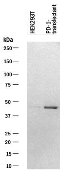
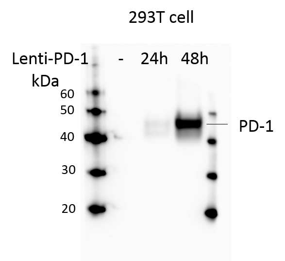
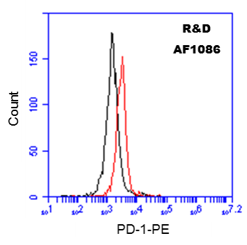
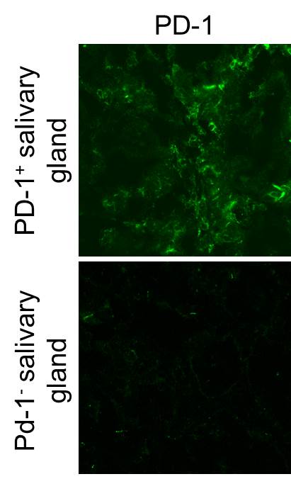
Programmed Death-1 (PD-1) is a type I transmembrane protein belonging to the CD28/CTLA-4 family of immunoreceptors that mediate signals for regulating immune responses (1). Members of the CD28/CTLA-4 family have been shown to either promote T cell activation (CD28 and ICOS) or down-regulate T cell activation (CTLA-4 and PD-1) (2). PD-1 is expressed on activated T cells, B cells, myeloid cells, and on a subset of thymocytes. In vitro, ligation of PD-1 inhibits TCR-mediated T-cell proliferation and production of IL-1, IL-4, IL-10, and IFN-gamma. In addition, PD-1 ligation also inhibits BCR mediated signaling. PD-1 deficient mice have a defect in peripheral tolerance and spontaneously develop autoimmune diseases (2, 3).

Two B7 family proteins, PD-L1 (also called B7-H1) and PD-L2 (also known as B7-DC), have been identified as PD-1 ligands. Unlike other B7 family proteins, both
PD‑L1 and PD-L2 are expressed in a wide variety of normal tissues including heart, placenta, and activated spleens (4). The wide expression of PD-L1 and PD-L2 and the inhibitor effects on PD-1 ligation indicate that PD-1 might be involved in the regulation of peripheral tolerance and may help prevent autoimmune diseases (2).

The human PD-1 gene encodes a 288 amino acid (aa) protein with a putative 20 aa signal peptide, a 148 aa extracellular region with one immunoglobulin-like V-type domain, a 24 aa transmembrane domain, and a 95 aa cytoplasmic region. The cytoplasmic tail contains two tyrosine residues that form the immuno-receptor
tyrosine-based inhibitory motif (ITIM) and immunoreceptor tyrosine-based switch motif (ITSM) that are important in mediating PD-1 signaling. Mouse and human PD-1 share approximately 60% aa sequence identity (4).

References

  1. Ishida, Y. et al. (1992) EMBO J. 11:3887.
  2. Nishimura, H. and T. Honjo (2001) Trends in Immunol. 22:265.
  3. Latchman, Y. et al. (2001) Nature Immun. 2:261.
  4. Carreno, B.M. and M. Collins (2002) Annu. Rev. Immunol. 20:29.

Long Name

Programmed Death-1

Alternate Names

CD279, PD1, PDCD1, SLEB2

Entrez Gene IDs

5133 (Human); 18566 (Mouse); 301626 (Rat); 100533201 (Porcine); 486213 (Canine); 102123659 (Cynomolgus Monkey)

Gene Symbol

PDCD1

UniProt

Additional PD-1 Products

Product Documents for Human PD‑1 Antibody

Certificate of Analysis

To download a Certificate of Analysis, please enter a lot or batch number in the search box below.

Note: Certificate of Analysis not available for kit components.

Product Specific Notices for Human PD‑1 Antibody

For research use only

Citations for Human PD‑1 Antibody

Customer Reviews for Human PD‑1 Antibody (8)

4.5 out of 5
8 Customer Ratings
5 Stars
50%
4 Stars
50%
3 Stars
0%
2 Stars
0%
1 Stars
0%

Have you used Human PD‑1 Antibody?

Submit a review and receive an Amazon gift card!

$25/€18/£15/$25CAN/¥2500 Yen for a review with an image

$10/€7/£6/$10CAN/¥1110 Yen for a review without an image

Submit a review
Amazon Gift Card

Customer Images


Showing  1 - 5 of 8 reviews Showing All
Filter By:
  • Human PD-1 Antibody
    Name: Negar FIROOZI
    Application: Western Blot
    Sample Tested: 293T human embryonic kidney cell line
    Species: Human
    Verified Customer | Posted 08/28/2023
    Letivirus carrying human PD-1 was used to infect 293T cells. The membrane was exposed to a concentration of 1 µg/mL Human PD‑1 Monoclonal Antibody. PD‑1 band was identified at approximately 45 kDa.
    Human PD‑1 Antibody AF1086
  • Human PD-1 Antibody
    Name: Anonymous
    Application: Immunocytochemistry/Immunofluorescence
    Sample Tested: Melanoma tissue
    Species: Human
    Verified Customer | Posted 11/23/2020
    Human PD‑1 Antibody AF1086
  • Human PD-1 Antibody
    Name: Anonymous
    Application: Western Blot
    Sample Tested: 293T human embryonic kidney cell line
    Species: Human
    Verified Customer | Posted 07/23/2018
    293T cells were infected by lentivirus overexpression of control or PD-1 for 24 and 48 h. Total cell lysates were subjected to western blot. PVDF membrane were probed with 1 um/ml Human PD-1 Antibody (AF1086). A specific band was detected for PD-1 at approximately 43 kDa. This experiment was conducted under reducing conditions
    Human PD‑1 Antibody AF1086
  • Human PD-1 Antibody
    Name: Casey Keyes
    Application: ELISA
    Sample Tested: Human recombinant antibody
    Species: Human
    Verified Customer | Posted 03/07/2018
  • Name: Anonymous
    Application: Simple Western
    Sample Tested: 2120Ep human embryonal carcinoma cell line
    Species: Human
    Verified Customer | Posted 11/24/2017
  • Human PD-1 Antibody
    Name: Anonymous
    Application: ELISA
    Sample Tested: Human recombinant test antibody
    Species: Human
    Verified Customer | Posted 09/29/2017
  • Human PD-1 Antibody
    Name: Anonymous
    Application: Flow Cytometry
    Sample Tested: Peripheral blood mononuclear cells (PBMCs)
    Species: Human
    Verified Customer | Posted 03/14/2017
    10^6 Human PBMCs were probed with 0.5 ug of Goat anti-Human PD-1 antibody (red), or Goat Isotype Control IgG (black), followed by PE-conjugated anti-Goat secondary antibody.
    Human PD‑1 Antibody AF1086
  • Name: Christopher England
    Application: Immunofluorescence
    Sample Tested: Mouse salivary gland (one injected with human T cells and one with no human T cells)
    Species: Human
    Verified Customer | Posted 07/09/2016
    Staining of PD-1 expression in Salivary Gland Tissue sections
    Human PD‑1 Antibody AF1086

There are no reviews that match your criteria.

Showing  1 - 5 of 8 reviews Showing All

Protocols

Find general support by application which include: protocols, troubleshooting, illustrated assays, videos and webinars.

FAQs

No product specific FAQs exist for this product.

View all FAQs for Antibodies