Interleukin 10 (IL-10) is a pleiotropic cytokine that can exert either immunostimulatory or immunosuppressive effects on a variety of cell types. It is produced predominantly by Th2 lymphocytes and other activated lymphoid cells.

Key Product Details
Validated by
Biological Validation
Species Reactivity
Validated:
Rat
Cited:
Rat
Applications
Validated:
Western Blot, ELISA Capture (Matched Antibody Pair)
Cited:
ELISA Development
Label
Unconjugated
Antibody Source
Monoclonal Mouse IgG2B Clone # 67232
Loading...
Product Specifications
Immunogen
E. coli-derived recombinant rat IL‑10
Ser19-Asn178
Accession # P29456
Ser19-Asn178
Accession # P29456
Specificity
Detects rat IL-10 in ELISAs and Western blots. In Western blots, shows approximately 15% cross-reactivity with recombinant mouse IL‑10 and less than 2% cross-reactivity with recombinant human IL-10.
Clonality
Monoclonal
Host
Mouse
Isotype
IgG2B
Scientific Data Images for Rat IL-10 Antibody (67232)
Detection of Recombinant Mouse and Rat IL‑10 by Western Blot.
Western blot shows 25 ng of Recombinant Rat IL-10 (Catalog # 522-RLB), Recombinant Human IL-10 (Catalog # 217-IL) and Recombinant Mouse IL-10 (Catalog # 417-ML). PVDF Membrane was probed with 1 µg/mL of Mouse Anti-Rat IL-10 Monoclonal Antibody (Catalog # MAB519) followed by HRP-conjugated Anti-Mouse IgG Secondary Antibody (Catalog # HAF007). A specific band was detected for IL-10 at approximately 15 kDa (as indicated). This experiment was conducted under reducing conditions and using Immunoblot Buffer Group 3.
Detection of Rat IL-10 by Immunohistochemistry
Nrg-1 promotes Treg cell response in the injured spinal cord. a, b Representative images of the gating strategy for flow cytometry after singlet selection are provided for vehicle- and Nrg-1-treated groups. c At 3-day post-injury, the number of CD3+CD4+ T cells in the spinal cord was significantly higher in vehicle-treated animals compared to uninjured control group. There was no significant difference in the population of infiltrated helper T cells and the number of FoxP3+ Treg cells (CD3+CD4+FoxP3+) between vehicle- and Nrg-1-treated groups. However, the population of IL-10 producing CD4+ T cells (CD3+CD4+IL-10+) was significantly increased in Nrg-1-treated animals in comparison to vehicle-treated group. d At 7 days post-SCI, despite no significant difference in the total helper and regulatory T cell populations between vehicle- and Nrg-1-treated groups, a significant reduction (1.9-fold) was observed in IFN gamma producing effector T cell population in Nrg-1-treated SCI rats compared to their vehicle-treated counterparts. e At 14 days post-SCI, infiltrated helper T cells reached their lowest level among all examined time-points, and Nrg-1 treatment had no significant effect on the total helper T cell population. IL-10 expressing Treg cells were significantly higher in both vehicle and Nrg-1 injured rats compared to uninjured animals. Nrg-1-treated animals showed a significantly higher number of Treg cells in their spinal cord at 14-day time-point compared to vehicle-treated rats. f However, at chronic (42-day) time-point, the number of T helper cells reached a maximum, with Nrg-1-treated animals harboring a significantly decreased population of CD4+ T-cells in their spinal cord compared to the vehicle-treated group. Most importantly, CD3+CD4+FoxP3+ and CD3+CD4+IL-10+ regulatory T cell populations were significantly increased in Nrg-1-treated groups. g Immunohistochemical images show the presence of Treg cells in the perilesional area (N = 5/group/time-point, *p < 0.05
Detection of Rat IL-10 by Immunohistochemistry
Nrg-1 treatment promotes Breg cell population following SCI. a Representative images of the gating strategy for flow cytometry of spinal cord are provided. b At 7 days post-injury, the number of CD45RA+ B cells was significantly increased in the spinal cord without any significant difference in the total and regulatory B cell populations between vehicle- and Nrg-1-treated groups. c At 14-day post-SCI, a significant increase in the number of Breg cells was observed in Nrg-1-treated animals compared to vehicle-treated group. d Chronically at 42 days post-SCI, the number of B cells in the spinal cord reached the highest level compared to all earlier time-points. Nrg-1 treatment resulted in a significant increase in the number of infiltrated B cells in the spinal cord and promoted IL-10 expressing Breg cells compared to vehicle treatment. e Immunohistochemical analysis verified the presence of Breg cells in the perilesional area of the injured spinal cord. f Representative images of the gating strategy for flow cytometry of blood are provided. g Analysis of the blood revealed a significant decline in B cell population at 7 days post-SCI without any significant change in the Breg cell population at this time-point. h, i No significant change in total and regulatory B cell populations were observed in the blood at 14- and 42-day time-points. (N = 5/group/time-point, *p < 0.05, **p < 0.01, ***p < 0.001, one-way ANOVA followed by Holm-Sidak post hoc test) Image collected and cropped by CiteAb from the following publication (https://jneuroinflammation.biomedcentral.com/articles/10.1186/s12974-01…), licensed under a CC-BY license. Not internally tested by R&D Systems.
Detection of Rat IL-10 by Immunohistochemistry
Comparison between P1 and P12 immunostaining intensities of IL-6 and IL-10 in brain. IL-6 and IL-10 expressions were increased only at P12 in brains of rat pups exposed to HI+/- LPS. Data are presented (A, C) as fold increase of IL-6 and IL-10 expressions compared to control (set at 1). (B) Increased IL-6 staining (arrowheads) in P12 spongiotic frontal cortex (pyknotic neurons with abnormal reduced contrast between nucleus and cytoplasm), and spongiotic underlying external capsule following LPS+HI exposure compared to control. (D) Increased IL-10 staining (arrowheads) in P12 lesioned frontal cortex and underlying external capsule exposed to LPS+HI compared to control. IHC was performed at 48 h post-HI, in 3 to 4 brains under each experimental condition. *p < 0.05, **p < 0.01, ***p < 0.001, one-way ANOVA with Newman-Keuls post test. Scale bars = 15 μm (B, D) Image collected and cropped by CiteAb from the following open publication (https://pubmed.ncbi.nlm.nih.gov/21599903), licensed under a CC-BY license. Not internally tested by R&D Systems.
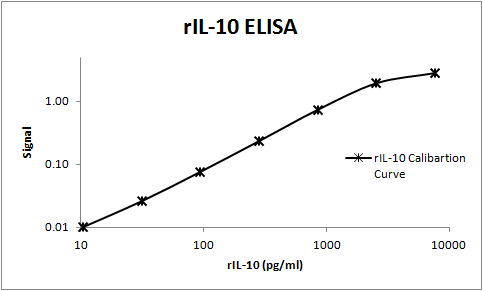
Rat IL-10 ELISA Standard Curve
Rat IL-10 was serially diluted and captured by Mouse Anti-Rat IL‑10 Monoclonal Antibody (Catalog # MAB519) coated on a Clear Polystyrene Microplate (Catalog # DY990). Goat Anti-Mouse/Rat IL‑10 Antigen Affinity-purified Polyclonal Antibody (Catalog # AF519) was biotinylated and incubated with the protein captured on the plate. Detection of the standard curve was achieved by incubating Streptavidin-HRP (Catalog # DY998)Applications for Rat IL-10 Antibody (67232)
Application
Recommended Usage
Western Blot
1 µg/mL
Sample: Recombinant Rat IL‑10 (Catalog # 522-RLB)
Sample: Recombinant Rat IL‑10 (Catalog # 522-RLB)
Rat IL-10 Sandwich Immunoassay
Please Note: Optimal dilutions of this antibody should be experimentally determined.
Reviewed Applications
Read 1 review rated 5 using MAB519 in the following applications:
Formulation, Preparation, and Storage
Purification
Protein A or G purified from hybridoma culture supernatant
Reconstitution
Reconstitute at 0.5 mg/mL in sterile PBS. For liquid material, refer to CoA for concentration.
Loading...
Formulation
Lyophilized from a 0.2 μm filtered solution in PBS with Trehalose. *Small pack size (SP) is supplied either lyophilized or as a 0.2 µm filtered solution in PBS.
Shipping
Lyophilized product is shipped at ambient temperature. Liquid small pack size (-SP) is shipped with polar packs. Upon receipt, store immediately at the temperature recommended below.
Stability & Storage
Use a manual defrost freezer and avoid repeated freeze-thaw cycles.
- 12 months from date of receipt, -20 to -70 °C as supplied.
- 1 month, 2 to 8 °C under sterile conditions after reconstitution.
- 6 months, -20 to -70 °C under sterile conditions after reconstitution.
Calculators
Background: IL-10
Long Name
Interleukin 10
Alternate Names
CSIF, GVHDS, IL10, IL10A, TGIF
Entrez Gene IDs
Gene Symbol
IL10
UniProt
Additional IL-10 Products
Product Documents for Rat IL-10 Antibody (67232)
Certificate of Analysis
To download a Certificate of Analysis, please enter a lot or batch number in the search box below.
Note: Certificate of Analysis not available for kit components.
Product Specific Notices for Rat IL-10 Antibody (67232)
For research use only
Related Research Areas
Citations for Rat IL-10 Antibody (67232)
 Powered by Bioz
Customer Reviews for Rat IL-10 Antibody (67232) (1)
5 out of 5
1 Customer Rating
Have you used Rat IL-10 Antibody (67232)?
Submit a review and receive an Amazon gift card!
$25/€18/£15/$25CAN/¥2500 Yen for a review with an image
$10/€7/£6/$10CAN/¥1110 Yen for a review without an image
Submit a review
Customer Images
Showing
1
-
1 of
1 review
Showing All
Filter By:
-
Application: ELISASample Tested: SerumSpecies: RatVerified Customer | Posted 11/08/2017This antibody was used to build an ELISA to measure rat IL-10. This antibody worked well as a capture antibody and BAF519 as the detection antibody.

There are no reviews that match your criteria.
Protocols
Find general support by application which include: protocols, troubleshooting, illustrated assays, videos and webinars.
- Cellular Response to Hypoxia Protocols
- R&D Systems Quality Control Western Blot Protocol
- Troubleshooting Guide: Western Blot Figures
- Western Blot Conditions
- Western Blot Protocol
- Western Blot Protocol for Cell Lysates
- Western Blot Troubleshooting
- Western Blot Troubleshooting Guide
- View all Protocols, Troubleshooting, Illustrated assays and Webinars
Loading...
Associated Pathways


