Interleukin 6 (IL-6) is a pleiotropic alpha -helical cytokine that plays important roles in acute phase reactions, inflammation, hematopoiesis, bone metabolism, and cancer progression. IL-6 activity is central to the transition from acute inflammation to either acquired immunity or chronic inflammatory disease. It is secreted by multiple cell types as a 22 kDa‑28 kDa phosphorylated and variably glycosylated molecule (1‑4). Mature canine IL-6 is 187 amino acids (aa) in length and shares 76%, 59%, 38%, and 40% aa sequence identity with feline, human, mouse, and rat IL-6, respectively (5). IL-6 induces signaling through a cell surface heterodimeric receptor complex composed of a ligand binding subunit (IL-6 R) and a signal transducing subunit (gp130). IL-6 binds to IL-6 R, triggering IL-6 R association with gp130 and gp130 dimerization (6). gp130 is also a component of the receptors for CLC, CNTF, CT-1, IL-11, IL-27, LIF, and OSM (7). Soluble forms of IL-6 R are generated by both alternate splicing and proteolytic cleavage (3). In a mechanism known as trans-signaling, complexes of soluble IL-6 and IL-6 R elicit responses from gp130‑expressing cells that lack cell surface IL-6 R (3). Trans-signaling enables a wider range of cell types to respond to IL-6, as the expression of gp130 is ubiquitous while that of IL-6 R is predominantly restricted to hepatocytes, leukocytes, and lymphocytes (3). Soluble splice forms of gp130 block trans-signaling from IL-6/IL-6 R but not from other cytokines that utilize gp130 as a co‑receptor (4, 8).

Key Product Details
Species Reactivity
Canine
Applications
Western Blot, Immunocytochemistry
Label
Unconjugated
Antibody Source
Monoclonal Mouse IgG2B Clone # 247002
Loading...
Product Specifications
Immunogen
E. coli-derived recombinant canine IL‑6
Thr23-Met207
Accession # P41323
Thr23-Met207
Accession # P41323
Specificity
Detects canine IL-6 in direct ELISAs and Western blots. In direct ELISAs and Western blots, approximately 10%‑50% cross‑reactivity with recombinant eqine IL-6, recombinant human (rh) IL-6, recombinant mouse (rm) IL‑6, recombinant porcine IL-6, and recombinant rat (rr) IL-6 is observed and no cross-reactivity with rmCardiotrophin-1, rhCLC, rrCNTF, recombinant cotton rat IL-6, rmIL-11, rmLIF, or rmOncostatin M is observed.
Clonality
Monoclonal
Host
Mouse
Isotype
IgG2B
Scientific Data Images for Canine IL-6 Antibody (247002)
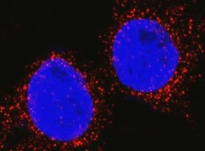
IL‑6 in Canine PBMCs.
IL-6 was detected in immersion fixed canine peripheral blood mononuclear cells (PBMCs) using Mouse Anti-Canine IL-6 Monoclonal Antibody (Catalog # MAB16091) at 25 µg/mL for 3 hours at room temperature. Cells were stained using the NorthernLights™ 557-conjugated Anti-Mouse IgG Secondary Antibody (red; Catalog # NL007) and counterstained with DAPI (blue). Specific staining was localized to cytoplasm. View our protocol for Fluorescent ICC Staining of Non-adherent Cells.Applications for Canine IL-6 Antibody (247002)
Application
Recommended Usage
Immunocytochemistry
10-30 µg/mL
Sample: Immersion fixed canine peripheral blood mononuclear cells
Sample: Immersion fixed canine peripheral blood mononuclear cells
Western Blot
1 µg/mL
Sample: Recombinant Canine IL‑6 (Catalog # 1609-CL)
Sample: Recombinant Canine IL‑6 (Catalog # 1609-CL)
Reviewed Applications
Read 1 review rated 5 using MAB16091 in the following applications:
Formulation, Preparation, and Storage
Purification
Protein A or G purified from hybridoma culture supernatant
Reconstitution
Reconstitute at 0.5 mg/mL in sterile PBS. For liquid material, refer to CoA for concentration.
Loading...
Formulation
Lyophilized from a 0.2 μm filtered solution in PBS with Trehalose. *Small pack size (SP) is supplied either lyophilized or as a 0.2 µm filtered solution in PBS.
Shipping
Lyophilized product is shipped at ambient temperature. Liquid small pack size (-SP) is shipped with polar packs. Upon receipt, store immediately at the temperature recommended below.
Stability & Storage
Use a manual defrost freezer and avoid repeated freeze-thaw cycles.
- 12 months from date of receipt, -20 to -70 °C as supplied.
- 1 month, 2 to 8 °C under sterile conditions after reconstitution.
- 6 months, -20 to -70 °C under sterile conditions after reconstitution.
Calculators
Background: IL-6
References
- Van Snick, J. (1990) Annu. Rev. Immunol. 8:253.
- Hodge, D.R. et al. (2005) Eur. J. Cancer 41:2502.
- Jones, S.A. (2005) J. Immunol. 175:3468.
- Rose-John, S. et al. (2006) J. Leukoc. Biol. 80:227.
- Kukielka, G.L. et al. (1995) Circulation 92:1866.
- Murakami, M. et al. (1993) Science 260:1808.
- Muller-Newen, G. (2003) Sci. STKE 2003:PE40.
- Mitsuyama, K. et al. (2006) Clin. Exp. Immunol. 143:125.
Long Name
Interleukin 6
Alternate Names
BSF-2, BSF2, IFNB2, IL6, MGI-2A
Entrez Gene IDs
Gene Symbol
IL6
UniProt
Additional IL-6 Products
Product Documents for Canine IL-6 Antibody (247002)
Certificate of Analysis
To download a Certificate of Analysis, please enter a lot or batch number in the search box below.
Note: Certificate of Analysis not available for kit components.
Product Specific Notices for Canine IL-6 Antibody (247002)
For research use only
Related Research Areas
Citations for Canine IL-6 Antibody (247002)
 Powered by Bioz
Customer Reviews for Canine IL-6 Antibody (247002) (1)
5 out of 5
1 Customer Rating
Have you used Canine IL-6 Antibody (247002)?
Submit a review and receive an Amazon gift card!
$25/€18/£15/$25CAN/¥2500 Yen for a review with an image
$10/€7/£6/$10CAN/¥1110 Yen for a review without an image
Submit a review
Customer Images
Showing
1
-
1 of
1 review
Showing All
Filter By:
-
Application: Immunocytochemistry/ImmunofluorescenceSample Tested: Peripheral blood mononuclear cells (PBMCs)Species: CanineVerified Customer | Posted 03/07/2022

There are no reviews that match your criteria.
Protocols
Find general support by application which include: protocols, troubleshooting, illustrated assays, videos and webinars.
- Appropriate Fixation of IHC/ICC Samples
- Cellular Response to Hypoxia Protocols
- ClariTSA™ Fluorophore Kits
- Detection & Visualization of Antibody Binding
- ICC Cell Smear Protocol for Suspension Cells
- ICC Immunocytochemistry Protocol Videos
- ICC for Adherent Cells
- Immunocytochemistry (ICC) Protocol
- Immunocytochemistry Troubleshooting
- Immunofluorescence of Organoids Embedded in Cultrex Basement Membrane Extract
- Immunohistochemistry (IHC) and Immunocytochemistry (ICC) Protocols
- Preparing Samples for IHC/ICC Experiments
- Preventing Non-Specific Staining (Non-Specific Binding)
- Primary Antibody Selection & Optimization
- Protocol for VisUCyte™ HRP Polymer Detection Reagent
- Protocol for the Fluorescent ICC Staining of Cell Smears - Graphic
- Protocol for the Fluorescent ICC Staining of Cultured Cells on Coverslips - Graphic
- Protocol for the Preparation and Fluorescent ICC Staining of Cells on Coverslips
- Protocol for the Preparation and Fluorescent ICC Staining of Non-adherent Cells
- Protocol for the Preparation and Fluorescent ICC Staining of Stem Cells on Coverslips
- Protocol for the Preparation of a Cell Smear for Non-adherent Cell ICC - Graphic
- R&D Systems Quality Control Western Blot Protocol
- TUNEL and Active Caspase-3 Detection by IHC/ICC Protocol
- The Importance of IHC/ICC Controls
- Troubleshooting Guide: Western Blot Figures
- Western Blot Conditions
- Western Blot Protocol
- Western Blot Protocol for Cell Lysates
- Western Blot Troubleshooting
- Western Blot Troubleshooting Guide
- View all Protocols, Troubleshooting, Illustrated assays and Webinars
Loading...
Associated Pathways


IL-21 Signaling Pathways and their Primary Biological Effects in Different Immune Cell Types
 Jak/STAT Signaling Pathway
 Mesenchymal Stem Cell Differentiation Pathways & Lineage-specific Markers
 NOD-like Receptor Signaling Pathways
 Th17 Differentiation Pathway
 Toll-Like Receptor Signaling Pathways
