Human CXCR5 Antibody (51505)
R&D Systems | Catalog # MAB190
Clone 51505 was used by HLDA to establish CD designation

Key Product Details
Validated by
Knockout/Knockdown
Species Reactivity
Validated:
Human
Cited:
Human, Rat, Rabbit
Applications
Validated:
Immunohistochemistry, Neutralization, Flow Cytometry, Immunocytochemistry, CyTOF-reported
Cited:
Immunohistochemistry, Immunohistochemistry-Paraffin, Immunohistochemistry-Frozen, Western Blot, Neutralization, Flow Cytometry, Immunocytochemistry, Assay Development, Biacore Binding Assay, Bioassay
Label
Unconjugated
Antibody Source
Monoclonal Mouse IgG2B Clone # 51505
Loading...
Product Specifications
Immunogen
NS0 mouse myeloma cell line transfected with human CXCR5
Met1-Phe372
Accession # P32302
Met1-Phe372
Accession # P32302
Specificity
Stains human CXCR5 transfectants but not the parental cell lines in flow cytometry. Does not cross-react with human CXCR2, CXCR3, or CXCR4 transfectants.
Clonality
Monoclonal
Host
Mouse
Isotype
IgG2B
Endotoxin Level
<0.10 EU per 1 μg of the antibody by the LAL method.
Scientific Data Images for Human CXCR5 Antibody (51505)
Detection of CXCR5 in CD19+ Human PBMCs by Flow Cytometry.
Human peripheral blood mononuclear cells (PBMCs) were stained with Mouse Anti-Human CD19 APC-conjugated Monoclonal Antibody (Catalog # FAB4867A) and either (A) Mouse Anti-Human CXCR5 Monoclonal Antibody (Catalog # MAB190) or (B) Mouse IgG2B control antibody (Catalog # MAB0041) followed by anti-Mouse IgG PE-conjugated secondary antibody (Catalog # F0102B). View our protocol for Staining Membrane-associated Proteins.
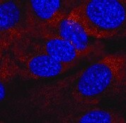
CXCR5 in Human PBMCs.
CXCR5 was detected in immersion fixed human peripheral blood mononuclear cells (PBMCs) using Mouse Anti-Human CXCR5 Monoclonal Antibody (Catalog # MAB190) at 10 µg/mL for 3 hours at room temperature. Cells were stained using the NorthernLights™ 637-conjugated Anti-Mouse IgG Secondary Antibody (red; Catalog # NL008) and counterstained with DAPI (blue). View our protocol for Fluorescent ICC Staining of Non-adherent Cells.
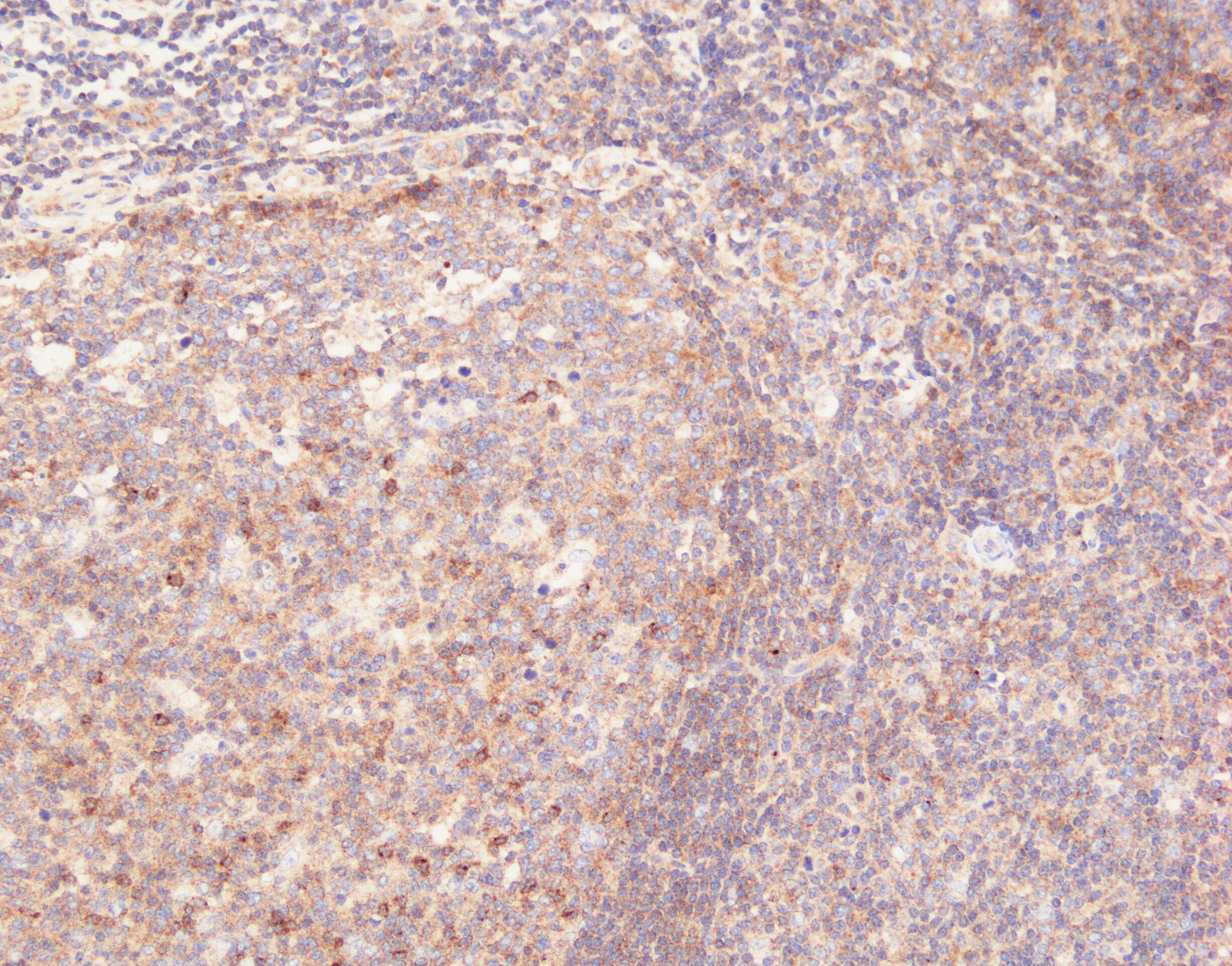
CXCR5 in Human Kidney.
CXCR5 was detected in immersion fixed paraffin-embedded sections of human kidney using Mouse Anti-Human CXCR5 Monoclonal Antibody (Catalog # MAB190) at 5 µg/mL overnight at 4 °C. Tissue was stained using the Anti-Mouse HRP-DAB Cell & Tissue Staining Kit (brown; Catalog # CTS002) and counterstained with hematoxylin (blue). Specific staining was localized to cell membranes. View our protocol for Chromogenic IHC Staining of Paraffin-embedded Tissue Sections.
Chemotaxis Induced by CXCL13/BLC/BCA‑1 and Neutralization by Human CXCR5 Antibody.
Recombinant Human CXCL13/BLC/BCA-1 (Catalog # 801-CX) chemoattracts the BaF3 mouse pro-B cell line transfected with human CXCR5 in a dose-dependent manner (orange line). The amount of cells that migrated through to the lower chemotaxis chamber was measured by Resazurin (Catalog # AR002). Chemotaxis elicited by Recombinant Human CXCL13/BLC/BCA-1 (0.05 µg/mL) is neutralized (green line) by increasing concentrations of Mouse Anti-Human CXCR5 Monoclonal Antibody (Catalog # MAB190). The ND50 is typically 0.25-1.5 µg/mL.
Detection of Human CXCR5 by Western Blot
Inverse correlation between p53 and CXCR5 expression and function in MCF-7 cells.(A) p53 knock-down results in an increase in the relative abundance of CXCR5 mRNA. The result of 10 experiments is shown. (B) Changes in p53 and CXCR5 protein levels correlate with the levels of corresponding mRNAs. mRNA levels were measured by RT-PCR in real time. Representative data are shown, the experiment was repeated 2 times for p53 and 3 times for CXCR5. Complete western blots are shown in Supplementary Figure S1. (C) CXCR5 stimulates of c-Jun mRNA expression in MCF-7 cells in p53-dependent manner. Cells were exposed to recombinant CXCL13 for 6 hours prior to RNA isolation. The result of three experiments is shown. *P < 0,01 versus MCF-7. Image collected and cropped by CiteAb from the following publication (https://pubmed.ncbi.nlm.nih.gov/25786345), licensed under a CC-BY license. Not internally tested by R&D Systems.Applications for Human CXCR5 Antibody (51505)
Application
Recommended Usage
CyTOF-reported
Ferrell, P.B., Jr. et al. (2016) PLoS ONE 11: e0153207. Ready to be labeled using established conjugation methods. No BSA or other carrier proteins that could interfere with conjugation.
Flow Cytometry
0.25 µg/106 cells
Sample: Human peripheral blood CD19+ B cells
Sample: Human peripheral blood CD19+ B cells
Immunocytochemistry
8-25 µg/mL
Sample: Immersion fixed human peripheral blood mononuclear cells (PBMCs)
Sample: Immersion fixed human peripheral blood mononuclear cells (PBMCs)
Immunohistochemistry
8-25 µg/mL
Sample: Immersion fixed paraffin-embedded sections of human lymph node and human kidney
Sample: Immersion fixed paraffin-embedded sections of human lymph node and human kidney
Neutralization
Measured by its ability to neutralize CXCL13/BLC/BCA‑1-induced chemotaxis in the BaF3 mouse pro‑B cell line transfected with human CXCR5. The Neutralization Dose (ND50) is typically 0.25-1.5 µg/mL in the presence of 0.05 µg/mL Recombinant Human CXCL13/BLC/BCA‑1.
Reviewed Applications
Read 3 reviews rated 4.7 using MAB190 in the following applications:
Flow Cytometry Panel Builder
Bio-Techne Knows Flow Cytometry
Save time and reduce costly mistakes by quickly finding compatible reagents using the Panel Builder Tool.
Advanced Features
- Spectra Viewer - Custom analysis of spectra from multiple fluorochromes
- Spillover Popups - Visualize the spectra of individual fluorochromes
- Antigen Density Selector - Match fluorochrome brightness with antigen density

Formulation, Preparation, and Storage
Purification
Protein A or G purified from hybridoma culture supernatant
Reconstitution
Reconstitute at 0.5 mg/mL in sterile PBS. For liquid material, refer to CoA for concentration.
Loading...
Formulation
Lyophilized from a 0.2 μm filtered solution in PBS with Trehalose. *Small pack size (SP) is supplied either lyophilized or as a 0.2 µm filtered solution in PBS.
Shipping
Lyophilized product is shipped at ambient temperature. Liquid small pack size (-SP) is shipped with polar packs. Upon receipt, store immediately at the temperature recommended below.
Stability & Storage
Use a manual defrost freezer and avoid repeated freeze-thaw cycles.
- 12 months from date of receipt, -20 to -70 °C as supplied.
- 1 month, 2 to 8 °C under sterile conditions after reconstitution.
- 6 months, -20 to -70 °C under sterile conditions after reconstitution.
Calculators
Background: CXCR5
Alternate Names
BLR1, CD185, CXCR5
Gene Symbol
CXCR5
UniProt
Additional CXCR5 Products
Product Documents for Human CXCR5 Antibody (51505)
Certificate of Analysis
To download a Certificate of Analysis, please enter a lot or batch number in the search box below.
Note: Certificate of Analysis not available for kit components.
Product Specific Notices for Human CXCR5 Antibody (51505)
For research use only
Citations for Human CXCR5 Antibody (51505)
 Powered by Bioz
Customer Reviews for Human CXCR5 Antibody (51505) (3)
4.7 out of 5
3 Customer Ratings
Have you used Human CXCR5 Antibody (51505)?
Submit a review and receive an Amazon gift card!
$25/€18/£15/$25CAN/¥2500 Yen for a review with an image
$10/€7/£6/$10CAN/¥1110 Yen for a review without an image
Submit a review
Customer Images
Showing
1
-
3 of
3 reviews
Showing All
Filter By:
-
Application: Immunocytochemistry/ImmunofluorescenceSample Tested: Human retinal pigment epithelium cellsSpecies: HumanVerified Customer | Posted 08/19/2021

-
Application: ImmunohistochemistrySample Tested: Tonsil tissueSpecies: HumanVerified Customer | Posted 09/11/2020retrieval in high pH buffer Ab was diluted 1:2000(0.25ug/mL)
-
Application: Western BlotSample Tested: See PMID 22330139Species: MouseVerified Customer | Posted 02/12/2015
There are no reviews that match your criteria.
Protocols
Find general support by application which include: protocols, troubleshooting, illustrated assays, videos and webinars.
- 7-Amino Actinomycin D (7-AAD) Cell Viability Flow Cytometry Protocol
- Antigen Retrieval Protocol (PIER)
- Antigen Retrieval for Frozen Sections Protocol
- Appropriate Fixation of IHC/ICC Samples
- Cellular Response to Hypoxia Protocols
- Chromogenic IHC Staining of Formalin-Fixed Paraffin-Embedded (FFPE) Tissue Protocol
- Chromogenic Immunohistochemistry Staining of Frozen Tissue
- ClariTSA™ Fluorophore Kits
- Detection & Visualization of Antibody Binding
- Extracellular Membrane Flow Cytometry Protocol
- Flow Cytometry Protocol for Cell Surface Markers
- Flow Cytometry Protocol for Staining Membrane Associated Proteins
- Flow Cytometry Staining Protocols
- Flow Cytometry Troubleshooting Guide
- Fluorescent IHC Staining of Frozen Tissue Protocol
- Graphic Protocol for Heat-induced Epitope Retrieval
- Graphic Protocol for the Preparation and Fluorescent IHC Staining of Frozen Tissue Sections
- Graphic Protocol for the Preparation and Fluorescent IHC Staining of Paraffin-embedded Tissue Sections
- Graphic Protocol for the Preparation of Gelatin-coated Slides for Histological Tissue Sections
- ICC Cell Smear Protocol for Suspension Cells
- ICC Immunocytochemistry Protocol Videos
- ICC for Adherent Cells
- IHC Sample Preparation (Frozen sections vs Paraffin)
- Immunocytochemistry (ICC) Protocol
- Immunocytochemistry Troubleshooting
- Immunofluorescence of Organoids Embedded in Cultrex Basement Membrane Extract
- Immunofluorescent IHC Staining of Formalin-Fixed Paraffin-Embedded (FFPE) Tissue Protocol
- Immunohistochemistry (IHC) and Immunocytochemistry (ICC) Protocols
- Immunohistochemistry Frozen Troubleshooting
- Immunohistochemistry Paraffin Troubleshooting
- Intracellular Flow Cytometry Protocol Using Alcohol (Methanol)
- Intracellular Flow Cytometry Protocol Using Detergents
- Intracellular Nuclear Staining Flow Cytometry Protocol Using Detergents
- Intracellular Staining Flow Cytometry Protocol Using Alcohol Permeabilization
- Intracellular Staining Flow Cytometry Protocol Using Detergents to Permeabilize Cells
- Preparing Samples for IHC/ICC Experiments
- Preventing Non-Specific Staining (Non-Specific Binding)
- Primary Antibody Selection & Optimization
- Propidium Iodide Cell Viability Flow Cytometry Protocol
- Protocol for Heat-Induced Epitope Retrieval (HIER)
- Protocol for Liperfluo
- Protocol for Making a 4% Formaldehyde Solution in PBS
- Protocol for VisUCyte™ HRP Polymer Detection Reagent
- Protocol for the Characterization of Human Th22 Cells
- Protocol for the Characterization of Human Th9 Cells
- Protocol for the Fluorescent ICC Staining of Cell Smears - Graphic
- Protocol for the Fluorescent ICC Staining of Cultured Cells on Coverslips - Graphic
- Protocol for the Preparation & Fixation of Cells on Coverslips
- Protocol for the Preparation and Chromogenic IHC Staining of Frozen Tissue Sections
- Protocol for the Preparation and Chromogenic IHC Staining of Frozen Tissue Sections - Graphic
- Protocol for the Preparation and Chromogenic IHC Staining of Paraffin-embedded Tissue Sections
- Protocol for the Preparation and Chromogenic IHC Staining of Paraffin-embedded Tissue Sections - Graphic
- Protocol for the Preparation and Fluorescent ICC Staining of Cells on Coverslips
- Protocol for the Preparation and Fluorescent ICC Staining of Non-adherent Cells
- Protocol for the Preparation and Fluorescent ICC Staining of Stem Cells on Coverslips
- Protocol for the Preparation and Fluorescent IHC Staining of Frozen Tissue Sections
- Protocol for the Preparation and Fluorescent IHC Staining of Paraffin-embedded Tissue Sections
- Protocol for the Preparation of Gelatin-coated Slides for Histological Tissue Sections
- Protocol for the Preparation of a Cell Smear for Non-adherent Cell ICC - Graphic
- Protocol: Annexin V and PI Staining by Flow Cytometry
- Protocol: Annexin V and PI Staining for Apoptosis by Flow Cytometry
- TUNEL and Active Caspase-3 Detection by IHC/ICC Protocol
- The Importance of IHC/ICC Controls
- Troubleshooting Guide: Fluorokine Flow Cytometry Kits
- Troubleshooting Guide: Immunohistochemistry
- View all Protocols, Troubleshooting, Illustrated assays and Webinars


