Human IFN-gamma Antibody (K3.53)
R&D Systems | Catalog # MAB2852

Key Product Details
Validated by
Biological Validation
Species Reactivity
Validated:
Human
Cited:
Human
Applications
Validated:
Western Blot, ELISA Capture (Matched Antibody Pair), Neutralization, Intracellular Staining by Flow Cytometry
Cited:
Neutralization, Flow Cytometry, Array Development, ELISA Capture, ELISA Development, ELISA Development (Capture), Functional Assay, Luminex Development
Label
Unconjugated
Antibody Source
Monoclonal Mouse IgG2A Clone # K3.53
Loading...
Product Specifications
Immunogen
E. coli-derived recombinant human IFN-gamma
Specificity
Detects human IFN‑ gamma in direct ELISAs and Western blots. In Western blots, no cross-reactivity with recombinant mouse IFN‑ gamma, recombinant rat IFN‑ gamma, recombinant porcine IFN‑ gamma, recombinant feline IFN‑ gamma, or recombinant cotton rat IFN‑ gamma is observed.
Clonality
Monoclonal
Host
Mouse
Isotype
IgG2A
Endotoxin Level
<0.10 EU per 1 μg of the antibody by the LAL method.
Scientific Data Images for Human IFN-gamma Antibody (K3.53)
Detection of IFN- gamma in Human PBMCs by Flow Cytometry.
Human peripheral blood mononuclear cells (PBMCs) treated with 50 ng/mL PMA, 1 ug/mL Ionomycin, and 3 uM Monensin overnight were stained with either (A) Mouse Anti-Human IFN- gamma Monoclonal Antibody (Catalog # MAB2852) or (B) Mouse IgG2A Isotype Control (Catalog # MAB003) followed by anti-Mouse IgG PE-conjugated secondary antibody (Catalog # F0102B) and Mouse Anti-Human CD3 epsilon APC-conjugated Monoclonal Antibody (Catalog # FAB100A). To facilitate intracellular staining, cells were fixed and permeabilized with FlowX FoxP3 Fixation & Permeabilization Buffer Kit (Catalog # FC012).View our protocol for Staining Intracellular Molecules.
IFN‑ gamma Inhibition of EMCV-induced Cytopathy and Neutralization by Human IFN‑ gamma Antibody.
Recombinant Human IFN-gammaamma (Catalog # 285-IF) reduces the Encephalomyocarditis Virus (EMCV)-induced cytopathy in the HeLa human cervical epithelial carcinoma cell line in a dose-dependent manner (orange line), as measured by crystal violet staining. Inhibition of EMCV activity elicited by Recombinant Human IFN-gammaamma (1 ng/mL) is neutralized (green line) by increasing concentrations of Mouse Anti-Human IFN-gammaamma Monoclonal Antibody (Catalog # MAB2852). The ND50 is typically ≤ 2 µg/mL.Applications for Human IFN-gamma Antibody (K3.53)
Application
Recommended Usage
Intracellular Staining by Flow Cytometry
0.25 µg/106 cells
Sample: Human peripheral blood mononuclear cells (PBMCs) treated with PMA, Ionomycin, and Monensin fixed and permeabilized with FlowX FoxP3 Fixation & Permeabilization Buffer Kit (Catalog # FC012)
Sample: Human peripheral blood mononuclear cells (PBMCs) treated with PMA, Ionomycin, and Monensin fixed and permeabilized with FlowX FoxP3 Fixation & Permeabilization Buffer Kit (Catalog # FC012)
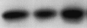
Western Blot
1 µg/mL
Sample: Recombinant Human IFN-gamma (Catalog # 285-IF)
Sample: Recombinant Human IFN-gamma (Catalog # 285-IF)
Neutralization
Measured by its ability to neutralize IFN‑ gamma inhibition of EMCV-induced cytopathy in the HeLa human cervical epithelial carcinoma cell line. Meager, A. (1987) in Lymphokines and Interferons, a Practical Approach. Clemens, M.J. et al. (eds): IRL Press. 129. The Neutralization Dose (ND50) is typically ≤2 µg/mL in the presence of 1 ng/mL Recombinant Human IFN‑ gamma.
Human IFN-gamma Sandwich Immunoassay
Please Note: Optimal dilutions of this antibody should be experimentally determined.
Reviewed Applications
Read 1 review rated 5 using MAB2852 in the following applications:
Flow Cytometry Panel Builder
Bio-Techne Knows Flow Cytometry
Save time and reduce costly mistakes by quickly finding compatible reagents using the Panel Builder Tool.
Advanced Features
- Spectra Viewer - Custom analysis of spectra from multiple fluorochromes
- Spillover Popups - Visualize the spectra of individual fluorochromes
- Antigen Density Selector - Match fluorochrome brightness with antigen density

Formulation, Preparation, and Storage
Purification
Protein A or G purified from hybridoma culture supernatant
Reconstitution
Reconstitute at 0.5 mg/mL in sterile PBS. For liquid material, refer to CoA for concentration.
Loading...
Formulation
Lyophilized from a 0.2 μm filtered solution in PBS and NaCl with Trehalose. *Small pack size (SP) is supplied either lyophilized or as a 0.2 µm filtered solution in PBS.
Shipping
Lyophilized product is shipped at ambient temperature. Liquid small pack size (-SP) is shipped with polar packs. Upon receipt, store immediately at the temperature recommended below.
Stability & Storage
Use a manual defrost freezer and avoid repeated freeze-thaw cycles.
- 12 months from date of receipt, -20 to -70 °C as supplied.
- 1 month, 2 to 8 °C under sterile conditions after reconstitution.
- 6 months, -20 to -70 °C under sterile conditions after reconstitution.
Calculators
Background: IFN-gamma
Long Name
Interferon gamma
Alternate Names
IFG, IFI, IFNG, IFNgamma
Entrez Gene IDs
Gene Symbol
IFNG
Additional IFN-gamma Products
Product Documents for Human IFN-gamma Antibody (K3.53)
Certificate of Analysis
To download a Certificate of Analysis, please enter a lot or batch number in the search box below.
Note: Certificate of Analysis not available for kit components.
Product Specific Notices for Human IFN-gamma Antibody (K3.53)
For research use only
Related Research Areas
Citations for Human IFN-gamma Antibody (K3.53)
 Powered by Bioz
Customer Reviews for Human IFN-gamma Antibody (K3.53) (1)
5 out of 5
1 Customer Rating
Have you used Human IFN-gamma Antibody (K3.53)?
Submit a review and receive an Amazon gift card!
$25/€18/£15/$25CAN/¥2500 Yen for a review with an image
$10/€7/£6/$10CAN/¥1110 Yen for a review without an image
Submit a review
Customer Images
Showing
1
-
1 of
1 review
Showing All
Filter By:
-
Application: Western BlotSample Tested: HeLa human cervical epithelial carcinoma cell lineSpecies: HumanVerified Customer | Posted 08/24/2021

There are no reviews that match your criteria.
Protocols
Find general support by application which include: protocols, troubleshooting, illustrated assays, videos and webinars.
- 7-Amino Actinomycin D (7-AAD) Cell Viability Flow Cytometry Protocol
- Cellular Response to Hypoxia Protocols
- Extracellular Membrane Flow Cytometry Protocol
- Flow Cytometry Protocol for Cell Surface Markers
- Flow Cytometry Protocol for Staining Membrane Associated Proteins
- Flow Cytometry Staining Protocols
- Flow Cytometry Troubleshooting Guide
- Intracellular Flow Cytometry Protocol Using Alcohol (Methanol)
- Intracellular Flow Cytometry Protocol Using Detergents
- Intracellular Nuclear Staining Flow Cytometry Protocol Using Detergents
- Intracellular Staining Flow Cytometry Protocol Using Alcohol Permeabilization
- Intracellular Staining Flow Cytometry Protocol Using Detergents to Permeabilize Cells
- Propidium Iodide Cell Viability Flow Cytometry Protocol
- Protocol for Liperfluo
- Protocol for the Characterization of Human Th22 Cells
- Protocol for the Characterization of Human Th9 Cells
- Protocol: Annexin V and PI Staining by Flow Cytometry
- Protocol: Annexin V and PI Staining for Apoptosis by Flow Cytometry
- R&D Systems Quality Control Western Blot Protocol
- Troubleshooting Guide: Fluorokine Flow Cytometry Kits
- Troubleshooting Guide: Western Blot Figures
- Western Blot Conditions
- Western Blot Protocol
- Western Blot Protocol for Cell Lysates
- Western Blot Troubleshooting
- Western Blot Troubleshooting Guide
- View all Protocols, Troubleshooting, Illustrated assays and Webinars
Loading...
Associated Pathways


