Lysosome-associated membrane protein-1 (LAMP1), also known as CD107a, is a 100‑130 kDa member of the LAMP family of glycoproteins. It is expressed in lysosomal and plasma membranes of macrophages, NK and T-cells, and with LAMP2, is essential for the formation of phagolysosomes. On the cell surface, it also presents carbohydrates to selectins. Mature human LAMP1 is a 389 amino acid (aa) type I transmembrane glycoprotein. It contains a 354 aa luminal/extracellular domain (ECD) (aa 28‑381) and a 12 aa cytoplasmic tail (aa 405‑416). The ECD has two large looping regions (aa 28‑193 and 227‑381) plus multiple N- and O-linked glycosylation sites. There is one potential splice variant that shows a 26 aa substitution in the signal sequence. Over aa 28‑380, human LAMP1 shares 64% aa identity with mouse LAMP1.
Human LAMP-1/CD107a Antibody (508921)
R&D Systems | Catalog # MAB4800

Key Product Details
Validated by
Biological Validation
Species Reactivity
Validated:
Human
Cited:
Human
Applications
Validated:
Immunohistochemistry, Intracellular Staining by Flow Cytometry, Immunocytochemistry, CyTOF-ready
Cited:
Immunohistochemistry, Western Blot, Immunocytochemistry, ELISA Capture
Label
Unconjugated
Antibody Source
Monoclonal Mouse IgG2B Clone # 508921
Loading...
Product Specifications
Immunogen
Mouse myeloma cell line NS0-derived recombinant human LAMP1/CD107a
Ala28-Asn380
Accession # P11279
Ala28-Asn380
Accession # P11279
Specificity
Detects human LAMP1/CD107a in direct ELISAs.
Clonality
Monoclonal
Host
Mouse
Isotype
IgG2B
Scientific Data Images for Human LAMP-1/CD107a Antibody (508921)
LAMP1/CD107a in Human Kidney.
LAMP1/CD107a was detected in immersion fixed paraffin-embedded sections of human kidney using Mouse Anti-Human LAMP1/CD107a Monoclonal Antibody (Catalog # MAB4800) at 15 µg/mL overnight at 4 °C. Before incubation with the primary antibody, tissue was subjected to heat-induced epitope retrieval using Antigen Retrieval Reagent-Basic (Catalog # CTS013). Tissue was stained using the Anti-Mouse HRP-DAB Cell & Tissue Staining Kit (brown; Catalog # CTS002) and counterstained with hematoxylin (blue). Specific staining was localized to lysosomes in epithelial cells. View our protocol for Chromogenic IHC Staining of Paraffin-embedded Tissue Sections.
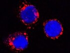
LAMP‑1/CD107a in THP‑1 Human Cell Line.
LAMP-1/CD107a was detected in immersion fixed THP-1 human acute monocytic leukemia cell line using Mouse Anti-Human LAMP-1/CD107a Monoclonal Antibody (Catalog # MAB4800) at 25 µg/mL for 3 hours at room temperature. Cells were stained using the NorthernLights™ 557-conjugated Anti-Mouse IgG Secondary Antibody (red; Catalog # NL007) and counterstained with DAPI(blue). Specific staining was localized to cytoplasmic. View our protocol for Fluorescent ICC Staining of Cells on Coverslips.
Detection of Human LAMP-1/CD107a by Western Blot
Increased SNX9 expression and co-localization with podocin are detectable in the cytoplasm of ADR-treated WT podocytes, whereas SNX9 KD podocytes exhibit little cytoplasmic expression of podocin.(a) Fluorescent micrographs of cultured human podocytes stained with SNX9 (green) and podocin (red) before and after ADR treatment (merged areas are in yellow). DAPI (blue) was used to indicate nuclei. Boxes indicate higher magnification areas presented in the lower panels. (b) Western blot analyses of the fractions from control podocytes or podocytes treated with ADR separated on linear OptiPrep gradients (5–25%). Distributions of SNX9 and podocin, as well as marker proteins of plasma membrane (caveolin), endosome/lysosome (LAMP1), mitochondria ( beta subunit of F1F0-ATPase), and endoplasmic reticulum (calnexin), were examined by western blot analysis. (c) Cultured human podocytes were transfected with nonfunctional control siRNA (upper panel) or SNX9 siRNA (middle and lower panels). Transfected cells, as decided by GFP expression, are indicated by arrowheads. Upper panel: Fluorescent micrographs of control siRNA-transfected podocyte stained with SNX9 (red). Middle panel: Fluorescent micrographs of SNX9 siRNA-transfected podocyte with ADR treatment stained with SNX9 (red). Lower panel: Fluorescent micrographs of SNX9 siRNA-transfected podocyte with ADR treatment stained with podocin (red). DAPI (blue) was used to indicate nuclei. Boxes indicate higher-magnification areas presented on the right. Image collected and cropped by CiteAb from the following publication (https://pubmed.ncbi.nlm.nih.gov/28266622), licensed under a CC-BY license. Not internally tested by R&D Systems.Applications for Human LAMP-1/CD107a Antibody (508921)
Application
Recommended Usage
CyTOF-ready
Ready to be labeled using established conjugation methods. No BSA or other carrier proteins that could interfere with conjugation.
Immunocytochemistry
8-25 µg/mL
Sample: Immersion fixed THP-1 human acute monocytic leukemia cell line
Sample: Immersion fixed THP-1 human acute monocytic leukemia cell line
Immunohistochemistry
8-25 µg/mL
Sample: Immersion fixed paraffin-embedded sections of human kidney
Sample: Immersion fixed paraffin-embedded sections of human kidney
Intracellular Staining by Flow Cytometry
2.5 µg/106 cells
Sample: THP‑1 human acute monocytic leukemia cell line fixed with paraformaldehyde and permeabilized with saponin
Sample: THP‑1 human acute monocytic leukemia cell line fixed with paraformaldehyde and permeabilized with saponin
Reviewed Applications
Read 1 review rated 5 using MAB4800 in the following applications:
Flow Cytometry Panel Builder
Bio-Techne Knows Flow Cytometry
Save time and reduce costly mistakes by quickly finding compatible reagents using the Panel Builder Tool.
Advanced Features
- Spectra Viewer - Custom analysis of spectra from multiple fluorochromes
- Spillover Popups - Visualize the spectra of individual fluorochromes
- Antigen Density Selector - Match fluorochrome brightness with antigen density

Formulation, Preparation, and Storage
Purification
Protein A or G purified from hybridoma culture supernatant
Reconstitution
Reconstitute at 0.5 mg/mL in sterile PBS. For liquid material, refer to CoA for concentration.
Loading...
Formulation
Lyophilized from a 0.2 μm filtered solution in PBS with Trehalose. *Small pack size (SP) is supplied either lyophilized or as a 0.2 µm filtered solution in PBS.
Shipping
Lyophilized product is shipped at ambient temperature. Liquid small pack size (-SP) is shipped with polar packs. Upon receipt, store immediately at the temperature recommended below.
Stability & Storage
Use a manual defrost freezer and avoid repeated freeze-thaw cycles.
- 12 months from date of receipt, -20 to -70 °C as supplied.
- 1 month, 2 to 8 °C under sterile conditions after reconstitution.
- 6 months, -20 to -70 °C under sterile conditions after reconstitution.
Calculators
Background: LAMP-1/CD107a
Long Name
Lysosome-associated Membrane Glycoprotein 1
Alternate Names
CD107a, LAMP1
Gene Symbol
LAMP1
UniProt
Additional LAMP-1/CD107a Products
Product Documents for Human LAMP-1/CD107a Antibody (508921)
Certificate of Analysis
To download a Certificate of Analysis, please enter a lot or batch number in the search box below.
Note: Certificate of Analysis not available for kit components.
Product Specific Notices for Human LAMP-1/CD107a Antibody (508921)
For research use only
Citations for Human LAMP-1/CD107a Antibody (508921)
 Powered by Bioz
Customer Reviews for Human LAMP-1/CD107a Antibody (508921) (1)
5 out of 5
1 Customer Rating
Have you used Human LAMP-1/CD107a Antibody (508921)?
Submit a review and receive an Amazon gift card!
$25/€18/£15/$25CAN/¥2500 Yen for a review with an image
$10/€7/£6/$10CAN/¥1110 Yen for a review without an image
Submit a review
Customer Images
Showing
1
-
1 of
1 review
Showing All
Filter By:
-
Application: Immunocytochemistry/ImmunofluorescenceSample Tested: THP‑1 Human Cell LineSpecies: HumanVerified Customer | Posted 02/10/2022

There are no reviews that match your criteria.
Protocols
Find general support by application which include: protocols, troubleshooting, illustrated assays, videos and webinars.
- 7-Amino Actinomycin D (7-AAD) Cell Viability Flow Cytometry Protocol
- Antigen Retrieval Protocol (PIER)
- Antigen Retrieval for Frozen Sections Protocol
- Appropriate Fixation of IHC/ICC Samples
- Cellular Response to Hypoxia Protocols
- Chromogenic IHC Staining of Formalin-Fixed Paraffin-Embedded (FFPE) Tissue Protocol
- Chromogenic Immunohistochemistry Staining of Frozen Tissue
- ClariTSA™ Fluorophore Kits
- Detection & Visualization of Antibody Binding
- Extracellular Membrane Flow Cytometry Protocol
- Flow Cytometry Protocol for Cell Surface Markers
- Flow Cytometry Protocol for Staining Membrane Associated Proteins
- Flow Cytometry Staining Protocols
- Flow Cytometry Troubleshooting Guide
- Fluorescent IHC Staining of Frozen Tissue Protocol
- Graphic Protocol for Heat-induced Epitope Retrieval
- Graphic Protocol for the Preparation and Fluorescent IHC Staining of Frozen Tissue Sections
- Graphic Protocol for the Preparation and Fluorescent IHC Staining of Paraffin-embedded Tissue Sections
- Graphic Protocol for the Preparation of Gelatin-coated Slides for Histological Tissue Sections
- ICC Cell Smear Protocol for Suspension Cells
- ICC Immunocytochemistry Protocol Videos
- ICC for Adherent Cells
- IHC Sample Preparation (Frozen sections vs Paraffin)
- Immunocytochemistry (ICC) Protocol
- Immunocytochemistry Troubleshooting
- Immunofluorescence of Organoids Embedded in Cultrex Basement Membrane Extract
- Immunofluorescent IHC Staining of Formalin-Fixed Paraffin-Embedded (FFPE) Tissue Protocol
- Immunohistochemistry (IHC) and Immunocytochemistry (ICC) Protocols
- Immunohistochemistry Frozen Troubleshooting
- Immunohistochemistry Paraffin Troubleshooting
- Intracellular Flow Cytometry Protocol Using Alcohol (Methanol)
- Intracellular Flow Cytometry Protocol Using Detergents
- Intracellular Nuclear Staining Flow Cytometry Protocol Using Detergents
- Intracellular Staining Flow Cytometry Protocol Using Alcohol Permeabilization
- Intracellular Staining Flow Cytometry Protocol Using Detergents to Permeabilize Cells
- Preparing Samples for IHC/ICC Experiments
- Preventing Non-Specific Staining (Non-Specific Binding)
- Primary Antibody Selection & Optimization
- Propidium Iodide Cell Viability Flow Cytometry Protocol
- Protocol for Heat-Induced Epitope Retrieval (HIER)
- Protocol for Liperfluo
- Protocol for Making a 4% Formaldehyde Solution in PBS
- Protocol for VisUCyte™ HRP Polymer Detection Reagent
- Protocol for the Characterization of Human Th22 Cells
- Protocol for the Characterization of Human Th9 Cells
- Protocol for the Fluorescent ICC Staining of Cell Smears - Graphic
- Protocol for the Fluorescent ICC Staining of Cultured Cells on Coverslips - Graphic
- Protocol for the Preparation & Fixation of Cells on Coverslips
- Protocol for the Preparation and Chromogenic IHC Staining of Frozen Tissue Sections
- Protocol for the Preparation and Chromogenic IHC Staining of Frozen Tissue Sections - Graphic
- Protocol for the Preparation and Chromogenic IHC Staining of Paraffin-embedded Tissue Sections
- Protocol for the Preparation and Chromogenic IHC Staining of Paraffin-embedded Tissue Sections - Graphic
- Protocol for the Preparation and Fluorescent ICC Staining of Cells on Coverslips
- Protocol for the Preparation and Fluorescent ICC Staining of Non-adherent Cells
- Protocol for the Preparation and Fluorescent ICC Staining of Stem Cells on Coverslips
- Protocol for the Preparation and Fluorescent IHC Staining of Frozen Tissue Sections
- Protocol for the Preparation and Fluorescent IHC Staining of Paraffin-embedded Tissue Sections
- Protocol for the Preparation of Gelatin-coated Slides for Histological Tissue Sections
- Protocol for the Preparation of a Cell Smear for Non-adherent Cell ICC - Graphic
- Protocol: Annexin V and PI Staining by Flow Cytometry
- Protocol: Annexin V and PI Staining for Apoptosis by Flow Cytometry
- TUNEL and Active Caspase-3 Detection by IHC/ICC Protocol
- The Importance of IHC/ICC Controls
- Troubleshooting Guide: Fluorokine Flow Cytometry Kits
- Troubleshooting Guide: Immunohistochemistry
- View all Protocols, Troubleshooting, Illustrated assays and Webinars
Loading...
Associated Pathways
