NKp46, along with NKp30 and NKp44, are activating receptors that have been collectively termed the natural cytotoxicity receptors (NCR) (1). These receptors lack significant sequence homology to one another. They are expressed almost exclusively by NK cells and play a major role in triggering some of the key lytic activities of NK cells. The CD56dimCD16+ subpopulation that makes up the majority of NK cells in the peripheral blood and spleen expresses NKp46 in both resting and activated states (2). The main NK cell population of the lymph node (CD56brightCD16-) expresses low levels of NKp46 in resting cells, but expression is up-regulated by IL-2. NKp46 is a type I transmembrane protein with two extracellular Ig-like domains followed by a short stalk region, a transmembrane domain containing a positively charged amino acid residue, and a short cytoplasmic tail. Through its positive charge in the transmembrane domain, NKp46 associates with the ITAM‑bearing signal adapter proteins, CD3 zeta and Fc epsilon R1 gamma, which are able to form disulfide-linked homodimers and heterodimers (3, 8). Studies with neutralizing antibodies indicate that the three NCRs are primarily responsible for triggering the NK-mediated lysis of many human tumor cell lines. Blocking any of the NCRs individually resulted in partial inhibition of tumor cell lysis, but nearly complete inhibition of lysis was observed if all three receptors were blocked simultaneously (4). NKp46 has also been implicated in recognition of virus-infected cells through its capacity to bind to viral hemagglutinins (5‑7). Human NKp46 shares 58% and 59% amino acid sequence identity with the mouse and rat proteins, respectively.
Best Seller
Human NKp46/NCR1 Antibody (195314)
R&D Systems | Catalog # MAB1850
Clone 195314 was used by HLDA to establish CD designation

Key Product Details
Species Reactivity
Validated:
Human
Cited:
Human, Mouse
Applications
Validated:
Immunohistochemistry, Western Blot, Flow Cytometry, Dual RNAscope ISH-IHC Compatible, Immunocytochemistry, Agonist Activity, CyTOF-reported
Cited:
Immunohistochemistry, Immunohistochemistry-Paraffin, Immunohistochemistry-Frozen, Neutralization, Flow Cytometry, Immunocytochemistry, Binding Assay, Bioassay, ELISA Capture, Epitope Mapping, FRET, Functional Assay
Label
Unconjugated
Antibody Source
Monoclonal Mouse IgG2B Clone # 195314
Loading...
Product Specifications
Immunogen
Mouse T cell hybridoma transfected with human NKp46/NCR1
Specificity
Detects human NKp46/NCR1 in direct ELISAs and Western blots.
Clonality
Monoclonal
Host
Mouse
Isotype
IgG2B
Endotoxin Level
<0.10 EU per 1 μg of the antibody by the LAL method.
Scientific Data Images for Human NKp46/NCR1 Antibody (195314)
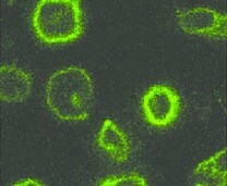
NKp46/NCR1 in NK-92 Human Cell Line and Human PBMCs.
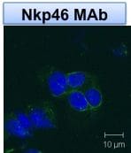
NKp46/NCR1 was detected in immersion fixed NK-92 human natural killer lymphoma cell line (left panel, positive stain) and human peripheral blood mononuclear cells (PBMCs; right panel, negative stain) using Mouse Anti-Human NKp46/NCR1 Monoclonal Antibody (Catalog # MAB1850) at 25 µg/mL for 3 hours at room temperature. Cells were stained using the NorthernLights™ 557-conjugated Anti-Mouse IgG Secondary Antibody (red; Catalog # NL007) and counterstained with DAPI (blue). Specific staining was localized to cytoplasm. View our protocol for Fluorescent ICC Staining of Non-adherent Cells.
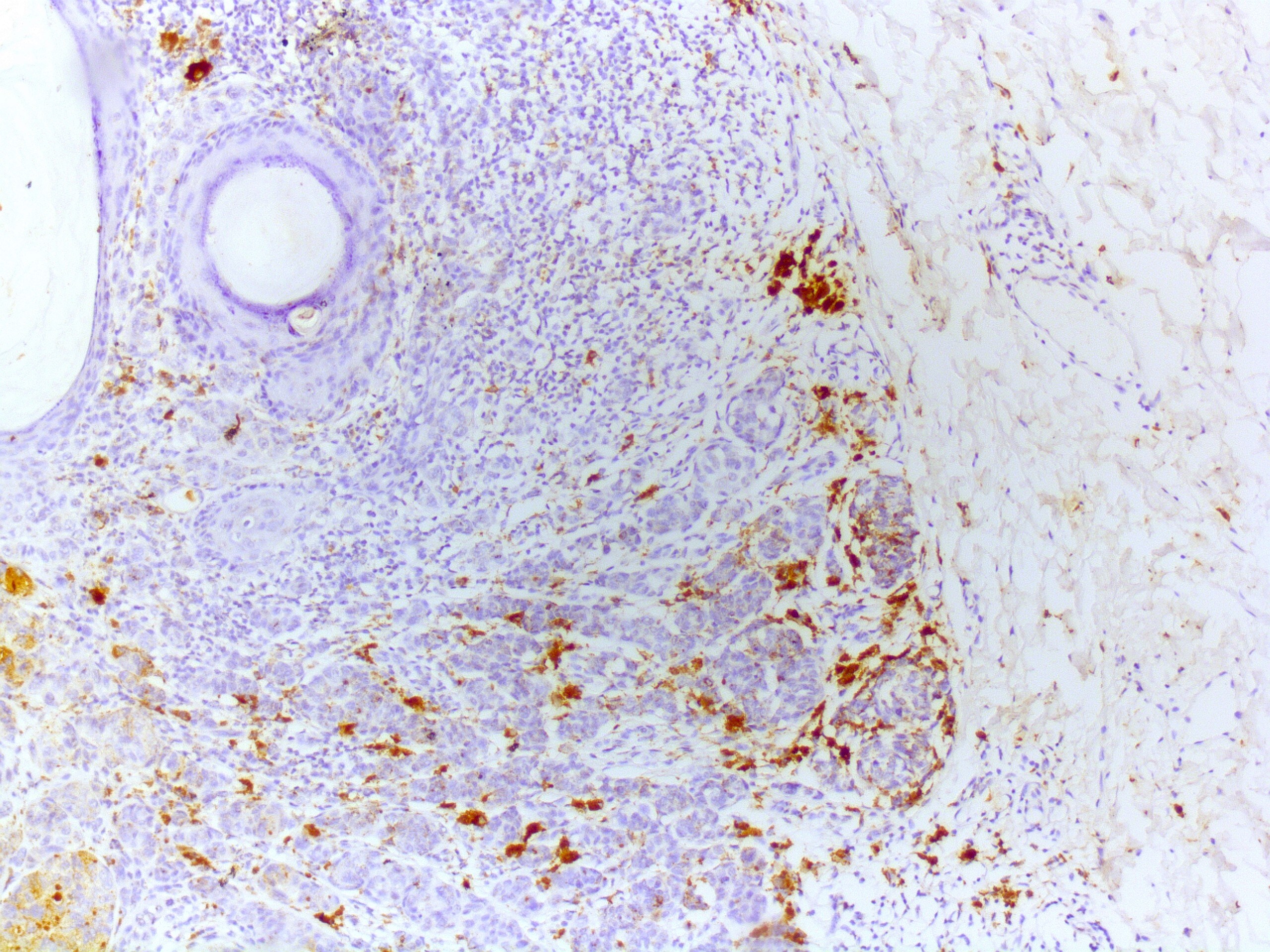
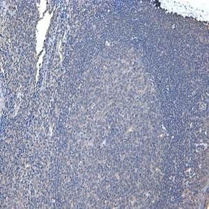
NKp46/NCR1 in Human Tonsil.
NKp46/NCR1 was detected in immersion fixed paraffin-embedded sections of human tonsil using Mouse Anti-Human NKp46/NCR1 Monoclonal Antibody (Catalog # MAB1850) at 5 µg/mL for 1 hour at room temperature followed by incubation with the Anti-Mouse IgG VisUCyte™ HRP Polymer Antibody (Catalog # VC001). Before incubation with the primary antibody, tissue was subjected to heat-induced epitope retrieval using Antigen Retrieval Reagent-Basic (Catalog # CTS013). Tissue was stained using DAB (brown) and counterstained with hematoxylin (blue). Specific staining was localized to natural killer cells in germinal center. View our protocol for IHC Staining with VisUCyte HRP Polymer Detection Reagents.
Detection of NKp46/NCR1 in Human PBMCs by Flow Cytometry.
Human peripheral blood mononuclear cells (PBMCs) were stained with (A) Mouse Anti-Human NKp46/NCR1 Monoclonal Antibody (Catalog # MAB1850) or (B) Mouse IgG2B Isotype Control (MAB0041) followed by Goat anti-Mouse IgG APC-conjugated Secondary Antibody (F0101B) and Rabbit Anti-Human NCAM-1/CD56 PE-conjugated Monoclonal Antibody (FAB24086P). Staining was performed using our Staining Membrane-associated Proteins protocol.
Detection of NKp46/NCR1 in Human Tonsil.
Formalin-fixed paraffin-embedded tissue sections of human tonsil were probed for NKp46 mRNA (ACD RNAScope Probe, catalog #312658; Fast Red chromogen, ACD catalog # 322750). Adjacent tissue section was processed for immunohistochemistry using mouse anti-human NKp46 monoclonal antibody (R&D Systems catalog # MAB1850) at 15ug/mL with overnight incubation at 4 degrees Celsius followed by incubation with anti-mouse IgG VisUCyte HRP Polymer Antibody (Catalog # VC001) and DAB chromogen (yellow-brown). Tissue was counterstained with hematoxylin (blue). Specific staining was localized to cell surface.Applications for Human NKp46/NCR1 Antibody (195314)
Application
Recommended Usage
Agonist Activity
Measured by its ability to induce IFN-gamma secretion by NK-92 human natural killer lymphoma cells.
The ED50 for this effect is typically ≤ 1 μg/mL.
The ED50 for this effect is typically ≤ 1 μg/mL.
CyTOF-reported
Horowitz, A. et al. Sci. Transl. Med. (2013) 208ra145. Ready to be labeled using established conjugation methods. No BSA or other carrier proteins that could interfere with conjugation.
Dual RNAscope ISH-IHC Compatible
3-25 µg/mL
Sample: Immersion fixed paraffin-embedded sections of human tonsil
Sample: Immersion fixed paraffin-embedded sections of human tonsil
Flow Cytometry
0.25 µg/106 cells
Sample: Human whole blood CD56+ natural killer cells
Sample: Human whole blood CD56+ natural killer cells
Immunocytochemistry
8-25 µg/mL
Sample: Immersion fixed NK‑92 human natural killer lymphoma cell line and human peripheral blood mononuclear cells (PBMCs)
Sample: Immersion fixed NK‑92 human natural killer lymphoma cell line and human peripheral blood mononuclear cells (PBMCs)
Immunohistochemistry
5-25 µg/mL
Sample: Immersion fixed paraffin-embedded sections of human tonsil
Sample: Immersion fixed paraffin-embedded sections of human tonsil
Western Blot
1 µg/mL
Sample: Recombinant Human NKp46/NCR1 Fc Chimera (Catalog # 1850-NK)
Sample: Recombinant Human NKp46/NCR1 Fc Chimera (Catalog # 1850-NK)
Reviewed Applications
Read 8 reviews rated 4.1 using MAB1850 in the following applications:
Flow Cytometry Panel Builder
Bio-Techne Knows Flow Cytometry
Save time and reduce costly mistakes by quickly finding compatible reagents using the Panel Builder Tool.
Advanced Features
- Spectra Viewer - Custom analysis of spectra from multiple fluorochromes
- Spillover Popups - Visualize the spectra of individual fluorochromes
- Antigen Density Selector - Match fluorochrome brightness with antigen density

Formulation, Preparation, and Storage
Purification
Protein A or G purified from hybridoma culture supernatant
Reconstitution
Reconstitute at 0.5 mg/mL in sterile PBS. For liquid material, refer to CoA for concentration.
Loading...
Formulation
Lyophilized from a 0.2 μm filtered solution in PBS with Trehalose. See Certificate of Analysis for details.
*Small pack size (-SP) is supplied either lyophilized or as a 0.2 µm filtered solution in PBS.
*Small pack size (-SP) is supplied either lyophilized or as a 0.2 µm filtered solution in PBS.
Shipping
Lyophilized product is shipped at ambient temperature. Liquid small pack size (-SP) is shipped with polar packs. Upon receipt, store immediately at the temperature recommended below.
Stability & Storage
Use a manual defrost freezer and avoid repeated freeze-thaw cycles.
- 12 months from date of receipt, -20 to -70 °C as supplied.
- 1 month, 2 to 8 °C under sterile conditions after reconstitution.
- 6 months, -20 to -70 °C under sterile conditions after reconstitution.
Calculators
Background: NKp46/NCR1
References
- Moretta, L. and A. Moretta (2004) EMBO J. 23:255.
- Ferlazzo, G. et al. (2004) J. Immunol. 172:1455.
- Augugliaro, R. et al. (2003) Eur. J. Immunol. 33:1235.
- Pende, D. et al. (1999) J. Exp. Med. 190:1505.
- Arnon, T. et al. (2004) Blood 103:664.
- Arnon, T. et al. (2001) Eur. J. Immunol. 31:2680.
- Mandelboim, O. et al. (2001) Nature 409:1055.
- Moretta, A. et al. (2001) Annu. Rev. Immunol. 19:197.
Alternate Names
CD335, Ly94, MAR-1, NCR1
Gene Symbol
NCR1
Additional NKp46/NCR1 Products
Product Documents for Human NKp46/NCR1 Antibody (195314)
Certificate of Analysis
To download a Certificate of Analysis, please enter a lot or batch number in the search box below.
Note: Certificate of Analysis not available for kit components.
Product Specific Notices for Human NKp46/NCR1 Antibody (195314)
For research use only
Citations for Human NKp46/NCR1 Antibody (195314)
 Powered by Bioz
Customer Reviews for Human NKp46/NCR1 Antibody (195314) (8)
4.1 out of 5
8 Customer Ratings
Have you used Human NKp46/NCR1 Antibody (195314)?
Submit a review and receive an Amazon gift card!
$25/€18/£15/$25CAN/¥2500 Yen for a review with an image
$10/€7/£6/$10CAN/¥1110 Yen for a review without an image
Submit a review
Customer Images
Showing
1
-
5 of
8 reviews
Showing All
Filter By:
-
Application: ImmunohistochemistrySample Tested: Cancer TissueSpecies: HumanVerified Customer | Posted 11/12/2025

-
Application: Immunocytochemistry/ImmunofluorescenceSample Tested: Natural killer cellsSpecies: HumanVerified Customer | Posted 08/09/2021NKp46/NCR1 in NK-92 human natural killer lymphoma cell line
-
Application: Imaging Mass CytometrySample Tested: FFPE and human cutaneous squamous cell carcinomaSpecies: HumanVerified Customer | Posted 10/05/2020Mass Cytometry - Image of human cutaneous squamous cell carcinoma tumor stained with Anti-NKp46/NCR1 Antibody [195314] (in red).
-
Application: ImmunohistochemistrySample Tested: Lymph node tissueSpecies: HumanVerified Customer | Posted 05/18/2018Paraffin embedded lymph node metastasis samples Antigen retrieval: Citrat buffer Antibody Dilution: 1:50 Buffer: TBS-T At this concentration, the isotype showed no specific staining, whereas MAB1850 showed clear surface staining of cells.
-
Application: Immunocytochemistry/ImmunofluorescenceSample Tested: Kidney (cortex) tissueSpecies: HumanVerified Customer | Posted 10/01/2017
-
Application: Immunocytochemistry/ImmunofluorescenceSample Tested: Natural killer cellsSpecies: HumanVerified Customer | Posted 10/26/2015Human NKp46/NCR1 MAb (Clone 195314), works well for detection of NKp46 on surface of NK cells. <br />Specificity: Reasonably specific<br />Sensitivity: Reasonably sensitive<br />Buffer: PBS<br />Dilution: 1:500
-
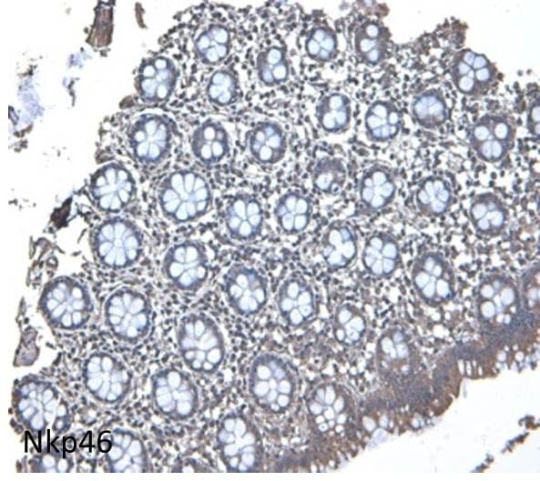
Application: ImmunohistochemistrySample Tested: Colon tissueSpecies: HumanVerified Customer | Posted 10/26/2015At this concentration there is little backgroud. <br />Specificity: Reasonably specific<br />Sensitivity: Reasonably sensitive<br />Buffer: PBS<br />Dilution: 1:500
-
Application: ImmunohistochemistrySample Tested: Colon tissueSpecies: HumanVerified Customer | Posted 10/26/2015Seems like the immune cells show a little staining. At 1:250 all cells showed staining. Dilutions are important. <br />Specificity: Reasonably specific<br />Sensitivity: Reasonably sensitive<br />Buffer: PBS<br />Dilution: 1:1000
There are no reviews that match your criteria.
Protocols
Find general support by application which include: protocols, troubleshooting, illustrated assays, videos and webinars.
- 7-Amino Actinomycin D (7-AAD) Cell Viability Flow Cytometry Protocol
- Antigen Retrieval Protocol (PIER)
- Antigen Retrieval for Frozen Sections Protocol
- Appropriate Fixation of IHC/ICC Samples
- Cellular Response to Hypoxia Protocols
- Chromogenic IHC Staining of Formalin-Fixed Paraffin-Embedded (FFPE) Tissue Protocol
- Chromogenic Immunohistochemistry Staining of Frozen Tissue
- ClariTSA™ Fluorophore Kits
- Detection & Visualization of Antibody Binding
- Extracellular Membrane Flow Cytometry Protocol
- Flow Cytometry Protocol for Cell Surface Markers
- Flow Cytometry Protocol for Staining Membrane Associated Proteins
- Flow Cytometry Staining Protocols
- Flow Cytometry Troubleshooting Guide
- Fluorescent IHC Staining of Frozen Tissue Protocol
- Graphic Protocol for Heat-induced Epitope Retrieval
- Graphic Protocol for the Preparation and Fluorescent IHC Staining of Frozen Tissue Sections
- Graphic Protocol for the Preparation and Fluorescent IHC Staining of Paraffin-embedded Tissue Sections
- Graphic Protocol for the Preparation of Gelatin-coated Slides for Histological Tissue Sections
- ICC Cell Smear Protocol for Suspension Cells
- ICC Immunocytochemistry Protocol Videos
- ICC for Adherent Cells
- IHC Sample Preparation (Frozen sections vs Paraffin)
- ISH-IHC Protocol for Chromogenic Detection on Formalin Fixed Paraffin Embedded (FFPE) Tissue
- Immunocytochemistry (ICC) Protocol
- Immunocytochemistry Troubleshooting
- Immunofluorescence of Organoids Embedded in Cultrex Basement Membrane Extract
- Immunofluorescent IHC Staining of Formalin-Fixed Paraffin-Embedded (FFPE) Tissue Protocol
- Immunohistochemistry (IHC) and Immunocytochemistry (ICC) Protocols
- Immunohistochemistry Frozen Troubleshooting
- Immunohistochemistry Paraffin Troubleshooting
- Intracellular Flow Cytometry Protocol Using Alcohol (Methanol)
- Intracellular Flow Cytometry Protocol Using Detergents
- Intracellular Nuclear Staining Flow Cytometry Protocol Using Detergents
- Intracellular Staining Flow Cytometry Protocol Using Alcohol Permeabilization
- Intracellular Staining Flow Cytometry Protocol Using Detergents to Permeabilize Cells
- Preparing Samples for IHC/ICC Experiments
- Preventing Non-Specific Staining (Non-Specific Binding)
- Primary Antibody Selection & Optimization
- Propidium Iodide Cell Viability Flow Cytometry Protocol
- Protocol for Heat-Induced Epitope Retrieval (HIER)
- Protocol for Liperfluo
- Protocol for Making a 4% Formaldehyde Solution in PBS
- Protocol for VisUCyte™ HRP Polymer Detection Reagent
- Protocol for the Characterization of Human Th22 Cells
- Protocol for the Characterization of Human Th9 Cells
- Protocol for the Fluorescent ICC Staining of Cell Smears - Graphic
- Protocol for the Fluorescent ICC Staining of Cultured Cells on Coverslips - Graphic
- Protocol for the Preparation & Fixation of Cells on Coverslips
- Protocol for the Preparation and Chromogenic IHC Staining of Frozen Tissue Sections
- Protocol for the Preparation and Chromogenic IHC Staining of Frozen Tissue Sections - Graphic
- Protocol for the Preparation and Chromogenic IHC Staining of Paraffin-embedded Tissue Sections
- Protocol for the Preparation and Chromogenic IHC Staining of Paraffin-embedded Tissue Sections - Graphic
- Protocol for the Preparation and Fluorescent ICC Staining of Cells on Coverslips
- Protocol for the Preparation and Fluorescent ICC Staining of Non-adherent Cells
- Protocol for the Preparation and Fluorescent ICC Staining of Stem Cells on Coverslips
- Protocol for the Preparation and Fluorescent IHC Staining of Frozen Tissue Sections
- Protocol for the Preparation and Fluorescent IHC Staining of Paraffin-embedded Tissue Sections
- Protocol for the Preparation of Gelatin-coated Slides for Histological Tissue Sections
- Protocol for the Preparation of a Cell Smear for Non-adherent Cell ICC - Graphic
- Protocol: Annexin V and PI Staining by Flow Cytometry
- Protocol: Annexin V and PI Staining for Apoptosis by Flow Cytometry
- R&D Systems Quality Control Western Blot Protocol
- TUNEL and Active Caspase-3 Detection by IHC/ICC Protocol
- The Importance of IHC/ICC Controls
- Troubleshooting Guide: Fluorokine Flow Cytometry Kits
- Troubleshooting Guide: Immunohistochemistry
- Troubleshooting Guide: Western Blot Figures
- Western Blot Conditions
- Western Blot Protocol
- Western Blot Protocol for Cell Lysates
- Western Blot Troubleshooting
- Western Blot Troubleshooting Guide
- View all Protocols, Troubleshooting, Illustrated assays and Webinars
Loading...
Associated Pathways

