VEGF Antibody (26503)
R&D Systems | Catalog # MAB293

Key Product Details
Validated by
Species Reactivity
Validated:
Cited:
Applications
Validated:
Cited:
Label
Antibody Source
Product Specifications
Immunogen
Ala27-Arg191
Accession # NP_001165097.1
Specificity
Clonality
Host
Isotype
Endotoxin Level
Scientific Data Images for VEGF Antibody (26503)
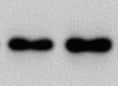
Detection of Recombinant Human VEGF by Western Blot.
Western blot shows 25 ng of Recombinant Human VEGF165 (293-VE), Recombinant Human VEGF111 (5336-VE), Recombinant Human VEGF121, aa 207-327 (4644-VS), Recombinant Human VEGF145 (aa 27-171) (7626-VE), Recombinant Human VEGF162 (2347-VE), Recombinant Human VEGF165b (3045-VE), Recombinant Human VEGF189 (aa 27-215) (8147-VE), Recombinant Human VEGF165 Extended Isoform (9018-VE), Recombinant Human VEGF-B167 (751-VE), Recombinant Mouse VEGF164 (493-MV), and Recombinant Rat VEGF164 (564-RV). PVDF Membrane was probed with 0.1 µg/mL of Mouse Anti-Human/Primate VEGF Monoclonal Antibody (Catalog # MAB293) followed by HRP-conjugated Anti-Mouse IgG Secondary Antibody (HAF007). A specific band was detected for VEGF at approximately 15-25 kDa (as indicated). This experiment was conducted under reducing conditions and using Immunoblot Buffer Group 3.
Cell Proliferation Induced by VEGF165and Neutralization by Human VEGF Antibody.
Recombinant Human VEGF165 (293-VE) stimulates proliferation in HUVEC human umbilical vein endothelial cells in a dose-dependent manner (orange line) as measured by Resazurin (AR002). Proliferation elicited by Recombinant Human VEGF165 (10 ng/mL) is neutralized (green line) by increasing concentrations of Mouse Anti-Human/Primate VEGF Monoclonal Antibody (Catalog # MAB293). The ND50 is typically 10-60 ng/mL.
Detection of Rat VEGF by Western Blot
Protein levels of VEGF (A), KDR (VEGF receptor 2, B), Angiopoietin 1 (C) and Tie2 (D) from retinal endothelial cells cultured in normoxia or hypoxia only and cells in each condition treated with Compound 49b. Hypoxia increased protein levels of all proteins, which were significantly reduced after treatment with Compound 49b. Data is mean ± SEM. *P<0.05 vs. normoxia. #P<0.05 vs. hypoxia. N = 4. Image collected and cropped by CiteAb from the following publication (https://pubmed.ncbi.nlm.nih.gov/27439004), licensed under a CC-BY license. Not internally tested by R&D Systems.
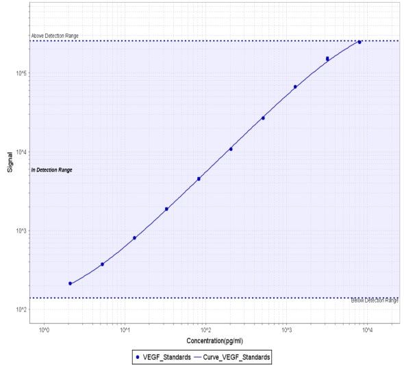
Human VEGF ELISA Standard Curve
Recombinant Human VEGF 165 (Catalog # 293-VE) was serially diluted and captured by Mouse Anti-Human/Primate VEGF Monoclonal Antibody (Catalog # MAB293) coated on a Clear Polystyrene Microplate (Catalog # DY990). Goat Anti-Human VEGF 165 Antigen Affinity-purified Polyclonal Antibody (Catalog # AF-293-NA) was biotinylated and incubated with the protein captured on the plate. Detection of the standard curve was achieved by incubating Streptavidin-HRP (Catalog # DY998)
Human VEGF DuoSet ELISA
Recombinant Human VEGF 165 (Catalog # 293-VE) was serially diluted and captured by Mouse Anti-Human/Primate VEGF Monoclonal Antibody (Catalog # MAB293) coated on a Clear Polystyrene Microplate (Catalog # DY990). Goat Anti-Human VEGF 165 Antigen Affinity-purified Polyclonal Antibody (Catalog # AF-293-NA) was biotinylated and incubated with the protein captured on the plate. Detection of the standard curve was achieved by incubating Streptavidin-HRP (Catalog # DY998)Applications for VEGF Antibody (26503)
Western Blot
Sample: Recombinant Human VEGF165 (Catalog # 293-VE)
Neutralization
Human/Primate VEGF Sandwich Immunoassay
Reviewed Applications
Read 8 reviews rated 4.8 using MAB293 in the following applications:
Formulation, Preparation, and Storage
Purification
Reconstitution
Reconstitute at 0.5 mg/mL in sterile PBS. For liquid material, refer to CoA for concentration.
Formulation
*Small pack size (-SP) is supplied either lyophilized or as a 0.2 µm filtered solution in PBS.
Shipping
Stability & Storage
Calculators
Background: VEGF
Long Name
Alternate Names
Entrez Gene IDs
Gene Symbol
UniProt
Additional VEGF Products
Product Documents for VEGF Antibody (26503)
Certificate of Analysis
To download a Certificate of Analysis, please enter a lot or batch number in the search box below.
Note: Certificate of Analysis not available for kit components.
Product Specific Notices for VEGF Antibody (26503)
For research use only
Citations for VEGF Antibody (26503)
 Powered by Bioz
Customer Reviews for VEGF Antibody (26503) (8)
Have you used VEGF Antibody (26503)?
Submit a review and receive an Amazon gift card!
$25/€18/£15/$25CAN/¥2500 Yen for a review with an image
$10/€7/£6/$10CAN/¥1110 Yen for a review without an image
Submit a review
Customer Images
-
Application: Western BlotSample Tested: THP-1 human acute monocytic leukemia cell lineSpecies: HumanVerified Customer | Posted 02/14/2022

-
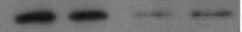
Application: Western BlotSample Tested: Metastatic breast cancerSpecies: HumanVerified Customer | Posted 08/09/2021
-
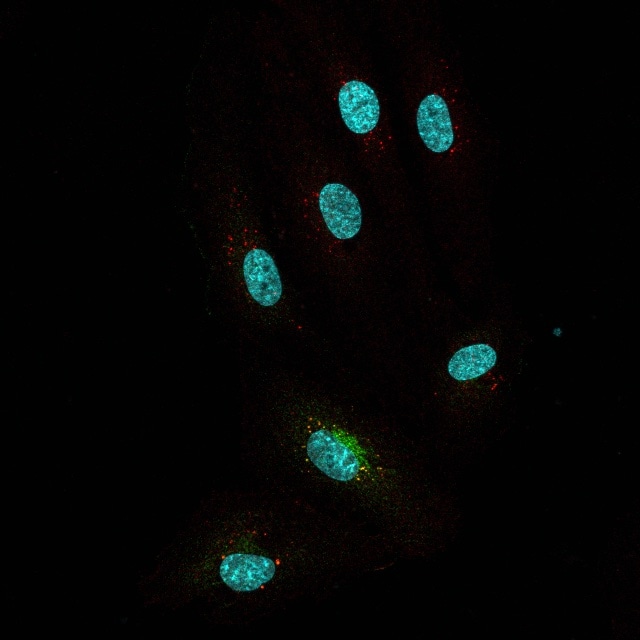
Application: Immunocytochemistry/ImmunofluorescenceSample Tested: HUVEC human umbilical vein endothelial cellsSpecies: HumanVerified Customer | Posted 07/16/2021IF of HUVEC cells with exogenous VEGF internalized (red) and colocalizing with VEGF receptor (green)
-
Application: Block/NeutralizeSample Tested: Serum-free Cell Culture Media and Breast cancer cellsSpecies: HumanVerified Customer | Posted 05/27/2019
-
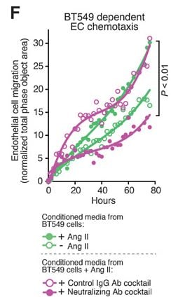
Application: Block/NeutralizeSample Tested: HUVEC human umbilical vein endothelial cellsSpecies: HumanVerified Customer | Posted 03/13/2019We used this VEGF-A neutralizing antibody (10 ng/mL) as part of an antibody cocktail containing several other antibodies to neutralize VEGF-A present in the Conditioned Medium (CM) from BT549 Cells treated with Angiotensin II CM (+/- VEGF-A Neutralizing ab Cocktail) was used to further look at Endothelial (HUVEC) Cell migration using Incucyte Chemotaxis Assay. Cancer Res; 78(5) March 1, 2018 (Fig 6F)
-
Application: Meso Scale DiscoverySample Tested: Retina/choroid, Vitreous humor and Aqueous humorSpecies: PorcineVerified Customer | Posted 06/22/2018After biotinylation, used as a capture reagent in MSD assay to detect ocular VEGF level in a swine model of laser-induced choroidal neovascularization. A standard curve with pig VEGF from Bio-Rad (Cat# PPP030) is shown (2-8,000 pg/ml).
-
Application: ELISASample Tested: CHO Chinese hamster ovary cell lineSpecies: HamsterVerified Customer | Posted 11/04/2017
-
Application: ELISASample Tested: THP-1 human acute monocytic leukemia cell lineSpecies: HumanVerified Customer | Posted 08/18/2017
There are no reviews that match your criteria.
Protocols
Find general support by application which include: protocols, troubleshooting, illustrated assays, videos and webinars.
- Cellular Response to Hypoxia Protocols
- R&D Systems Quality Control Western Blot Protocol
- Troubleshooting Guide: Western Blot Figures
- Western Blot Conditions
- Western Blot Protocol
- Western Blot Protocol for Cell Lysates
- Western Blot Troubleshooting
- Western Blot Troubleshooting Guide
- View all Protocols, Troubleshooting, Illustrated assays and Webinars
FAQs for VEGF Antibody (26503)
-
Q: What are the differences between MAB293 and MAB293R?
A: MAB293 is produced by culturing a hybridoma cell line, whereas MAB293R is produced via recombinant DNA technology, based on the known, proprietary sequence of MAB293. The benefit of recombinant antibodies is the ability to offer a more consistent antibody, with an indefinite supply. As far as research applications, both products will work the same.
Associated Pathways


