
Key Product Details
Validated by
Biological Validation
Species Reactivity
Validated:
Human
Cited:
Human, Mouse, Rat, Porcine, Avian - Chicken, Avian - Quail, Bovine, Guinea Pig, Primate - Macaca mulatta (Rhesus Macaque), Rabbit
Applications
Validated:
Immunohistochemistry, Western Blot, Neutralization, Dual RNAscope ISH-IHC Compatible, Immunocytochemistry
Cited:
Immunohistochemistry, Immunohistochemistry-Paraffin, Immunohistochemistry-Frozen, Western Blot, ELISA, Neutralization, Flow Cytometry, Immunocytochemistry, Immunoprecipitation, Affinity Assay, Bioassay, Column, ELISA Capture, ELISA Development, ELISA Development (Capture), ELISA Development (Detection), Immunoassay Development
Label
Unconjugated
Antibody Source
Polyclonal Goat IgG
Loading...
Product Specifications
Immunogen
S. frugiperda insect ovarian cell line Sf 21-derived recombinant human VEGF165
Ala27-Arg191
Accession # NP_001165097.1
Ala27-Arg191
Accession # NP_001165097.1
Specificity
Detects human VEGF in direct ELISAs and Western blots.
Clonality
Polyclonal
Host
Goat
Isotype
IgG
Endotoxin Level
<0.10 EU per 1 μg of the antibody by the LAL method.
Scientific Data Images for Human VEGF 165 Antibody
Detection of Human VEGF by Western Blot.
Western blot shows lysates of human lung tissue, human kidney tissue, and human liver tissue. PVDF membrane was probed with 5-10 µg/mL of Goat Anti-Human VEGF 165Antigen Affinity-purified Polyclonal Antibody (Catalog # AF-293-NA) followed by HRP-conjugated Anti-Goat IgG Secondary Antibody (HAF017). A specific band was detected for VEGF at approximately 27 kDa (as indicated). This experiment was conducted under reducing conditions and using Immunoblot Buffer Group 1.
VEGF in HUVEC Cells.
VEGF was detected in immersion fixed human umbilical vein endothelial cells (HUVECs) using Goat Anti-Human VEGF165 Antigen Affinity-purified Polyclonal Antibody (Catalog # AF-293-NA) at 10 µg/mL for 3 hours at room temperature. Cells were stained using the NorthernLights™ 557-conjugated Anti-Goat IgG Secondary Antibody (yellow; NL001) and counter-stained with DAPI (blue). View our protocol for Fluorescent ICC Staining of Non-adherent Cells.
VEGF in Human Liver Cancer Tissue.
VEGF was detected in immersion fixed paraffin-embedded sections of human liver cancer tissue using Goat Anti-Human VEGF165 Antigen Affinity-purified Polyclonal Antibody (Catalog # AF-293-NA) at 3 µg/mL for 1 hour at room temperature followed by incubation with the Anti-Goat IgG VisUCyte™ HRP Polymer Antibody (VC004). Before incubation with the primary antibody, tissue was subjected to heat-induced epitope retrieval using Antigen Retrieval Reagent-Basic (CTS013). Tissue was stained using DAB (brown) and counterstained with hematoxylin (blue). Specific staining was localized to cytoplasm in cancer cells. View our protocol for IHC Staining with VisUCyte HRP Polymer Detection Reagents.
Cell Proliferation Induced by VEGF165and Neutralization by Human VEGF Antibody.
Recombinant Human VEGF165 (293-VE) stimulates proliferation in HUVEC human umbilical vein endothelial cells in a dose-dependent manner (orange line). Proliferation elicited by Recombinant Human VEGF165 (10 ng/mL) is neutralized (green line) by increasing concentrations of Goat Anti-Human VEGF165 Antigen Affinity-purified Polyclonal Antibody (Catalog # AF-293-NA). The ND50 is typically 0.02‑0.12 µg/mL.
Detection of Human VEGF by Western Blot
VEGF expression determined by Western blot and immunoprecipitation.A. Western blot using LiCor Odyssey to simultaneously image pan-VEGF and VEGF-A165b probed western blot. Two different podocyte samples, and a primary RPE sample were run on a gel and probed with antibodies to VEGF-A165b (mouse monoclonal anti-CTRSLTRKD, and 680nm-donkey anti-mouse, top image) and pan-VEGF (rabbit polyclonal anti-VEGF, and 800nm-donkey anti-rabbit, middle image). The bottom image is the pseudocoloured combined image (600nm green, 800nm red). Note the red VEGF165, but yellow VEGF-A165b. MWM = molecular weight marker. d = dimer, m = monomer. B. Protein extracted from human cell lines (adenoma and adenocarcinoma(AC)) subjected to immunoprecipitation (IP) for VEGF-A165b and immunoblotting (IB) for total VEGF-A. A clear strong band was seen in the IP for both cell types at ∼23kDa and ∼46kDa, consistent with the IP for recombinant human VEGF-A165b. A weaker band was seen in the input protein (not subjected to IP), and a second band slightly higher in the AC. A weak band at approximately 56kDa and 28kDa was seen in all lanes subjected to IP, including the VEGF-A165a band, but not seen in the recombinant human VEGF-A165b not subjected to IP, indicating that this is cross reactivity with the IgG. This band was clearly above the VEGF-A165b bands. C. Protein extracted from human cell lines (adenoma and adenocarcinoma(AC)) subjected to immunoprecipitation (IP) for VEGF-A and immunoblotting (IB) for VEGF-A165b. A clear strong band was seen in the IP for both cell types at ∼23kDa, the same size as recombinant human VEGF-A165b. In the input a band at ∼46Da was seen predominantly, for both cell types, labelled as VEGF-A165b dimers. D. Mouse tissues probed with VEGF-A165b antibody detect mouse IgG due to the secondary antibody. Top image, western blot of mouse tissues, recombinant mouse IgG or human VEGF-A165b or VEGF-A165b probed with mouse anti-CTRSLTRKD, and 680nm-donkey anti-mouse IgG. Bottom ima
Detection of Human VEGF by Western Blot
Inhibitor of differentiation 4 (ID4) expression in breast cancer cells leads to the activation of an angiogenic programme in macrophages. a Expression matrix representing a panel of angiogenic factors evaluated using TaqMan Low-Density Arrays (TLDA) in macrophages obtained by 1,25-dihydroxyvitamin D3 (VitD3)-mediated differentiation of HL60 cells and subsequently cultured in RPMI medium or in conditioned media (CM) from control (EV) or ID4-overexpressing (ID4) MDA-MB-468 breast cancer cells. b Western blot showing ID4-HA overexpression in MDA-MB-468 cells. c Selected genes modulated in the arrays were evaluated by RT-qPCR in macrophages obtained from VitD3-mediated differentiation of U937 cells and subsequently cultivated in RPMI medium (CTR) or in CM from control (CM si-SCR) or ID4-depleted (CM si-ID4) MDA-MB-468 cells. d Western blot analysis showing the level of ID4 protein after transfection of the indicated small interfering RNAs (siRNAs) in MDA-MB-468 cells. e–g Western blot analysis of ephrin B2 (EphB2), granulin (GRN) and hypoxia-inducible factor (HIF)-1A proteins in differentiated U937 cells cultured in CM si-SCR or CM si-ID4 from MDA-MB-468 cells. h Immunofluorescence analysis of HIF-1A protein performed in differentiated U937 cells cultured in the presence of CM si-SCR or CM si-ID4 from MDA-MB-468 cells. i Western blotting showing the efficiency of vascular endothelial growth factor A (VEGFA) depletion by siRNA transfection in MDA-MB-468 cells used to prepare CM used in experiments shown in (j). j RT-qPCR analysis of the indicated messenger RNAs in U937 macrophages cultivated in the presence of CM from control (si-SCR) or VEGFA-depleted (si-VEGFA) MDA-MB-468 cells. k RT-qPCR analysis of the indicated genes in differentiated U937 cells cultivated in RPMI medium or in CM from MDA-MB-468 cells in the presence of VEGFA blocking antibody (Ab) or a control Ab. Specifically, VEGFA blocking Ab or control Ab were incubated with CM for 30 minutes at room temperatur
Detection of Human VEGF by Western Blot
Western blot analysis of the effects of RNA interference on VEGF expression in HTF cells. (a) Relative expression of VEGF in cells treated with shRNA-2 and shRNA-3; (b) protein expression levels in treated groups and naïve HTF controls. ∗∗∗p < 0.01. Image collected and cropped by CiteAb from the following open publication (https://pubmed.ncbi.nlm.nih.gov/28168047), licensed under a CC-BY license. Not internally tested by R&D Systems.
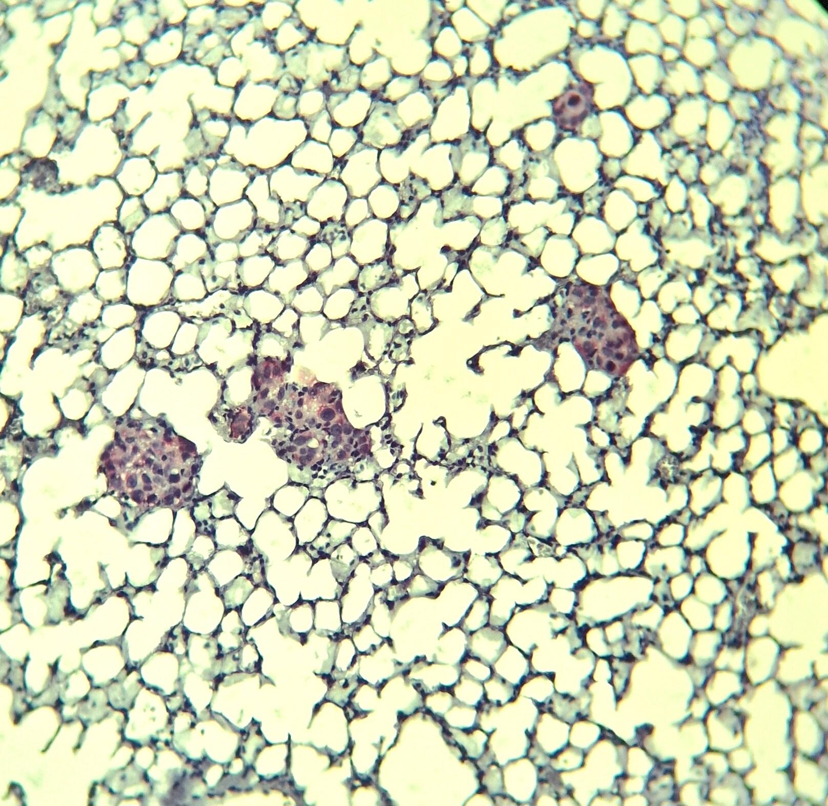
Detection of VEGF in Human Liver.
Formalin-fixed paraffin-embedded tissue sections of human liver were probed for VEGF mRNA (ACD RNAScope Probe, catalog #423168, Fast Red chromogen, ACD catalog # 322750). Adjacent tissue section was processed for immunohistochemistry using goat anti-human VEGF polyclonal antibody (R&D Systems catalog # AF-293-NA) at 5ug/mL with overnight incubation at 4 degrees Celsius followed by incubation with anti-goat IgG VisUCyte HRP Polymer Antibody (Catalog # VC004) and DAB chromogen (yellow-brown). Tissue was counterstained with hematoxylin (blue). Specific staining was localized to hepatocytes.
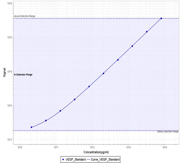
Human VEGF ELISA Standard Curve
Recombinant Human VEGF 165 (Catalog # 293-VE) was serially diluted and captured by Mouse Anti-Human/Primate VEGF Monoclonal Antibody (Catalog # MAB293) coated on a Clear Polystyrene Microplate (Catalog # DY990). Goat Anti-Human VEGF 165 Antigen Affinity-purified Polyclonal Antibody (Catalog # AF-293-NA) was biotinylated and incubated with the protein captured on the plate. Detection of the standard curve was achieved by incubating Streptavidin-HRP (Catalog # DY998)
Human VEGF DuoSet ELISA
Recombinant Human VEGF 165 (Catalog # 293-VE) was serially diluted and captured by Mouse Anti-Human/Primate VEGF Monoclonal Antibody (Catalog # MAB293) coated on a Clear Polystyrene Microplate (Catalog # DY990). Goat Anti-Human VEGF 165 Antigen Affinity-purified Polyclonal Antibody (Catalog # AF-293-NA) was biotinylated and incubated with the protein captured on the plate. Detection of the standard curve was achieved by incubating Streptavidin-HRP (Catalog # DY998)Applications for Human VEGF 165 Antibody
Application
Recommended Usage
Dual RNAscope ISH-IHC Compatible
5-15 µg/mL
Sample: Immersion fixed paraffin-embedded sections of human liver
Sample: Immersion fixed paraffin-embedded sections of human liver
Immunocytochemistry
5-15 µg/mL
Sample: Immersion fixed human umbilical vein endothelial cells
Sample: Immersion fixed human umbilical vein endothelial cells
Immunohistochemistry
3-15 µg/mL
Sample: Immersion fixed paraffin-embedded sections of human liver cancer tissue
Sample: Immersion fixed paraffin-embedded sections of human liver cancer tissue
Western Blot
5-10 µg/mL
Sample: Human lung tissue, human kidney tissue, and human liver tissue
Sample: Human lung tissue, human kidney tissue, and human liver tissue
Neutralization
Measured by its ability to neutralize VEGF165-induced proliferation in HUVEC human umbilical vein endothelial cells. Conn, G. et al. (1990) Proc. Natl. Acad. Sci. USA 87:1323. The Neutralization Dose (ND50) is typically 0.02‑0.12 µg/mL in the presence of 10 ng/mL Recombinant Human VEGF165.
Reviewed Applications
Read 5 reviews rated 4.6 using AF-293-NA in the following applications:
Formulation, Preparation, and Storage
Purification
Antigen Affinity-purified
Reconstitution
Reconstitute at 0.2 mg/mL in sterile PBS. For liquid material, refer to CoA for concentration.
Loading...
Formulation
Lyophilized from a 0.2 μm filtered solution in PBS with Trehalose. See Certificate of Analysis for details.
*Small pack size (-SP) is supplied either lyophilized or as a 0.2 µm filtered solution in PBS.
*Small pack size (-SP) is supplied either lyophilized or as a 0.2 µm filtered solution in PBS.
Shipping
Lyophilized product is shipped at ambient temperature. Liquid small pack size (-SP) is shipped with polar packs. Upon receipt, store immediately at the temperature recommended below.
Stability & Storage
Use a manual defrost freezer and avoid repeated freeze-thaw cycles.
- 12 months from date of receipt, -20 to -70 °C as supplied.
- 1 month, 2 to 8 °C under sterile conditions after reconstitution.
- 6 months, -20 to -70 °C under sterile conditions after reconstitution.
Calculators
Background: VEGF
Long Name
Vascular Endothelial Growth Factor
Alternate Names
MVCD1, VAS, Vasculotropin, VEGF-A, VEGFA, VPF
Entrez Gene IDs
Gene Symbol
VEGFA
UniProt
Additional VEGF Products
Product Documents for Human VEGF 165 Antibody
Certificate of Analysis
To download a Certificate of Analysis, please enter a lot or batch number in the search box below.
Note: Certificate of Analysis not available for kit components.
Product Specific Notices for Human VEGF 165 Antibody
For research use only
Citations for Human VEGF 165 Antibody
 Powered by Bioz
Customer Reviews for Human VEGF 165 Antibody (5)
4.6 out of 5
5 Customer Ratings
Have you used Human VEGF 165 Antibody?
Submit a review and receive an Amazon gift card!
$25/€18/£15/$25CAN/¥2500 Yen for a review with an image
$10/€7/£6/$10CAN/¥1110 Yen for a review without an image
Submit a review
Customer Images
Showing
1
-
5 of
5 reviews
Showing All
Filter By:
-
Application: ImmunohistochemistrySample Tested: DU145 human prostate carcinoma cell lineSpecies: HumanVerified Customer | Posted 11/12/2025

-
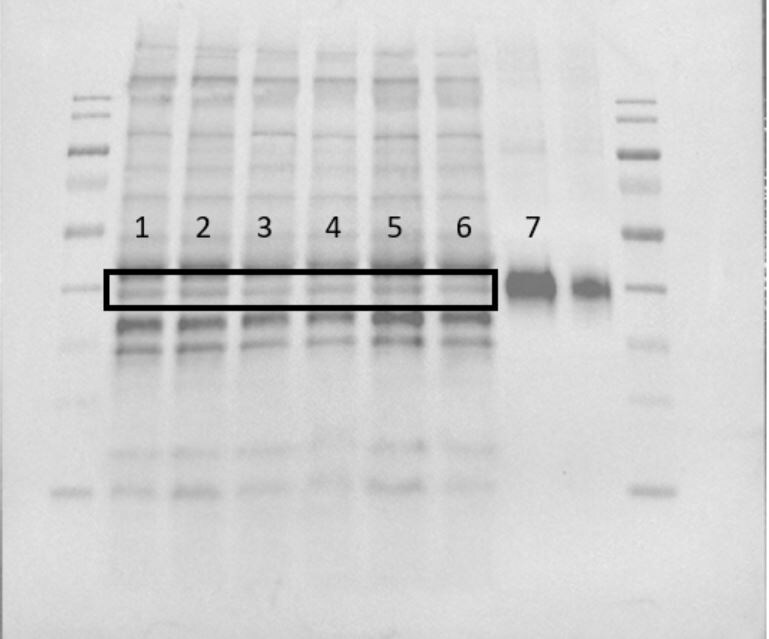
Application: Western BlotSample Tested: Skeletal muscleSpecies: HumanVerified Customer | Posted 02/25/2020Lane 7 shows the VEGF positive control 293-VE-010.
-
Application: Meso Scale DiscoverySample Tested: Vitreous humor, Aqueous humor and Retina/choroidSpecies: PorcineVerified Customer | Posted 06/22/2018After labeling with Sulfo-Tag, used as a detection reagent in MSD assay to detect ocular VEGF level in a swine model of laser-induced choroidal neovascularization. A standard curve with pig VEGF from Bio-Rad (Cat# PPP030) is shown (2-8,000 pg/ml).
-
Application: Block/NeutralizeSample Tested: Primary pumonary arterial endothelial cell lysateSpecies: HumanVerified Customer | Posted 11/23/2015Neutralization of recombinant VEGF-induced VEGFR2 phosphorylation.
-
Application: Western BlotSample Tested: See PMID 22897854Species: HumanVerified Customer | Posted 01/07/2015
There are no reviews that match your criteria.
Protocols
Find general support by application which include: protocols, troubleshooting, illustrated assays, videos and webinars.
- Antigen Retrieval Protocol (PIER)
- Antigen Retrieval for Frozen Sections Protocol
- Appropriate Fixation of IHC/ICC Samples
- Cellular Response to Hypoxia Protocols
- Chromogenic IHC Staining of Formalin-Fixed Paraffin-Embedded (FFPE) Tissue Protocol
- Chromogenic Immunohistochemistry Staining of Frozen Tissue
- ClariTSA™ Fluorophore Kits
- Detection & Visualization of Antibody Binding
- Fluorescent IHC Staining of Frozen Tissue Protocol
- Graphic Protocol for Heat-induced Epitope Retrieval
- Graphic Protocol for the Preparation and Fluorescent IHC Staining of Frozen Tissue Sections
- Graphic Protocol for the Preparation and Fluorescent IHC Staining of Paraffin-embedded Tissue Sections
- Graphic Protocol for the Preparation of Gelatin-coated Slides for Histological Tissue Sections
- ICC Cell Smear Protocol for Suspension Cells
- ICC Immunocytochemistry Protocol Videos
- ICC for Adherent Cells
- IHC Sample Preparation (Frozen sections vs Paraffin)
- ISH-IHC Protocol for Chromogenic Detection on Formalin Fixed Paraffin Embedded (FFPE) Tissue
- Immunocytochemistry (ICC) Protocol
- Immunocytochemistry Troubleshooting
- Immunofluorescence of Organoids Embedded in Cultrex Basement Membrane Extract
- Immunofluorescent IHC Staining of Formalin-Fixed Paraffin-Embedded (FFPE) Tissue Protocol
- Immunohistochemistry (IHC) and Immunocytochemistry (ICC) Protocols
- Immunohistochemistry Frozen Troubleshooting
- Immunohistochemistry Paraffin Troubleshooting
- Preparing Samples for IHC/ICC Experiments
- Preventing Non-Specific Staining (Non-Specific Binding)
- Primary Antibody Selection & Optimization
- Protocol for Heat-Induced Epitope Retrieval (HIER)
- Protocol for Making a 4% Formaldehyde Solution in PBS
- Protocol for VisUCyte™ HRP Polymer Detection Reagent
- Protocol for the Fluorescent ICC Staining of Cell Smears - Graphic
- Protocol for the Fluorescent ICC Staining of Cultured Cells on Coverslips - Graphic
- Protocol for the Preparation & Fixation of Cells on Coverslips
- Protocol for the Preparation and Chromogenic IHC Staining of Frozen Tissue Sections
- Protocol for the Preparation and Chromogenic IHC Staining of Frozen Tissue Sections - Graphic
- Protocol for the Preparation and Chromogenic IHC Staining of Paraffin-embedded Tissue Sections
- Protocol for the Preparation and Chromogenic IHC Staining of Paraffin-embedded Tissue Sections - Graphic
- Protocol for the Preparation and Fluorescent ICC Staining of Cells on Coverslips
- Protocol for the Preparation and Fluorescent ICC Staining of Non-adherent Cells
- Protocol for the Preparation and Fluorescent ICC Staining of Stem Cells on Coverslips
- Protocol for the Preparation and Fluorescent IHC Staining of Frozen Tissue Sections
- Protocol for the Preparation and Fluorescent IHC Staining of Paraffin-embedded Tissue Sections
- Protocol for the Preparation of Gelatin-coated Slides for Histological Tissue Sections
- Protocol for the Preparation of a Cell Smear for Non-adherent Cell ICC - Graphic
- R&D Systems Quality Control Western Blot Protocol
- TUNEL and Active Caspase-3 Detection by IHC/ICC Protocol
- The Importance of IHC/ICC Controls
- Troubleshooting Guide: Immunohistochemistry
- Troubleshooting Guide: Western Blot Figures
- Western Blot Conditions
- Western Blot Protocol
- Western Blot Protocol for Cell Lysates
- Western Blot Troubleshooting
- Western Blot Troubleshooting Guide
- View all Protocols, Troubleshooting, Illustrated assays and Webinars
FAQs for Human VEGF 165 Antibody
Showing
1
-
1 of
1 FAQ
Showing All
-
Q: Does Human VEGF165 Antibody, Catalog # AF-293-NA, recognize VEGF189?
A: Yes, AF-293-NA detects VEGF189
Loading...
Associated Pathways


