
Key Product Details
Species Reactivity
Validated:
Multi-Species
Cited:
Human, Mouse, Xenograft
Applications
Validated:
Immunohistochemistry, Western Blot, ELISA Capture (Matched Antibody Pair)
Cited:
Western Blot, Neutralization, Immunocytochemistry, ELISA Capture, ELISA Development
Label
Unconjugated
Antibody Source
Monoclonal Mouse IgG2B Clone # 8607
Loading...
Product Specifications
Immunogen
Porcine TGF-beta 1.2
Specificity
Detects TGF-beta 2 from multiple species in ELISAs and Western blots. In ELISAs, no cross-reactivity or interfereence with recombinant human (rh) TGF-alpha, recombinant porcineTGF-beta 1, rhTGF-beta 1, recombinant canine TGF-beta 3, recombinant amphibian TGF-beta 5, rhTGF-beta RII, or rhTGF-beta RIII is observed.
Clonality
Monoclonal
Host
Mouse
Isotype
IgG2B
Scientific Data Images for TGF‑ beta 2 Antibody
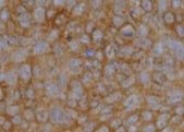
TGF‑ beta 2 in Human Prostate Cancer.
TGF-beta 2 was detected in immersion fixed paraffin-embedded sections of human prostate cancer tissue using Mouse Anti-TGF-beta 2 Monoclonal Antibody (Catalog # MAB612) at 15 µg/mL overnight at 4 °C. Tissue was stained using the Anti-Mouse HRP-DAB Cell & Tissue Staining Kit (brown; Catalog # CTS002) and counterstained with hematoxylin (blue). Specific staining was localized to cytoplasm in cancer cells. View our protocol for Chromogenic IHC Staining of Paraffin-embedded Tissue Sections.
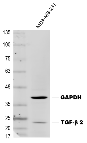
Detection of TGF-beta 2 by Western Blot
Effects of transforming growth factor (TGF)-beta 2 silencing on metastatic phenotypes of sonic-hedgehog medulloblastoma cells with high LOXL1-AS1 expression. A RT-qPCR analysis of TGF-beta 2 expression in Daoy-LOXL1-AS1 and Daoy-MYCN cells. B RT-qPCR analysis of four stemness markers in Daoy-LOXL1-AS1 and Daoy-MYCN cells relative to the parental group. C Western blot analysis of four stemness markers in Daoy-LOXL1-AS1 and Daoy-MYCN cells. Quantification values below each band are relative to the parental group. D, G Sphere-formation assay of (D) Daoy-LOXL1-AS1 and (G) Daoy-MYCN cells, including microscopic images (left images), and the number and sizes of spheres of ≥150 μm in diameter (right bar chart). Scale bars, 500 μm in 4× and 100 μm in 20× magnification. E, H Wound-healing assay of (E) Daoy-LOXL1-AS1 and (H) Daoy-MYCN cells. F, I Transwell migration and invasion assays of (F) Daoy-LOXL1-AS1 and (I) Daoy-MYCN cells including quantification of cell migration (left bar chart) and invasion (right bar chart). Quantitative data are presented as mean ± SD of replicates from representative of three independent experiments. NC, negative control; ns, non-significant. * p < 0.05, ** p < 0.01, *** p < 0.001 Image collected and cropped by CiteAb from the following open publication (https://jeccr.biomedcentral.com/articles/10.1186/s13046-024-03057-0), licensed under a CC-BY license. Not internally tested by R&D Systems.Applications for TGF‑ beta 2 Antibody
Application
Recommended Usage
Immunohistochemistry
8-25 µg/mL
Sample: Immersion fixed paraffin-embedded sections of human prostate cancer tissue
Sample: Immersion fixed paraffin-embedded sections of human prostate cancer tissue
Western Blot
1 µg/mL
Sample: Recombinant Human TGF-beta 2 (Catalog # 302-B2)
under non-reducing conditions only
Sample: Recombinant Human TGF-beta 2 (Catalog # 302-B2)
under non-reducing conditions only
Human TGF-beta 2 Sandwich Immunoassay
Please Note: Optimal dilutions of this antibody should be experimentally determined.
Reviewed Applications
Read 3 reviews rated 5 using MAB612 in the following applications:
Formulation, Preparation, and Storage
Purification
Protein A or G purified from ascites
Reconstitution
Reconstitute at 0.5 mg/mL in sterile PBS. For liquid material, refer to CoA for concentration.
Loading...
Formulation
Lyophilized from a 0.2 μm filtered solution in PBS with Trehalose. *Small pack size (SP) is supplied either lyophilized or as a 0.2 µm filtered solution in PBS.
Shipping
Lyophilized product is shipped at ambient temperature. Liquid small pack size (-SP) is shipped with polar packs. Upon receipt, store immediately at the temperature recommended below.
Stability & Storage
Use a manual defrost freezer and avoid repeated freeze-thaw cycles.
- 12 months from date of receipt, -20 to -70 °C as supplied.
- 1 month, 2 to 8 °C under sterile conditions after reconstitution.
- 6 months, -20 to -70 °C under sterile conditions after reconstitution.
Calculators
Background: TGF-beta 2
Long Name
Transforming Growth Factor beta 2
Alternate Names
TGFB2, TGFbeta 2
Gene Symbol
TGFB2
Additional TGF-beta 2 Products
Product Documents for TGF‑ beta 2 Antibody
Certificate of Analysis
To download a Certificate of Analysis, please enter a lot or batch number in the search box below.
Note: Certificate of Analysis not available for kit components.
Product Specific Notices for TGF‑ beta 2 Antibody
For research use only
Citations for TGF‑ beta 2 Antibody
 Powered by Bioz
Customer Reviews for TGF‑ beta 2 Antibody (3)
5 out of 5
3 Customer Ratings
Have you used TGF‑ beta 2 Antibody?
Submit a review and receive an Amazon gift card!
$25/€18/£15/$25CAN/¥2500 Yen for a review with an image
$10/€7/£6/$10CAN/¥1110 Yen for a review without an image
Submit a review
Customer Images
Showing
1
-
3 of
3 reviews
Showing All
Filter By:
-
Application: ImmunohistochemistrySample Tested: Skin tissueSpecies: HumanVerified Customer | Posted 11/07/2021

-
Application: ELISASample Tested: Recombinant proteinSpecies: HumanVerified Customer | Posted 03/28/2019I used this antibody as a capture antibody to generate an in-house ELISA for TGF beta. I coated the plate overnight with the antibody at 2ug/mL in PBS 100uL/well
-
Application: Western BlotSample Tested: MDA-MB-231 Cell LysateSpecies: HumanVerified Customer | Posted 04/25/2017MDA-MB-231 cell extract probed with Mouse anti-Human TGF-beta 2 antibody (#MAB612) at 1 ug/mL, followed by Goat anti-Mouse secondary antibody at 1:5,000 dilution. PVDF membrane blocked with 5% BSA-PBS.
There are no reviews that match your criteria.
Protocols
Find general support by application which include: protocols, troubleshooting, illustrated assays, videos and webinars.
- Antigen Retrieval Protocol (PIER)
- Antigen Retrieval for Frozen Sections Protocol
- Appropriate Fixation of IHC/ICC Samples
- Cellular Response to Hypoxia Protocols
- Chromogenic IHC Staining of Formalin-Fixed Paraffin-Embedded (FFPE) Tissue Protocol
- Chromogenic Immunohistochemistry Staining of Frozen Tissue
- ClariTSA™ Fluorophore Kits
- Detection & Visualization of Antibody Binding
- Fluorescent IHC Staining of Frozen Tissue Protocol
- Graphic Protocol for Heat-induced Epitope Retrieval
- Graphic Protocol for the Preparation and Fluorescent IHC Staining of Frozen Tissue Sections
- Graphic Protocol for the Preparation and Fluorescent IHC Staining of Paraffin-embedded Tissue Sections
- Graphic Protocol for the Preparation of Gelatin-coated Slides for Histological Tissue Sections
- IHC Sample Preparation (Frozen sections vs Paraffin)
- Immunofluorescent IHC Staining of Formalin-Fixed Paraffin-Embedded (FFPE) Tissue Protocol
- Immunohistochemistry (IHC) and Immunocytochemistry (ICC) Protocols
- Immunohistochemistry Frozen Troubleshooting
- Immunohistochemistry Paraffin Troubleshooting
- Preparing Samples for IHC/ICC Experiments
- Preventing Non-Specific Staining (Non-Specific Binding)
- Primary Antibody Selection & Optimization
- Protocol for Heat-Induced Epitope Retrieval (HIER)
- Protocol for Making a 4% Formaldehyde Solution in PBS
- Protocol for VisUCyte™ HRP Polymer Detection Reagent
- Protocol for the Preparation & Fixation of Cells on Coverslips
- Protocol for the Preparation and Chromogenic IHC Staining of Frozen Tissue Sections
- Protocol for the Preparation and Chromogenic IHC Staining of Frozen Tissue Sections - Graphic
- Protocol for the Preparation and Chromogenic IHC Staining of Paraffin-embedded Tissue Sections
- Protocol for the Preparation and Chromogenic IHC Staining of Paraffin-embedded Tissue Sections - Graphic
- Protocol for the Preparation and Fluorescent IHC Staining of Frozen Tissue Sections
- Protocol for the Preparation and Fluorescent IHC Staining of Paraffin-embedded Tissue Sections
- Protocol for the Preparation of Gelatin-coated Slides for Histological Tissue Sections
- R&D Systems Quality Control Western Blot Protocol
- TUNEL and Active Caspase-3 Detection by IHC/ICC Protocol
- The Importance of IHC/ICC Controls
- Troubleshooting Guide: Immunohistochemistry
- Troubleshooting Guide: Western Blot Figures
- Western Blot Conditions
- Western Blot Protocol
- Western Blot Protocol for Cell Lysates
- Western Blot Troubleshooting
- Western Blot Troubleshooting Guide
- View all Protocols, Troubleshooting, Illustrated assays and Webinars
Loading...
Associated Pathways


