
Key Product Details
Validated by
Orthogonal Validation
Species Reactivity
Validated:
Human, Mouse
Cited:
Human, Mouse
Applications
Validated:
Multiplex Immunofluorescence, Immunohistochemistry, Western Blot, Dual RNAscope ISH-IHC Compatible, Immunocytochemistry, Simple Western, COMET
Cited:
Immunohistochemistry, Immunohistochemistry-Paraffin, Western Blot, Flow Cytometry, Immunocytochemistry, Chromatin Immunoprecipitation (ChIP)
Label
Unconjugated
Antibody Source
Monoclonal Mouse IgG2B Clone # 634913
Loading...
Product Specifications
Immunogen
E. coli-derived recombinant human GATA-3
Pro135-Ser258
Accession # P23771
Pro135-Ser258
Accession # P23771
Specificity
Detects human and mouse GATA-3 in Western blots.
Clonality
Monoclonal
Host
Mouse
Isotype
IgG2B
Scientific Data Images for GATA-3 Antibody (634913)
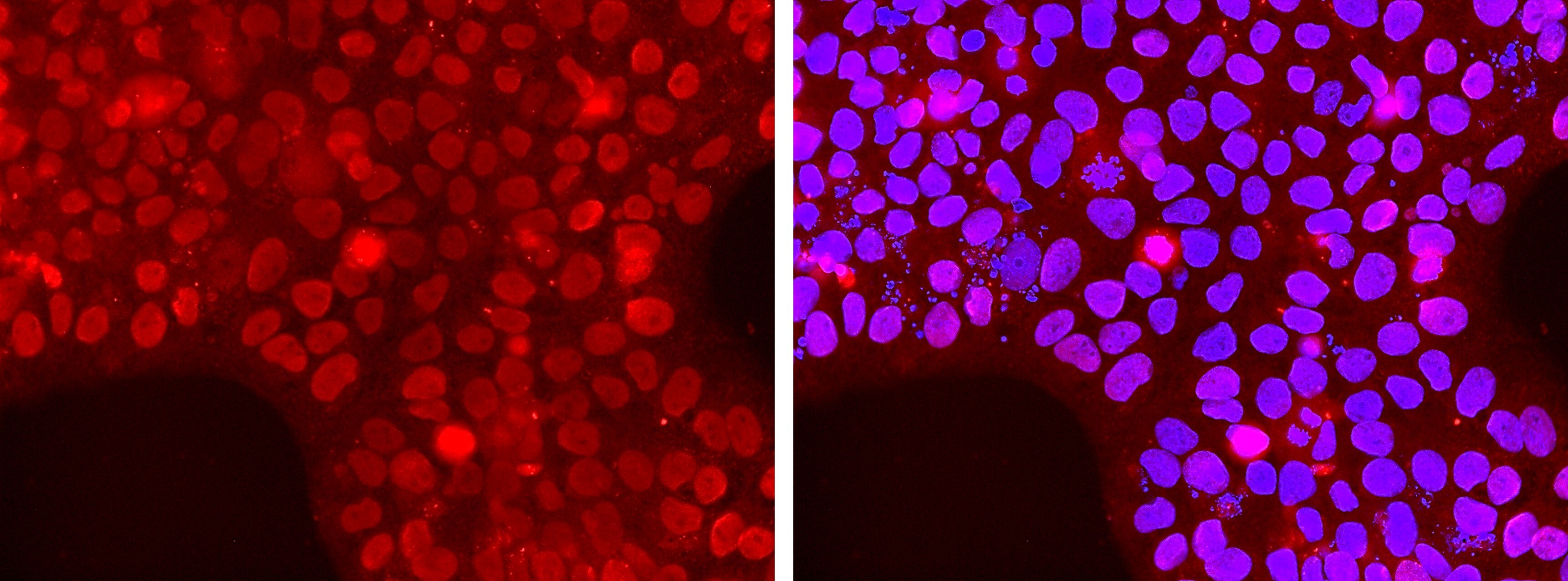
Detection of GATA-3 in Human Breast Cancer via Multiplex Immunofluorescence staining on COMET™
GATA-3 was detected in immersion fixed paraffin-embedded sections of human breast cancer using Mouse Anti-Human GATA‑3 Monoclonal Antibody (MAB6330) at 20 µg/mL at 37 ° Celsius for 4 minutes. Before incubation with the primary antibody, tissue underwent an all-in-one dewaxing and antigen retrieval preprocessing using PreTreatment Module (PT Module) and Dewax and HIER Buffer H (pH 9). Tissue was stained using the Alexa Fluor™ 647 Goat anti-Mouse IgG Secondary Antibody at 1:200 at 37 ° Celsius for 2 minutes. (Yellow; Lunaphore Catalog # DR647MS) and counterstained with DAPI (blue; Lunaphore Catalog # DR100). Specific staining was localized to the nucleus with dim cytoplasmic background signal. Protocol available in COMET™ Panel Builder.
Detection of Human and Mouse GATA‑3 by Western Blot.
Western blot shows lysates of HeLa human cervical epithelial carcinoma cell line, MCF-7 human breast cancer cell line, Jurkat human acute T cell leukemia cell line, and EL-4 mouse lymphoblast cell line. PVDF Membrane was probed with 0.1 µg/mL of Moue Anti-Human/Mouse GATA-3 Monoclonal Antibody (Catalog # MAB6330) followed by HRP-conjugated Anti-Mouse IgG Secondary Antibody (HAF007). Specific bands were detected for full length (FL) GATA-3 at approximately 52 kDa and the splice form (SF) found in MCF-7 cells at approximately 40 kDa (as indicated). This experiment was conducted under reducing conditions and using Immunoblot Buffer Group 1.
GATA‑3 in MCF‑7 Human Cell Line.
GATA-3 was detected in immersion fixed MCF-7 human breast cancer cell line using Mouse Anti-Human/Mouse GATA-3 Monoclonal Antibody (Catalog # MAB6330) at 3 µg/mL for 3 hours at room temperature. Cells were stained using the NorthernLights™ 557-conjugated Anti-Mouse IgG Secondary Antibody (red; NL007) and counterstained with DAPI (blue). Specific staining was localized to nuclei. View our protocol for Fluorescent ICC Staining of Cells on Coverslips.
GATA‑3 in Human Breast Cancer Tissue.
GATA-3 was detected in immersion fixed paraffin-embedded sections of human breast cancer tissue using Mouse Anti-Human/Mouse GATA-3 Monoclonal Antibody (Catalog # MAB6330) at 15 µg/mL overnight at 4 °C. Before incubation with the primary antibody, tissue was subjected to heat-induced epitope retrieval using Antigen Retrieval Reagent-Basic (CTS013). Tissue was stained using the Anti-Mouse HRP-DAB Cell & Tissue Staining Kit (brown CTS002) and counterstained with hematoxylin (blue). Specific staining was localized to nuclei. View our protocol for Chromogenic IHC Staining of Paraffin-embedded Tissue Sections.
Detection of Human GATA‑3 by Simple WesternTM.
Simple Western lane view shows lysates of Jurkat human acute T cell leukemia cell line and MCF‑7 human breast cancer cell line, loaded at 0.5 mg/mL. Specific bands were detected for GATA‑3 at approximately 53 (splice variant) and 61 kDa (full length) as indicated, using 10 µg/mL of Mouse Anti-Human/Mouse GATA‑3 Monoclonal Antibody (Catalog # MAB6330). This experiment was conducted under reducing conditions and using the 12-230 kDa separation system.Non-specific interaction with the 230 kDa Simple Western standard may be seen with this antibody.

GATA-3 in Human Breast Cancer Tissue Using Dual RNAscope® ISH and IHC.
GATA-3 mRNA (red) and protein (green) was detected in formalin-fixed paraffin-embedded tissue sections of human breast cancer tissue probed with ACD RNAScope® Probe (Catalog # 403551) followed by immunohistochemistry using R&D Systems Mouse Anti-Human/Mouse GATA-3 Monoclonal Antibody (Catalog# MAB6330) at 5 μg/mL for 1 hour at room temperature followed by incubation with the Anti-Mouse IgG VisUCyte HRP Polymer Antibody (VC001). Tissue was stained using ACD RNAscope® 2.5 HD Duplex Detection Reagents (Catalog # 322500).Applications for GATA-3 Antibody (634913)
Application
Recommended Usage
COMET
Optimal dilutions of this antibody should be experimentally determined.
Dual RNAscope ISH-IHC Compatible
5-25 µg/mL
Sample: Immersion fixed paraffin-embedded sections of human breast cancer tissue
Sample: Immersion fixed paraffin-embedded sections of human breast cancer tissue
Immunocytochemistry
3-25 µg/mL
Sample: Immersion fixed MCF-7 human breast cancer cell line
Sample: Immersion fixed MCF-7 human breast cancer cell line
Immunohistochemistry
8-25 µg/mL
Sample: Immersion fixed paraffin-embedded sections of human breast cancer tissue
Sample: Immersion fixed paraffin-embedded sections of human breast cancer tissue
Multiplex Immunofluorescence
20 µg/mL
Sample: Paraffin embedded tissue sections of Human Breast Cancer
Sample: Paraffin embedded tissue sections of Human Breast Cancer
Simple Western
10 µg/mL
Sample: Jurkat human acute T cell leukemia cell line and MCF‑7 human breast cancer cell line
Sample: Jurkat human acute T cell leukemia cell line and MCF‑7 human breast cancer cell line
Western Blot
0.1 µg/mL
Sample: HeLa human cervical epithelial carcinoma cell line, MCF‑7 human breast cancer cell line, Jurkat human acute T cell leukemia cell line, and EL‑4 mouse lymphoblast cell line
Sample: HeLa human cervical epithelial carcinoma cell line, MCF‑7 human breast cancer cell line, Jurkat human acute T cell leukemia cell line, and EL‑4 mouse lymphoblast cell line
Reviewed Applications
Read 1 review rated 4 using MAB6330 in the following applications:
Formulation, Preparation, and Storage
Purification
Protein A or G purified from hybridoma culture supernatant
Reconstitution
Reconstitute at 0.5 mg/mL in sterile PBS. For liquid material, refer to CoA for concentration.
Loading...
Formulation
Lyophilized from a 0.2 μm filtered solution in PBS with Trehalose. See Certificate of Analysis for details.
*Small pack size (-SP) is supplied either lyophilized or as a 0.2 µm filtered solution in PBS.
*Small pack size (-SP) is supplied either lyophilized or as a 0.2 µm filtered solution in PBS.
Shipping
Lyophilized product is shipped at ambient temperature. Liquid small pack size (-SP) is shipped with polar packs. Upon receipt, store immediately at the temperature recommended below.
Stability & Storage
Use a manual defrost freezer and avoid repeated freeze-thaw cycles.
- 12 months from date of receipt, -20 to -70 °C as supplied.
- 1 month, 2 to 8 °C under sterile conditions after reconstitution.
- 6 months, -20 to -70 °C under sterile conditions after reconstitution.
Calculators
Background: GATA-3
Additional GATA-3 Products
Product Documents for GATA-3 Antibody (634913)
Certificate of Analysis
To download a Certificate of Analysis, please enter a lot or batch number in the search box below.
Note: Certificate of Analysis not available for kit components.
Product Specific Notices for GATA-3 Antibody (634913)
For research use only
Related Research Areas
Citations for GATA-3 Antibody (634913)
 Powered by Bioz
Customer Reviews for GATA-3 Antibody (634913) (1)
4 out of 5
1 Customer Rating
Have you used GATA-3 Antibody (634913)?
Submit a review and receive an Amazon gift card!
$25/€18/£15/$25CAN/¥2500 Yen for a review with an image
$10/€7/£6/$10CAN/¥1110 Yen for a review without an image
Submit a review
Customer Images
Showing
1
-
1 of
1 review
Showing All
Filter By:
-
Application: Immunocytochemistry/ImmunofluorescenceSample Tested: BeWo human choriocarcinoma cell lineSpecies: HumanVerified Customer | Posted 03/20/2023BeWo Cell. Gata3 & Merge with DAPI.

There are no reviews that match your criteria.
Protocols
Find general support by application which include: protocols, troubleshooting, illustrated assays, videos and webinars.
- Antigen Retrieval Protocol (PIER)
- Antigen Retrieval for Frozen Sections Protocol
- Appropriate Fixation of IHC/ICC Samples
- Cellular Response to Hypoxia Protocols
- Chromogenic IHC Staining of Formalin-Fixed Paraffin-Embedded (FFPE) Tissue Protocol
- Chromogenic Immunohistochemistry Staining of Frozen Tissue
- ClariTSA™ Fluorophore Kits
- Detection & Visualization of Antibody Binding
- Fluorescent IHC Staining of Frozen Tissue Protocol
- Graphic Protocol for Heat-induced Epitope Retrieval
- Graphic Protocol for the Preparation and Fluorescent IHC Staining of Frozen Tissue Sections
- Graphic Protocol for the Preparation and Fluorescent IHC Staining of Paraffin-embedded Tissue Sections
- Graphic Protocol for the Preparation of Gelatin-coated Slides for Histological Tissue Sections
- ICC Cell Smear Protocol for Suspension Cells
- ICC Immunocytochemistry Protocol Videos
- ICC for Adherent Cells
- IHC Sample Preparation (Frozen sections vs Paraffin)
- ISH-IHC Protocol for Chromogenic Detection on Formalin Fixed Paraffin Embedded (FFPE) Tissue
- Immunocytochemistry (ICC) Protocol
- Immunocytochemistry Troubleshooting
- Immunofluorescence of Organoids Embedded in Cultrex Basement Membrane Extract
- Immunofluorescent IHC Staining of Formalin-Fixed Paraffin-Embedded (FFPE) Tissue Protocol
- Immunohistochemistry (IHC) and Immunocytochemistry (ICC) Protocols
- Immunohistochemistry Frozen Troubleshooting
- Immunohistochemistry Paraffin Troubleshooting
- Preparing Samples for IHC/ICC Experiments
- Preventing Non-Specific Staining (Non-Specific Binding)
- Primary Antibody Selection & Optimization
- Protocol for Heat-Induced Epitope Retrieval (HIER)
- Protocol for Making a 4% Formaldehyde Solution in PBS
- Protocol for VisUCyte™ HRP Polymer Detection Reagent
- Protocol for the Fluorescent ICC Staining of Cell Smears - Graphic
- Protocol for the Fluorescent ICC Staining of Cultured Cells on Coverslips - Graphic
- Protocol for the Preparation & Fixation of Cells on Coverslips
- Protocol for the Preparation and Chromogenic IHC Staining of Frozen Tissue Sections
- Protocol for the Preparation and Chromogenic IHC Staining of Frozen Tissue Sections - Graphic
- Protocol for the Preparation and Chromogenic IHC Staining of Paraffin-embedded Tissue Sections
- Protocol for the Preparation and Chromogenic IHC Staining of Paraffin-embedded Tissue Sections - Graphic
- Protocol for the Preparation and Fluorescent ICC Staining of Cells on Coverslips
- Protocol for the Preparation and Fluorescent ICC Staining of Non-adherent Cells
- Protocol for the Preparation and Fluorescent ICC Staining of Stem Cells on Coverslips
- Protocol for the Preparation and Fluorescent IHC Staining of Frozen Tissue Sections
- Protocol for the Preparation and Fluorescent IHC Staining of Paraffin-embedded Tissue Sections
- Protocol for the Preparation of Gelatin-coated Slides for Histological Tissue Sections
- Protocol for the Preparation of a Cell Smear for Non-adherent Cell ICC - Graphic
- R&D Systems Quality Control Western Blot Protocol
- TUNEL and Active Caspase-3 Detection by IHC/ICC Protocol
- The Importance of IHC/ICC Controls
- Troubleshooting Guide: Immunohistochemistry
- Troubleshooting Guide: Western Blot Figures
- Western Blot Conditions
- Western Blot Protocol
- Western Blot Protocol for Cell Lysates
- Western Blot Troubleshooting
- Western Blot Troubleshooting Guide
- View all Protocols, Troubleshooting, Illustrated assays and Webinars


