
Key Product Details
Species Reactivity
Validated:
Human
Cited:
Human, Mouse, Primate - Papio cynocephalus (Yellow Baboon)
Applications
Validated:
Western Blot, Neutralization, Immunocytochemistry
Cited:
Immunohistochemistry, Western Blot, Neutralization, Immunocytochemistry, Affinity Assay, Bioassay, ELISA Development, Functional Assay
Label
Unconjugated
Antibody Source
Polyclonal Goat IgG
Loading...
Product Specifications
Immunogen
E. coli-derived recombinant human TNF-alpha
Val77-Leu233
Accession # P01375
Val77-Leu233
Accession # P01375
Specificity
Detects human TNF‑ alpha in direct ELISAs and Western blots.
Clonality
Polyclonal
Host
Goat
Isotype
IgG
Endotoxin Level
<0.10 EU per 1 μg of the antibody by the LAL method.
Scientific Data Images for Human TNF-alpha Antibody
Cytotoxicity Induced by TNF‑ alpha and Neutralization by Human TNF-alpha Antibody.
Recombinant Human TNF-a (Catalog # 210-TA) induces cytotoxicity in the L-929 mouse fibroblast cell line in a dose-dependent manner (orange line). Cytotoxicity elicited by Recombinant Human TNF-a (1.5 ng/mL) is neutralized (green line) by increasing concentrations of Goat Anti-Human TNF-a Antigen Affinity-purified Polyclonal Antibody (Catalog # AF-210-NA). The ND50 is typically 0.01-0.06 µg/mL.
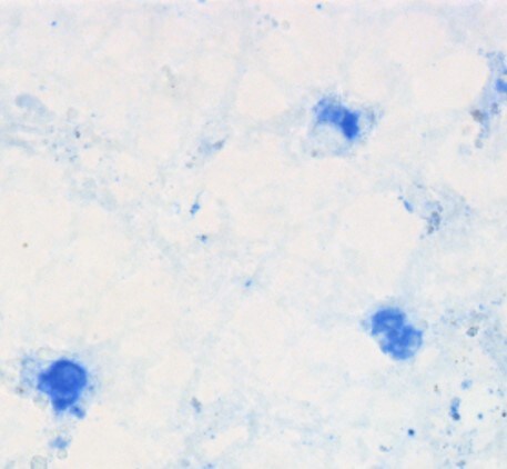
TNF‑ alpha in Human PBMCs.
TNF-a was detected in immersion fixed human peripheral blood mononuclear cells (PBMCs) using Goat Anti-Human TNF-a Antigen Affinity-purified Polyclonal Antibody (Catalog # AF-210-NA) at 5 µg/mL for 3 hours at room temperature. Cells were stained using the NorthernLights™ 557-conjugated Anti-Goat IgG Secondary Antibody (red; Catalog # NL001) and counterstained with DAPI (blue). Specific staining was localized to cytoplasm. View our protocol for Fluorescent ICC Staining of Non-adherent Cells.
Detection of Human TNF-alpha by Functional
TPM1 and TPM4 expression in response to TNF alpha.High molecular weight tropomyosin isoform expression in THP1 cells in response to elevated concentrations of TNF alpha. Image collected and cropped by CiteAb from the following publication (https://pubmed.ncbi.nlm.nih.gov/27649540), licensed under a CC-BY license. Not internally tested by R&D Systems.
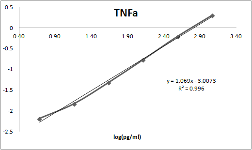
Human TNF-alpha ELISA Standard Curve
Human Granzyme B was serially diluted and captured by Mouse Anti-Human TNF‑ alpha Monoclonal Antibody (Catalog # MAB610) coated on a Clear Polystyrene Microplate (Catalog # DY990). Goat Anti-Human TNF‑ alpha Antigen Affinity-purified Polyclonal Antibody (Catalog # AF-210-NA) was biotinylated and incubated with the protein captured on the plate. Detection of the standard curve was achieved by incubating Streptavidin-HRP (Catalog # DY998)Applications for Human TNF-alpha Antibody
Application
Recommended Usage
Immunocytochemistry
5-15 µg/mL
Sample: Immersion fixed human peripheral blood mononuclear cells
Sample: Immersion fixed human peripheral blood mononuclear cells
Western Blot
0.1 µg/mL
Sample: Recombinant Human TNF‑ alpha (Catalog # 210-TA)
Sample: Recombinant Human TNF‑ alpha (Catalog # 210-TA)
Neutralization
Measured by its ability to neutralize TNF‑ alpha -induced cytotoxicity in the L‑929 mouse fibroblast cell line. Matthews, N. and M. L. Neale (1987) in Lymphokines and Interferons, A Practical Approach. Clemens, M. J. et al. (eds): IRL Press. 221. The Neutralization Dose (ND50) is typically 0.01-0.06 μg/ml in the presence of 1.5 ng/mL Recombinant Human TNF‑ alpha.
Reviewed Applications
Read 3 reviews rated 5 using AF-210-NA in the following applications:
Formulation, Preparation, and Storage
Purification
Antigen Affinity-purified
Reconstitution
Reconstitute at 0.2 mg/mL in sterile PBS. For liquid material, refer to CoA for concentration.
Loading...
Formulation
Lyophilized from a 0.2 μm filtered solution in PBS with Trehalose. *Small pack size (SP) is supplied either lyophilized or as a 0.2 µm filtered solution in PBS.
Shipping
Lyophilized product is shipped at ambient temperature. Liquid small pack size (-SP) is shipped with polar packs. Upon receipt, store immediately at the temperature recommended below.
Stability & Storage
Use a manual defrost freezer and avoid repeated freeze-thaw cycles.
- 12 months from date of receipt, -20 to -70 °C as supplied.
- 1 month, 2 to 8 °C under sterile conditions after reconstitution.
- 6 months, -20 to -70 °C under sterile conditions after reconstitution.
Calculators
Background: TNF-alpha
Long Name
Tumor Necrosis Factor alpha
Alternate Names
Cachetin, DIF, TNF, TNF-A, TNFA, TNFalpha, TNFG1F, TNFSF1A, TNFSF2
Entrez Gene IDs
Gene Symbol
TNF
UniProt
Additional TNF-alpha Products
Product Documents for Human TNF-alpha Antibody
Certificate of Analysis
To download a Certificate of Analysis, please enter a lot or batch number in the search box below.
Note: Certificate of Analysis not available for kit components.
Product Specific Notices for Human TNF-alpha Antibody
For research use only
Related Research Areas
Citations for Human TNF-alpha Antibody
 Powered by Bioz
Customer Reviews for Human TNF-alpha Antibody (3)
5 out of 5
3 Customer Ratings
Have you used Human TNF-alpha Antibody?
Submit a review and receive an Amazon gift card!
$25/€18/£15/$25CAN/¥2500 Yen for a review with an image
$10/€7/£6/$10CAN/¥1110 Yen for a review without an image
Submit a review
Customer Images
Showing
1
-
3 of
3 reviews
Showing All
Filter By:
-
Application: ImmunohistochemistrySample Tested: Adult brainSpecies: HumanVerified Customer | Posted 04/13/2018Published in https://www.ncbi.nlm.nih.gov/pubmed/28169287 Used at 10ug/ml. Briefly, frozen brain sections were fixed in 4% PFA (Fisher Scientific), followed by antigen retrieval using heating in acid citric buffer (Vector, Burlingame, CA, USA). Endogenous avidin-biotin was blocked for 15 min (Vector). Sections were incubated with 10% horse serum in PBS (Biosera, Boussens, France) and Fc Receptor Blocking Solution was added (Human TruStain FcX Biolegend, London, UK). Primary antibody was added overnight at 4 °C and detected with donkey anti-goat-biotin (ab6578, Abcam), followed by streptavidin-alkaline phosphatase (SA-5100, Vector) and visualised with the Vector Blue Alkaline Phosphatase Substrate Kit III (Vector).

-
Application: ELISASample Tested: Heparin Plasma, EDTA Plasma and Citrate PlasmaSpecies: HumanVerified Customer | Posted 01/18/2018
-
Application: ELISASample Tested: Serum and PlasmaSpecies: HumanVerified Customer | Posted 11/16/2017This antibody was used with MAB610 to build an ELISA to measure TNFa. This antibody was used as the detection while MAB610 was used as the capture.
There are no reviews that match your criteria.
Protocols
Find general support by application which include: protocols, troubleshooting, illustrated assays, videos and webinars.
- Appropriate Fixation of IHC/ICC Samples
- Cellular Response to Hypoxia Protocols
- ClariTSA™ Fluorophore Kits
- Detection & Visualization of Antibody Binding
- ICC Cell Smear Protocol for Suspension Cells
- ICC Immunocytochemistry Protocol Videos
- ICC for Adherent Cells
- Immunocytochemistry (ICC) Protocol
- Immunocytochemistry Troubleshooting
- Immunofluorescence of Organoids Embedded in Cultrex Basement Membrane Extract
- Immunohistochemistry (IHC) and Immunocytochemistry (ICC) Protocols
- Preparing Samples for IHC/ICC Experiments
- Preventing Non-Specific Staining (Non-Specific Binding)
- Primary Antibody Selection & Optimization
- Protocol for VisUCyte™ HRP Polymer Detection Reagent
- Protocol for the Fluorescent ICC Staining of Cell Smears - Graphic
- Protocol for the Fluorescent ICC Staining of Cultured Cells on Coverslips - Graphic
- Protocol for the Preparation and Fluorescent ICC Staining of Cells on Coverslips
- Protocol for the Preparation and Fluorescent ICC Staining of Non-adherent Cells
- Protocol for the Preparation and Fluorescent ICC Staining of Stem Cells on Coverslips
- Protocol for the Preparation of a Cell Smear for Non-adherent Cell ICC - Graphic
- R&D Systems Quality Control Western Blot Protocol
- TUNEL and Active Caspase-3 Detection by IHC/ICC Protocol
- The Importance of IHC/ICC Controls
- Troubleshooting Guide: Western Blot Figures
- Western Blot Conditions
- Western Blot Protocol
- Western Blot Protocol for Cell Lysates
- Western Blot Troubleshooting
- Western Blot Troubleshooting Guide
- View all Protocols, Troubleshooting, Illustrated assays and Webinars
Loading...
Associated Pathways


IL-15 Signaling Pathways and their Primary Biological Effects in Different Immune Cell Types
 Innate Lymphoid Cell Differentiation Pathways
 mTOR Signaling Pathway
 NOD-like Receptor Signaling Pathways
 Th1 Differentiation Pathway
 TNF Superfamily Pathway: Human Ligand-Receptor Interactions & their Associated Functions
 Toll-Like Receptor Signaling Pathways
