
Key Product Details
Validated by
Biological Validation
Species Reactivity
Validated:
Mouse
Cited:
Human, Mouse, Rat, Rabbit
Applications
Validated:
Immunohistochemistry, Western Blot, Neutralization, Immunocytochemistry
Cited:
Immunohistochemistry, Immunohistochemistry-Paraffin, Immunohistochemistry-Frozen, Western Blot, Neutralization, Immunocytochemistry, ELISA Development (Capture)
Label
Unconjugated
Antibody Source
Polyclonal Goat IgG
Loading...
Product Specifications
Immunogen
E. coli-derived recombinant mouse IL-6
Phe25-Thr211
Accession # P08505
Phe25-Thr211
Accession # P08505
Specificity
Detects mouse IL-6 in direct ELISAs and Western blots. In direct ELISAs, approximately 100% cross-reactivity with recombinant rat IL‑6 and recombinant cotton rat IL-6 is observed and less than 15% cross-reactivity with recombinant human IL-6 is observed.
Clonality
Polyclonal
Host
Goat
Isotype
IgG
Endotoxin Level
<0.10 EU per 1 μg of the antibody by the LAL method.
Scientific Data Images for Mouse IL-6 Antibody
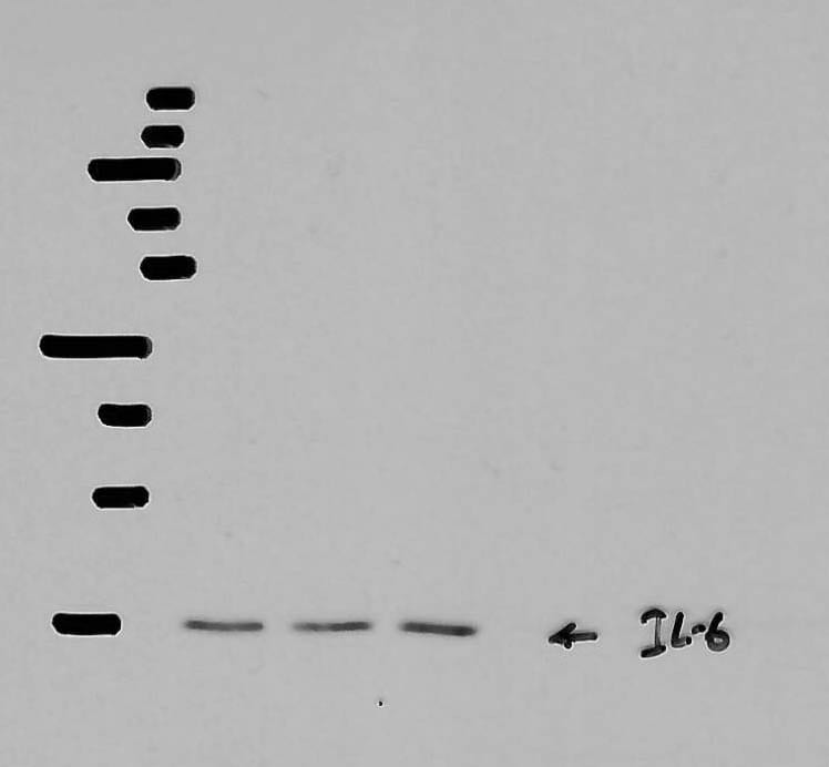
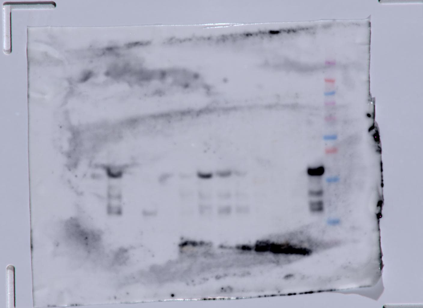
Detection of Mouse IL‑6 by Western Blot.
Western blot shows lysates of RAW 264.7 mouse monocyte/macrophage cell line untreated (-) or treated (+) with LPS. PVDF membrane was probed with 0.5 µg/mL of Goat Anti-Mouse IL-6 Antigen Affinity-purified Polyclonal Antibody (Catalog # AF-406-NA) followed by HRP-conjugated Anti-Goat IgG Secondary Antibody (Catalog # HAF017). A specific band was detected for IL-6 at approximately 22 kDa (as indicated). This experiment was conducted under reducing conditions and using Immunoblot Buffer Group 1.
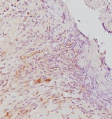
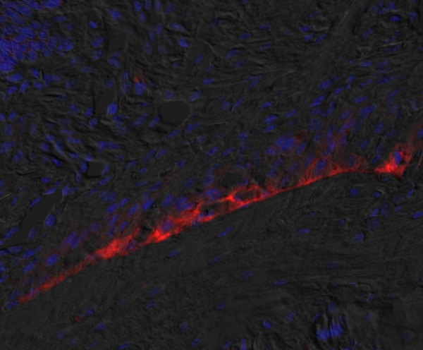
IL‑6 in Mouse Thymus.
IL-6 was detected in perfusion fixed frozen sections of mouse thymus using Goat Anti-Mouse IL-6 Antigen Affinity-purified Polyclonal Antibody (Catalog # AF-406-NA) at 5 µg/mL for 1 hour at room temperature followed by incubation with the Anti-Goat IgG VisUCyte™ HRP Polymer Antibody (Catalog # VC004). Tissue was stained using DAB (brown) and counterstained with hematoxylin (blue). Specific staining was localized to cell surface in lymphocytes. View our protocol for IHC Staining with VisUCyte HRP Polymer Detection Reagents.
Cell Proliferation Induced by IL‑6 and Neutralization by Mouse IL‑6 Antibody.
Recombinant Mouse IL-6 (Catalog # 406-ML) stimulates proliferation in the T1165.85.2.1 mouse plasmacytoma cell line in a dose-dependent manner (orange line). Proliferation elicited by Recombinant Mouse IL-6 (0.25 ng/mL) is neutralized (green line) by increasing concentrations of Goat Anti-Mouse IL-6 Antigen Affinity-purified Polyclonal Antibody (Catalog # AF-406-NA). The ND50 is typically 0.01-0.03 µg/mL.
Detection of Mouse IL-6 by Western Blot
EphA2‐Dependent tumor induction of osteoclast differentiation requires IL‐6. (A) ELISA analysis for IL‐6 protein levels in conditioned media harvested from 4T1. delta C cells relative to 4T1.V controls (p < 0.05, Mann–Whitney test). (B) Graph shows the % tartrate‐resistant acid phosphatase (TRAP)+ osteoclasts in indirect cocultures of osteoclast progenitors with 4T1.V + IgG, 4T1.V + anti‐mouse IL‐6 neutralizing antibody, 4T1. delta C + PBS, and 4T1. delta C + recombinant murine IL‐6. (p < 0.05, Mann–Whitney test). (C) Graph shows the % TRAP+ osteoclasts in direct cocultures of osteoclast progenitors with 4T1.V + IgG, 4T1.V + anti‐mouse IL‐6 neutralizing antibody, 4T1. delta C + PBS, and 4T1. delta C + recombinant murine IL‐6, (p < 0.05, Mann–Whitney test). There were five to eight fields per condition from three independent experiments. (D) Immunoblots show phosphorylated Stat3 levels in osteoclast progenitor cells treated with IL‐6 in the presence or absence of neutralizing anti‐mouse IL‐6 antibody. Uniform loading was confirmed by probing blots for total Stat3 and actin. Image collected and cropped by CiteAb from the following open publication (https://pubmed.ncbi.nlm.nih.gov/33869989), licensed under a CC-BY license. Not internally tested by R&D Systems.
Detection of Mouse IL-6 by Western Blot
Inflammatory cytokines. (a) Representative band image and Coomassie brilliant blue staining of the total amount of protein; (b) TNF‐ alpha ; (c) IL‐6; and (d) IL‐1 beta. The data are present in each plot as the mean ± standard deviation (n = 6 per group). dpi, days post‐injury; DW, distilled water group; SP, spermidine group. Image collected and cropped by CiteAb from the following open publication (https://pubmed.ncbi.nlm.nih.gov/39448391), licensed under a CC-BY license. Not internally tested by R&D Systems.
Detection of IL-6 by Western Blot
Inflammatory cytokines. (a) Representative images of the bands and Coomassie brilliant blue staining of the total amount of protein; (b) TNF‐ alpha ; (c) IL‐6. The data are presented as the mean ± standard deviation (n = 6 per group). DW, Distilled water group; SP, Spermidine group. Image collected and cropped by CiteAb from the following open publication (https://pubmed.ncbi.nlm.nih.gov/39910742), licensed under a CC-BY license. Not internally tested by R&D Systems.
Immersion fixed paraffin-embedded sections of mouse spleen
IL‑6 was detected in immersion fixed paraffin-embedded sections of mouse spleen using Goat Anti-Mouse IL‑6 Antigen Affinity-purified Polyclonal Antibody (Catalog # AF-406-NA) at 1 µg/ml overnight at 4 °C. Before incubation with the primary antibody, tissue was subjected to heat-induced epitope retrieval using VisUCyte Antigen Retrieval Reagent-Basic (Catalog # VCTS021). Tissue was stained using the HRP-conjugated Anti-Goat IgG Secondary Antibody (Catalog # HAF017) and counterstained with hematoxylin (blue). Specific staining was localized to the cytoplasm. View our protocol for Chromogenic IHC Staining of Paraffin-embedded Tissue Sections.
Mouse IL-6 ELISA Standard Curve
Recombinant Mouse IL‑6 (Catalog # 406-ML) was serially diluted and captured by Rat Anti-Mouse IL‑6 Monoclonal Antibody (Catalog # MAB406) coated on a Clear Polystyrene Microplate (Catalog # DY990). Goat Anti-Mouse IL‑6 Antigen Affinity-purified Polyclonal Antibody (Catalog # AF-406-NA) was biotinylated and incubated with the protein captured on the plate. Detection of the standard curve was achieved by incubating Streptavidin-HRP (Catalog # DY998)Applications for Mouse IL-6 Antibody
Application
Recommended Usage
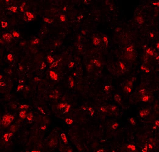
Immunocytochemistry
5-15 µg/mL
Sample: Immersion fixed mouse splenocytes treated with PMA and ionomycin
Sample: Immersion fixed mouse splenocytes treated with PMA and ionomycin
Immunohistochemistry
0.25-25 µg/mL
Sample: Perfusion fixed frozen sections of mouse thymus and Immersion fixed paraffin-embedded sections of mouse spleen
Sample: Perfusion fixed frozen sections of mouse thymus and Immersion fixed paraffin-embedded sections of mouse spleen
Western Blot
0.5 µg/mL
Sample: RAW 264.7 mouse monocyte/macrophage cell line treated with LPS
Sample: RAW 264.7 mouse monocyte/macrophage cell line treated with LPS
Neutralization
Measured by its ability to neutralize IL‑6-induced proliferation in the T1165.85.2.1 mouse plasmacytoma cell line. Nordan, R.P. and M. Potter (1986) Science 233:566. The Neutralization Dose (ND50) is typically 0.01-0.03 µg/mL in the presence of 0.25 ng/mL Recombinant Mouse IL‑6.
Reviewed Applications
Read 7 reviews rated 3.9 using AF-406-NA in the following applications:
Formulation, Preparation, and Storage
Purification
Antigen Affinity-purified
Reconstitution
Reconstitute at 0.2 mg/mL in sterile PBS. For liquid material, refer to CoA for concentration.
Loading...
Formulation
Lyophilized from a 0.2 μm filtered solution in PBS with Trehalose. See Certificate of Analysis for details.
*Small pack size (-SP) is supplied either lyophilized or as a 0.2 µm filtered solution in PBS.
*Small pack size (-SP) is supplied either lyophilized or as a 0.2 µm filtered solution in PBS.
Shipping
Lyophilized product is shipped at ambient temperature. Liquid small pack size (-SP) is shipped with polar packs. Upon receipt, store immediately at the temperature recommended below.
Stability & Storage
Use a manual defrost freezer and avoid repeated freeze-thaw cycles.
- 12 months from date of receipt, -20 to -70 °C as supplied.
- 1 month, 2 to 8 °C under sterile conditions after reconstitution.
- 6 months, -20 to -70 °C under sterile conditions after reconstitution.
Calculators
Background: IL-6
Long Name
Interleukin 6
Alternate Names
BSF-2, BSF2, IFNB2, IL6, MGI-2A
Entrez Gene IDs
Gene Symbol
IL6
UniProt
Additional IL-6 Products
Product Documents for Mouse IL-6 Antibody
Certificate of Analysis
To download a Certificate of Analysis, please enter a lot or batch number in the search box below.
Note: Certificate of Analysis not available for kit components.
Product Specific Notices for Mouse IL-6 Antibody
For research use only
Related Research Areas
Citations for Mouse IL-6 Antibody
 Powered by Bioz
Customer Reviews for Mouse IL-6 Antibody (7)
3.9 out of 5
7 Customer Ratings
Have you used Mouse IL-6 Antibody?
Submit a review and receive an Amazon gift card!
$25/€18/£15/$25CAN/¥2500 Yen for a review with an image
$10/€7/£6/$10CAN/¥1110 Yen for a review without an image
Submit a review
Customer Images
Showing
1
-
5 of
7 reviews
Showing All
Filter By:
-
Application: Immunocytochemistry/ImmunofluorescenceSample Tested: A20 mouse B cell lymphoma cell lineSpecies: MouseVerified Customer | Posted 02/14/2022

-
Application: Immunocytochemistry/ImmunofluorescenceSample Tested: Connective tissueSpecies: RatVerified Customer | Posted 07/06/2021
-
Application: Western BlotSample Tested: Adipose tissueSpecies: Human Endothelial cell and HumanVerified Customer | Posted 04/06/2021Protein from adipose tissue that was isolated with WT and KO mice, were subjected to immunoblotting for IL-6.
-
Application: ELISASample Tested: SerumSpecies: MouseVerified Customer | Posted 12/18/2020
-
Application: ImmunohistochemistrySample Tested: mouse bone tissueSpecies: MouseVerified Customer | Posted 11/23/2020
-
Application: Western BlotSample Tested: Pancreas tissueSpecies: MouseVerified Customer | Posted 10/13/2020
Bio-Techne ResponseThank you for reviewing our product. We are sorry to hear that this product did not perform as expected. We have been in touch with the customer to resolve this issue according to our Product Guarantee and to the customer’s satisfaction.
-
Application: Immunohistochemistry-FrozenSample Tested: See PMID 20683902Species: MouseVerified Customer | Posted 01/08/2015
There are no reviews that match your criteria.
Protocols
Find general support by application which include: protocols, troubleshooting, illustrated assays, videos and webinars.
- Antigen Retrieval Protocol (PIER)
- Antigen Retrieval for Frozen Sections Protocol
- Appropriate Fixation of IHC/ICC Samples
- Cellular Response to Hypoxia Protocols
- Chromogenic IHC Staining of Formalin-Fixed Paraffin-Embedded (FFPE) Tissue Protocol
- Chromogenic Immunohistochemistry Staining of Frozen Tissue
- ClariTSA™ Fluorophore Kits
- Detection & Visualization of Antibody Binding
- Fluorescent IHC Staining of Frozen Tissue Protocol
- Graphic Protocol for Heat-induced Epitope Retrieval
- Graphic Protocol for the Preparation and Fluorescent IHC Staining of Frozen Tissue Sections
- Graphic Protocol for the Preparation and Fluorescent IHC Staining of Paraffin-embedded Tissue Sections
- Graphic Protocol for the Preparation of Gelatin-coated Slides for Histological Tissue Sections
- ICC Cell Smear Protocol for Suspension Cells
- ICC Immunocytochemistry Protocol Videos
- ICC for Adherent Cells
- IHC Sample Preparation (Frozen sections vs Paraffin)
- Immunocytochemistry (ICC) Protocol
- Immunocytochemistry Troubleshooting
- Immunofluorescence of Organoids Embedded in Cultrex Basement Membrane Extract
- Immunofluorescent IHC Staining of Formalin-Fixed Paraffin-Embedded (FFPE) Tissue Protocol
- Immunohistochemistry (IHC) and Immunocytochemistry (ICC) Protocols
- Immunohistochemistry Frozen Troubleshooting
- Immunohistochemistry Paraffin Troubleshooting
- Preparing Samples for IHC/ICC Experiments
- Preventing Non-Specific Staining (Non-Specific Binding)
- Primary Antibody Selection & Optimization
- Protocol for Heat-Induced Epitope Retrieval (HIER)
- Protocol for Making a 4% Formaldehyde Solution in PBS
- Protocol for VisUCyte™ HRP Polymer Detection Reagent
- Protocol for the Fluorescent ICC Staining of Cell Smears - Graphic
- Protocol for the Fluorescent ICC Staining of Cultured Cells on Coverslips - Graphic
- Protocol for the Preparation & Fixation of Cells on Coverslips
- Protocol for the Preparation and Chromogenic IHC Staining of Frozen Tissue Sections
- Protocol for the Preparation and Chromogenic IHC Staining of Frozen Tissue Sections - Graphic
- Protocol for the Preparation and Chromogenic IHC Staining of Paraffin-embedded Tissue Sections
- Protocol for the Preparation and Chromogenic IHC Staining of Paraffin-embedded Tissue Sections - Graphic
- Protocol for the Preparation and Fluorescent ICC Staining of Cells on Coverslips
- Protocol for the Preparation and Fluorescent ICC Staining of Non-adherent Cells
- Protocol for the Preparation and Fluorescent ICC Staining of Stem Cells on Coverslips
- Protocol for the Preparation and Fluorescent IHC Staining of Frozen Tissue Sections
- Protocol for the Preparation and Fluorescent IHC Staining of Paraffin-embedded Tissue Sections
- Protocol for the Preparation of Gelatin-coated Slides for Histological Tissue Sections
- Protocol for the Preparation of a Cell Smear for Non-adherent Cell ICC - Graphic
- R&D Systems Quality Control Western Blot Protocol
- TUNEL and Active Caspase-3 Detection by IHC/ICC Protocol
- The Importance of IHC/ICC Controls
- Troubleshooting Guide: Immunohistochemistry
- Troubleshooting Guide: Western Blot Figures
- Western Blot Conditions
- Western Blot Protocol
- Western Blot Protocol for Cell Lysates
- Western Blot Troubleshooting
- Western Blot Troubleshooting Guide
- View all Protocols, Troubleshooting, Illustrated assays and Webinars
Loading...
Associated Pathways


IL-21 Signaling Pathways and their Primary Biological Effects in Different Immune Cell Types
 Jak/STAT Signaling Pathway
 Mesenchymal Stem Cell Differentiation Pathways & Lineage-specific Markers
 NOD-like Receptor Signaling Pathways
 Th17 Differentiation Pathway
 Toll-Like Receptor Signaling Pathways
