Human IL-1 beta/IL-1F2 Antibody
R&D Systems | Catalog # AF-201-NA

Key Product Details
Validated by
Orthogonal Validation, Biological Validation
Species Reactivity
Validated:
Human
Cited:
Human, Mouse, Amphibian, Canine, Lizard, Primate - Macaca mulatta (Rhesus Macaque)
Applications
Validated:
Immunohistochemistry, Western Blot, Neutralization, Dual RNAscope ISH-IHC Compatible, Immunocytochemistry, Simple Western
Cited:
Immunohistochemistry, Immunohistochemistry-Paraffin, Western Blot, Neutralization, Immunocytochemistry, Bioassay, Blocking/Neutralizing, Cell Culture, Dot Blot, Functional Assay
Label
Unconjugated
Antibody Source
Polyclonal Goat IgG
Loading...
Product Specifications
Immunogen
E. coli-derived recombinant human IL-1 beta /IL-1F2
Ala117-Ser269
Accession # NP_000567
Ala117-Ser269
Accession # NP_000567
Specificity
Detects human IL-1 beta /IL-1F2 in direct ELISAs and Western blots.
Clonality
Polyclonal
Host
Goat
Isotype
IgG
Endotoxin Level
<0.10 EU per 1 μg of the antibody by the LAL method.
Scientific Data Images for Human IL-1 beta/IL-1F2 Antibody
IL-1 beta /IL-1F2 in Human Tonsil Using Dual RNAscope®ISH and IHC.
IL-1 beta /IL-1F2 mRNA was detected in formalin-fixed paraffin-embedded tissue sections of human tonsil probed with ACD RNAScope®Probe (Catalog # 310361) and stained using ACD RNAscope®2.5 HD Detection Reagents-Red (top image, Catalog # 32260). Adjacent tissue section was processed for immunohistochemistry using R&D Systems Goat Anti-Human IL-1 beta /IL-1F2 Antigen Affinity-purified Polyclonal Antibody (Catalog # AF-201-NA) at 1 ug/mL for 1 hour at room temperature followed by incubation with the Anti-Goat IgG VisUCyte HRP Polymer Antibody (R&D Systems, Catalog # VC004) and DAB chromogen (lower image, yellow-brown). Tissues were counterstained with hematoxylin (blue).
Detection of Human IL‑1 beta /IL‑1F2 by Western Blot.
Western blot shows lysates of THP-1 human acute monocytic leukemia cell line untreated (-) or treated (+) with 200 nM PMA for 24 hours and 10 µg/mL LPS and 3 hours. PVDF membrane was probed with 0.1 µg/mL of Goat Anti-Human IL-1 beta /IL-1F2 Antigen Affinity-purified Polyclonal Antibody (Catalog # AF-201-NA) followed by HRP-conjugated Anti-Goat IgG Secondary Antibody (Catalog # HAF017). A specific band was detected for IL-1 beta /IL-1F2 at approximately 36 kDa (as indicated). This experiment was conducted under reducing conditions and using Immunoblot Buffer Group 1.
Detection of IL‑1 beta /IL‑1F2 in THP‑1 Human Cell Line.
IL‑1 beta /IL‑1F2 was detected in immersion fixed THP‑1 human acute monocytic leukemia cell line using Mouse Anti-Human IL‑1 beta /IL‑1F2 Monoclonal Antibody (Catalog # MAB601) at 5 µg/ml for 3 hours at room temperature. Cells were stained using the NorthernLights™ 557-conjugated Anti-Goat IgG Secondary Antibody (red; Catalog # NL001) and counterstained with DAPI (blue). Specific staining was localized to the cytoplasm of THP-1 cells treated with 200nM PMA for 24 hours then 10ug/mL LPS for 24 hours. View our protocol for Fluorescent ICC Staining of Cells on Coverslips.
IL‑1 beta /IL‑1F2 in Human PBMCs.
IL-1 beta /IL-1F2 was detected in immersion fixed human peripheral blood mononuclear cells (PBMCs) treated with LPS and monensin using Goat Anti-Human IL-1 beta /IL-1F2 Antigen Affinity-purified Polyclonal Antibody (Catalog # AF-201-NA) at 10 µg/mL for 3 hours at room temperature. Cells were stained using the NorthernLights™ 557-conjugated Anti-Goat IgG Secondary Antibody (red; Catalog # NL001) and counterstained with DAPI (blue). View our protocol for Fluorescent ICC Staining of Cells on Coverslips.
IL‑1 beta /IL‑1F2 in Human Tonsil.
IL-1 beta /IL-1F2 was detected in immersion fixed paraffin-embedded sections of human tonsil using Goat Anti-Human IL-1 beta /IL-1F2 Antigen Affinity-purified Polyclonal Antibody (Catalog # AF-201-NA) at 1 µg/mL for 1 hour at room temperature followed by incubation with the Anti-Goat IgG VisUCyte™ HRP Polymer Antibody (Catalog # VC004). Tissue was stained using DAB (brown) and counterstained with hematoxylin (blue). Specific staining was localized to cytoplasm in lymphocytes. View our protocol for IHC Staining with VisUCyte HRP Polymer Detection Reagents.
Detection of Human IL‑1 beta /IL‑1F2 by Simple WesternTM.
Simple Western lane view shows lysates of THP‑1 human acute monocytic leukemia cell line untreated (-) or treated (+) with 200 nm PMA and 10 ug/ml LPS for 24 hrs and 3 hrs, respectively, and loaded at 0.2 mg/mL. A specific band was detected for IL‑1 beta /IL‑1F2 at approximately 39 kDa (as indicated) using 1 µg/mL of Goat Anti-Human IL‑1 beta /IL‑1F2 Antigen Affinity-purified Polyclonal Antibody (Catalog # AF-201-NA). This experiment was conducted under reducing conditions and using the 12-230 kDa separation system.
Cell Proliferation Induced by IL‑1 beta /IL‑1F2 and Neutral-ization by Human IL‑1 beta /IL‑1F2 Antibody.
Recombinant Human IL-1 beta /IL-1F2 (Catalog # 201-LB) stimulates proliferation in the the D10.G4.1 mouse helper T cell line in a dose-dependent manner (orange line). Proliferation elicited by Recombinant Human IL-1 beta /IL-1F2 (50 pg/mL) is neutralized (green line) by increasing concentrations of Goat Anti-Human IL-1 beta /IL-1F2 Antigen Affinity-purified Polyclonal Antibody (Catalog # AF-201-NA). The ND50 is typically 6-42 ng/mL.
Detection of Mouse IL-1 beta/IL-1F2 by Western Blot
BTK inhibitors and its dysfunctional mutation suppress NLRP3 inflammasome activation.(a) Enzyme-linked immunosorbent assay (ELISA) of human IL-1 beta in supernatants and immunoblot analysis of human IL-1 beta p17, caspase-1 p20/p22 in supernatants and pro-IL-1 beta in cell lysates of THP-1-Mφs that were pretreated with the indicated inhibitors for 30 min and then stimulated with alum for 6 h. ELISA of murine IL-1 beta (b,c) and TNF-alpha (b) in supernatants of LPS-primed murine peritoneal macrophages that were pretreated with LFM-A13 and then stimulated with alum for 3 h. (d,e) Immunoblot analysis of the indicated proteins (d) and ELISA of murine IL-1 beta and IL-6 in supernatants of LPS-primed peritoneal macrophages from Xid and WT mice stimulated with alum for 6 h. (f) ELISA of murine IL-1 beta in supernatants of LPS-primed murine peritoneal macrophages pretreated with LFM-A13 and/or Syk inhibitor (R406), then stimulated with alum for 3 h. (g) ELISA of murine IL-1 beta and immunoblot analysis of murine caspase-1 p20 in supernatant of LPS-primed murine peritoneal macrophages stimulated with the indicated NLRP3 inflammasome activators for 3 h. Immunoblot analysis of murine IL-1 beta p17, caspase-1 p20 in supernatants, pro-IL-1 beta, pro-caspase-1 and ASC in cell lysates (h), and ELISA of murine IL-1 beta in supernatants (i) of LPS-primed murine peritoneal macrophages that were pretreated with LFM-A13 and then stimulated with alum or poly(dA:dT) for 3 h. Data are representative of three independent experiments. Data are presented as mean±s.d. (triplicate). **P<0.01; ***P<0.003. Two-sided Student's t-test. Image collected and cropped by CiteAb from the following publication (https://pubmed.ncbi.nlm.nih.gov/26059659), licensed under a CC-BY license. Not internally tested by R&D Systems.
Detection of Mouse IL-1 beta/IL-1F2 by Immunohistochemistry
Pyroptosis/IL-1 beta pathway is enhanced in vivo and can be modulated by anakinra. a In the PI model, phosphorylated-p65 NF-kappa B, NLRP3, 8-OHdG, IL-1 beta, and TUNEL were detected by IHC (Bar, 100 μm). b IL-1 beta was immuno-localized in gum tissues of mice with PI alone and with orthotopic mammary tumor introduction (Bar, 100 μm). c The mRNA expression of IL-1 beta was measured in the gingiva of the indicated groups. Serum IL-1 beta was detected by ELISA (n ≥ 5; Student’s t test). d The size of primary tumors was measured (n ≥ 7). e Early metastasis in lymph nodes were measured by luciferase assay and normalized by protein concentration, where anakinra reduced the metastasis to the axillary and cervical lymph nodes. MDSC content was also decreased by anakinra in cervical lymph node. (n ≥ 7; Ank anakinra; ANOVA, *P < 0.05; **P < 0.01; ***P < 0.001; ****P < 0.0001) Image collected and cropped by CiteAb from the following publication (https://pubmed.ncbi.nlm.nih.gov/31685946), licensed under a CC-BY license. Not internally tested by R&D Systems.
Detection of Human IL-1 beta/IL-1F2 by Immunohistochemistry
TMZ inhibits tumor growth and enhances the expression of NLRP1, IL-1 beta, and Notch1 in 1205Lu tumors in vivo. (A) Tumor growth curve of 1205Lu parental cells injected subcutaneously in mice, intraperitoneally treated with a TMZ cycle or 10% dimethyl sulfoxide (DMSO) (Control) (indicated by arrows). Tumor growth was monitored for 26 days. Data are expressed as the mean ± SEM. n = 10 tumors (control) or 12 tumors (TMZ). (B) qRT-PCR analysis of NLRP1 expression in single tumor cells isolated from the tumor tissues studied in (A). Data are expressed as the mean ± SEM. n = 3 samples. * p < 0.05 and *** p < 0.001 vs the corresponding tumors. (C–E) Immunohistochemical staining of tumor tissues with NLRP1 (C), IL-1 beta (D), and Notch1 (E). Samples MB656, MB657, and MB658 were from the control group, whereas MB659, MB660, and MB661 were from the TMZ group. Bar = 50 μm (left panels, 20×) or 20 μm (right panels, 63×). Representative images are shown. Image collected and cropped by CiteAb from the following publication (https://pubmed.ncbi.nlm.nih.gov/32899791), licensed under a CC-BY license. Not internally tested by R&D Systems.
Detection of Human IL-1 beta/IL-1F2 by Western Blot
BTK inhibitors and its dysfunctional mutation suppress NLRP3 inflammasome activation.(a) Enzyme-linked immunosorbent assay (ELISA) of human IL-1 beta in supernatants and immunoblot analysis of human IL-1 beta p17, caspase-1 p20/p22 in supernatants and pro-IL-1 beta in cell lysates of THP-1-Mφs that were pretreated with the indicated inhibitors for 30 min and then stimulated with alum for 6 h. ELISA of murine IL-1 beta (b,c) and TNF-alpha (b) in supernatants of LPS-primed murine peritoneal macrophages that were pretreated with LFM-A13 and then stimulated with alum for 3 h. (d,e) Immunoblot analysis of the indicated proteins (d) and ELISA of murine IL-1 beta and IL-6 in supernatants of LPS-primed peritoneal macrophages from Xid and WT mice stimulated with alum for 6 h. (f) ELISA of murine IL-1 beta in supernatants of LPS-primed murine peritoneal macrophages pretreated with LFM-A13 and/or Syk inhibitor (R406), then stimulated with alum for 3 h. (g) ELISA of murine IL-1 beta and immunoblot analysis of murine caspase-1 p20 in supernatant of LPS-primed murine peritoneal macrophages stimulated with the indicated NLRP3 inflammasome activators for 3 h. Immunoblot analysis of murine IL-1 beta p17, caspase-1 p20 in supernatants, pro-IL-1 beta, pro-caspase-1 and ASC in cell lysates (h), and ELISA of murine IL-1 beta in supernatants (i) of LPS-primed murine peritoneal macrophages that were pretreated with LFM-A13 and then stimulated with alum or poly(dA:dT) for 3 h. Data are representative of three independent experiments. Data are presented as mean±s.d. (triplicate). **P<0.01; ***P<0.003. Two-sided Student's t-test. Image collected and cropped by CiteAb from the following publication (https://pubmed.ncbi.nlm.nih.gov/26059659), licensed under a CC-BY license. Not internally tested by R&D Systems.
Detection of Human IL-1 beta/IL-1F2 by Immunohistochemistry
IL-1 and IL-1R1 expression is enriched in the melanoma stroma. (A) Real-time qPCR analysis of IL1A and IL1B expression in stage-III and stage-IV melanoma tumor samples (n = 39) relative to expression in human skin samples (n = 8). ***, P < 0.001; Mann-Whitney test. (B) Analysis of IL1B expression in normal skin and benign nevi samples (nonmalignant; n = 25) and cutaneous melanoma samples (malignant; n = 45) from an available gene expression dataset (Talantov et al., 2005) accessed through the Oncomine platform. (C) Sections from a case of primary cutaneous melanoma stained for IL-1 beta, CD163, and CD68 expression as indicated by labels. Bars: (i) 200 µm; (ii) 50 µm; (iii) 33 µm. (D) Serial sections from two skin metastases (i–iii and iv–vi, respectively), stained for IL-1R1 and SMA expression as indicated by the labels. Bars: (i and iv) 200 µm; (ii, iii, v, and vi) 33 µm. (C and D) Arrowheads indicate cells that are clearly double stained. (E) Western blot analysis of IL-1R1 and IL-1 beta precursor protein expression in a panel of cell lines. Data are representative of three independent experiments. (F) Secreted IL-1 beta in conditioned media from a panel of melanoma cell lines detected by ELISA. Data are represented as mean ± SEM for three independent samples in each group. **, P < 0.01; Dunn’s multiple comparisons test. (E and F) Macrophages (Mφ) were stimulated with 100 ng/ml IFN-gamma and 20 ng/ml LPS. Image collected and cropped by CiteAb from the following publication (https://pubmed.ncbi.nlm.nih.gov/28450382), licensed under a CC-BY license. Not internally tested by R&D Systems.
Detection of IL-1 beta /IL-1F2 by Western Blot
LPS, type I and type II interferons increase Pyrin expression and enable Pyrin activation in human macrophages.(A) Pyrin and IL-1 beta expression in hMDM treated with either IFN-gamma (200 U/ml), LPS (10 ng/ml), TNF alpha (50 ng/ml), IL-10 (100 ng/ml), IFN-beta (5,000 U/ml), IL-4 (1,000 U/ml), or Pam3CSK4 (20 ng/ml) for either 5 or 18 h. Representative of 3 independent experiments. (B) Pyrin (MEFV) or IL-1 beta (IL1 beta ) transcript from hMDM-treated LPS (10 ng/ml) or Pam3CSK4 (20 ng/ml) for 12 h. Mean and SEM of the fold change of 3 experimental replicates shown. The underlying data can be found in the summary data file in the tab Fig 5B. Image collected and cropped by CiteAb from the following open publication (https://pubmed.ncbi.nlm.nih.gov/36342970), licensed under a CC-BY license. Not internally tested by R&D Systems.
Detection of IL-1 beta /IL-1F2 by Western Blot
TcdB triggers a NLRP3-independent inflammasome response in murine macrophages.IL-1 beta release from WT and NLRP3-deficient BMDM (A) or PMs (B) primed with LPS (200 ng/ml, 3 h), then activated with nigericin and TcdB for 2 h or dA:dT for 4 h. (C) Caspase-1 and IL-1 beta immunoblots of precipitated supernatant or cell lysate from WT and NLRP3-deficient BMDM treated as in (A). (D) Caspase-1 and IL-1 beta immunoblots of precipitated supernatant or cell lysate from LPS primed WT BMDM either untreated or pretreated with CP-456,773 (2.5 μM, 30 min), then stimulated as in (A). Mean and SEM of 3 independent experiments shown, immunoblots are representative of 3 independent experiments. The underlying data can be found in the summary data file in the tab Fig 4A and 4B. BMDM, bone marrow–derived macrophage; LPS, lipopolysaccharide; PM, peritoneal macrophage; WT, wild-type. Image collected and cropped by CiteAb from the following open publication (https://pubmed.ncbi.nlm.nih.gov/36342970), licensed under a CC-BY license. Not internally tested by R&D Systems.
Detection of IL-1 beta /IL-1F2 by Western Blot
LPS, type I and type II interferons increase Pyrin expression and enable Pyrin activation in human macrophages.(A) Pyrin and IL-1 beta expression in hMDM treated with either IFN-gamma (200 U/ml), LPS (10 ng/ml), TNF alpha (50 ng/ml), IL-10 (100 ng/ml), IFN-beta (5,000 U/ml), IL-4 (1,000 U/ml), or Pam3CSK4 (20 ng/ml) for either 5 or 18 h. Representative of 3 independent experiments. (B) Pyrin (MEFV) or IL-1 beta (IL1 beta ) transcript from hMDM-treated LPS (10 ng/ml) or Pam3CSK4 (20 ng/ml) for 12 h. Mean and SEM of the fold change of 3 experimental replicates shown. The underlying data can be found in the summary data file in the tab Fig 5B. Image collected and cropped by CiteAb from the following open publication (https://pubmed.ncbi.nlm.nih.gov/36342970), licensed under a CC-BY license. Not internally tested by R&D Systems.
Detection of IL-1 beta /IL-1F2 by Western Blot
TcdB triggers a NLRP3-independent inflammasome response in murine macrophages.IL-1 beta release from WT and NLRP3-deficient BMDM (A) or PMs (B) primed with LPS (200 ng/ml, 3 h), then activated with nigericin and TcdB for 2 h or dA:dT for 4 h. (C) Caspase-1 and IL-1 beta immunoblots of precipitated supernatant or cell lysate from WT and NLRP3-deficient BMDM treated as in (A). (D) Caspase-1 and IL-1 beta immunoblots of precipitated supernatant or cell lysate from LPS primed WT BMDM either untreated or pretreated with CP-456,773 (2.5 μM, 30 min), then stimulated as in (A). Mean and SEM of 3 independent experiments shown, immunoblots are representative of 3 independent experiments. The underlying data can be found in the summary data file in the tab Fig 4A and 4B. BMDM, bone marrow–derived macrophage; LPS, lipopolysaccharide; PM, peritoneal macrophage; WT, wild-type. Image collected and cropped by CiteAb from the following open publication (https://pubmed.ncbi.nlm.nih.gov/36342970), licensed under a CC-BY license. Not internally tested by R&D Systems.
Detection of IL-1 beta /IL-1F2 by Western Blot
TcdB triggers a NLRP3-independent inflammasome response in murine macrophages.IL-1 beta release from WT and NLRP3-deficient BMDM (A) or PMs (B) primed with LPS (200 ng/ml, 3 h), then activated with nigericin and TcdB for 2 h or dA:dT for 4 h. (C) Caspase-1 and IL-1 beta immunoblots of precipitated supernatant or cell lysate from WT and NLRP3-deficient BMDM treated as in (A). (D) Caspase-1 and IL-1 beta immunoblots of precipitated supernatant or cell lysate from LPS primed WT BMDM either untreated or pretreated with CP-456,773 (2.5 μM, 30 min), then stimulated as in (A). Mean and SEM of 3 independent experiments shown, immunoblots are representative of 3 independent experiments. The underlying data can be found in the summary data file in the tab Fig 4A and 4B. BMDM, bone marrow–derived macrophage; LPS, lipopolysaccharide; PM, peritoneal macrophage; WT, wild-type. Image collected and cropped by CiteAb from the following open publication (https://pubmed.ncbi.nlm.nih.gov/36342970), licensed under a CC-BY license. Not internally tested by R&D Systems.
Human IL-1 beta / IL-1F2 ELISA Standard Curve
Recombinant Human IL‑1 beta /IL‑1F2 (Catalog # 201-LB) was serially diluted and captured by Mouse Anti-Human IL‑1 beta /IL‑1F2 Monoclonal Antibody (Catalog # MAB601) coated on a Clear Polystyrene Microplate (Catalog # DY990). Goat Anti-Human IL‑1 beta /IL‑1F2 Antigen Affinity-purified Polyclonal Antibody (Catalog # AF-201-NA) was biotinylated and incubated with the protein captured on the plate. Detection of the standard curve was achieved by incubating Streptavidin-HRP (Catalog # DY998)Applications for Human IL-1 beta/IL-1F2 Antibody
Application
Recommended Usage
Dual RNAscope ISH-IHC Compatible
1-15 µg/mL
Sample: Immersion fixed paraffin-embedded sections of human tonsil
Sample: Immersion fixed paraffin-embedded sections of human tonsil
Immunocytochemistry
5-15 µg/mL
Sample: Immersion fixed human peripheral blood lymphocytes and immersion fixed human peripheral blood mononuclear cells treated with LPS and monensin and THP-1 human acute monocytic leukemia cell line untreated and treated with PMA and LPS
Sample: Immersion fixed human peripheral blood lymphocytes and immersion fixed human peripheral blood mononuclear cells treated with LPS and monensin and THP-1 human acute monocytic leukemia cell line untreated and treated with PMA and LPS
Immunohistochemistry
1-15 µg/mL
Sample: Immersion fixed paraffin-embedded sections of human tonsil
Sample: Immersion fixed paraffin-embedded sections of human tonsil
Simple Western
1 µg/mL
Sample: TF‑1 human erythroleukemic cell line
Sample: TF‑1 human erythroleukemic cell line
Western Blot
0.1 µg/mL
Sample: THP‑1 human acute monocytic leukemia cell line treated with PMA and LPS
Sample: THP‑1 human acute monocytic leukemia cell line treated with PMA and LPS
Neutralization
Measured by its ability to neutralize IL‑1 beta /IL‑1F2-induced proliferation in the D10.G4.1 mouse helper T cell line. Symons, J.A. et al. (1987) in Lymphokines and Interferons, a Practical Approach. Clemens, M.J. et al. (eds): IRL Press. 272. The Neutralization Dose (ND50) is typically 6-42 ng/mL in the presence of 50 pg/mL Recombinant Human IL‑1 beta /IL‑1F2.
Reviewed Applications
Read 5 reviews rated 4.2 using AF-201-NA in the following applications:
Formulation, Preparation, and Storage
Purification
Antigen Affinity-purified
Reconstitution
Reconstitute at 0.2 mg/mL in sterile PBS. For liquid material, refer to CoA for concentration.
Loading...
Formulation
Lyophilized from a 0.2 μm filtered solution in PBS with Trehalose. See Certificate of Analysis for details.
*Small pack size (-SP) is supplied either lyophilized or as a 0.2 µm filtered solution in PBS.
*Small pack size (-SP) is supplied either lyophilized or as a 0.2 µm filtered solution in PBS.
Shipping
Lyophilized product is shipped at ambient temperature. Liquid small pack size (-SP) is shipped with polar packs. Upon receipt, store immediately at the temperature recommended below.
Stability & Storage
Use a manual defrost freezer and avoid repeated freeze-thaw cycles.
- 12 months from date of receipt, -20 to -70 °C as supplied.
- 1 month, 2 to 8 °C under sterile conditions after reconstitution.
- 6 months, -20 to -70 °C under sterile conditions after reconstitution.
Calculators
Background: IL-1 beta/IL-1F2
Long Name
Interleukin 1 beta
Alternate Names
IL-1b, IL-1F2, IL1 beta, IL1B
Entrez Gene IDs
Gene Symbol
IL1B
UniProt
Additional IL-1 beta/IL-1F2 Products
Product Documents for Human IL-1 beta/IL-1F2 Antibody
Certificate of Analysis
To download a Certificate of Analysis, please enter a lot or batch number in the search box below.
Note: Certificate of Analysis not available for kit components.
Product Specific Notices for Human IL-1 beta/IL-1F2 Antibody
For research use only
Related Research Areas
Citations for Human IL-1 beta/IL-1F2 Antibody
 Powered by Bioz
Customer Reviews for Human IL-1 beta/IL-1F2 Antibody (5)
4.2 out of 5
5 Customer Ratings
Have you used Human IL-1 beta/IL-1F2 Antibody?
Submit a review and receive an Amazon gift card!
$25/€18/£15/$25CAN/¥2500 Yen for a review with an image
$10/€7/£6/$10CAN/¥1110 Yen for a review without an image
Submit a review
Customer Images
Showing
1
-
5 of
5 reviews
Showing All
Filter By:
-
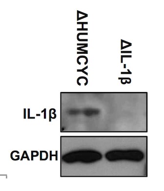
Application: Western BlotSample Tested: KO cellsSpecies: HumanVerified Customer | Posted 02/19/2020Western Blot showing THP-1 IL-1B KO cells using CRISPR technology. humcyc pseudogene used as control for the KO.

-
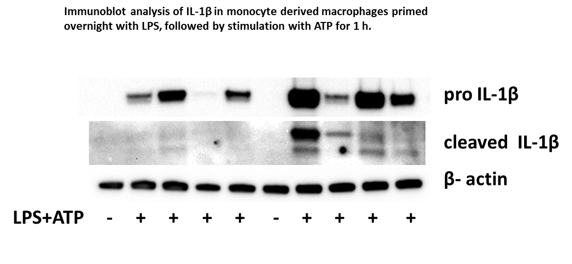
Application: Western BlotSample Tested: THP-1 human acute monocytic leukemia cell line and monocyte derived macrophagesSpecies: HumanVerified Customer | Posted 04/13/2017These Abs recognize both pro-IL1b (p31) and active, cleaved form (p17) in cell lysates and conditioned media of activated human monocyte derived macrophages and THP-1 derived macrophages. To see p17 IL-1b I used 10% tricine gel due to small size of detected protein. I use antibody at dilution 1:500
-
Application: Acoustic AssaySample Tested: Human Plasma and PlasmaSpecies: HumanVerified Customer | Posted 11/24/2015Acoustic Assay on the BioScale ViBE Platform
-
Application: Western BlotSample Tested: See PMID 24006511Species: HumanVerified Customer | Posted 01/06/2015
-
Application: Western BlotSample Tested: See PMID 23221073Species: HumanVerified Customer | Posted 01/06/2015
There are no reviews that match your criteria.
Protocols
Find general support by application which include: protocols, troubleshooting, illustrated assays, videos and webinars.
- Antigen Retrieval Protocol (PIER)
- Antigen Retrieval for Frozen Sections Protocol
- Appropriate Fixation of IHC/ICC Samples
- Cellular Response to Hypoxia Protocols
- Chromogenic IHC Staining of Formalin-Fixed Paraffin-Embedded (FFPE) Tissue Protocol
- Chromogenic Immunohistochemistry Staining of Frozen Tissue
- ClariTSA™ Fluorophore Kits
- Detection & Visualization of Antibody Binding
- Fluorescent IHC Staining of Frozen Tissue Protocol
- Graphic Protocol for Heat-induced Epitope Retrieval
- Graphic Protocol for the Preparation and Fluorescent IHC Staining of Frozen Tissue Sections
- Graphic Protocol for the Preparation and Fluorescent IHC Staining of Paraffin-embedded Tissue Sections
- Graphic Protocol for the Preparation of Gelatin-coated Slides for Histological Tissue Sections
- ICC Cell Smear Protocol for Suspension Cells
- ICC Immunocytochemistry Protocol Videos
- ICC for Adherent Cells
- IHC Sample Preparation (Frozen sections vs Paraffin)
- ISH-IHC Protocol for Chromogenic Detection on Formalin Fixed Paraffin Embedded (FFPE) Tissue
- Immunocytochemistry (ICC) Protocol
- Immunocytochemistry Troubleshooting
- Immunofluorescence of Organoids Embedded in Cultrex Basement Membrane Extract
- Immunofluorescent IHC Staining of Formalin-Fixed Paraffin-Embedded (FFPE) Tissue Protocol
- Immunohistochemistry (IHC) and Immunocytochemistry (ICC) Protocols
- Immunohistochemistry Frozen Troubleshooting
- Immunohistochemistry Paraffin Troubleshooting
- Preparing Samples for IHC/ICC Experiments
- Preventing Non-Specific Staining (Non-Specific Binding)
- Primary Antibody Selection & Optimization
- Protocol for Heat-Induced Epitope Retrieval (HIER)
- Protocol for Making a 4% Formaldehyde Solution in PBS
- Protocol for VisUCyte™ HRP Polymer Detection Reagent
- Protocol for the Fluorescent ICC Staining of Cell Smears - Graphic
- Protocol for the Fluorescent ICC Staining of Cultured Cells on Coverslips - Graphic
- Protocol for the Preparation & Fixation of Cells on Coverslips
- Protocol for the Preparation and Chromogenic IHC Staining of Frozen Tissue Sections
- Protocol for the Preparation and Chromogenic IHC Staining of Frozen Tissue Sections - Graphic
- Protocol for the Preparation and Chromogenic IHC Staining of Paraffin-embedded Tissue Sections
- Protocol for the Preparation and Chromogenic IHC Staining of Paraffin-embedded Tissue Sections - Graphic
- Protocol for the Preparation and Fluorescent ICC Staining of Cells on Coverslips
- Protocol for the Preparation and Fluorescent ICC Staining of Non-adherent Cells
- Protocol for the Preparation and Fluorescent ICC Staining of Stem Cells on Coverslips
- Protocol for the Preparation and Fluorescent IHC Staining of Frozen Tissue Sections
- Protocol for the Preparation and Fluorescent IHC Staining of Paraffin-embedded Tissue Sections
- Protocol for the Preparation of Gelatin-coated Slides for Histological Tissue Sections
- Protocol for the Preparation of a Cell Smear for Non-adherent Cell ICC - Graphic
- R&D Systems Quality Control Western Blot Protocol
- TUNEL and Active Caspase-3 Detection by IHC/ICC Protocol
- The Importance of IHC/ICC Controls
- Troubleshooting Guide: Immunohistochemistry
- Troubleshooting Guide: Western Blot Figures
- Western Blot Conditions
- Western Blot Protocol
- Western Blot Protocol for Cell Lysates
- Western Blot Troubleshooting
- Western Blot Troubleshooting Guide
- View all Protocols, Troubleshooting, Illustrated assays and Webinars
Loading...
Associated Pathways


Innate Lymphoid Cell Differentiation Pathways
 NOD-like Receptor Signaling Pathways
 Th17 Differentiation Pathway
 Toll-Like Receptor Signaling Pathways
