
Key Product Details
Species Reactivity
Validated:
Human, Primate
Cited:
Human, Mouse
Applications
Validated:
Immunohistochemistry, ELISA Capture (Matched Antibody Pair), Neutralization, Dual RNAscope ISH-IHC Compatible, Immunoprecipitation
Cited:
Immunohistochemistry, Immunohistochemistry-Paraffin, Neutralization, Flow Cytometry, ELISA Capture, ELISA Development, ELISA Development (Capture), Luminex Development
Label
Unconjugated
Antibody Source
Monoclonal Mouse IgG2B Clone # 41809
Loading...
Product Specifications
Immunogen
E. coli-derived recombinant human IL-17
Ile20-Ala155
Accession # Q16552
Ile20-Ala155
Accession # Q16552
Specificity
Detects human and primate IL-17 in direct ELISAs. In direct ELISAs, approximately 12% cross-reactivity with recombinant canine IL-17 is observed and 25%-50% reactivity with recombinant human (rh) IL-17A/IL-17F heterodimer is observed. No cross-reactivity with recombinant mouse IL-17, rhIL-17B, rhIL-17C, rhIL-17D, rhIL-17E, or rhIL-17F is observed.
Clonality
Monoclonal
Host
Mouse
Isotype
IgG2B
Endotoxin Level
<0.10 EU per 1 μg of the antibody by the LAL method.
Scientific Data Images for IL-17/IL-17A Antibody (41809)
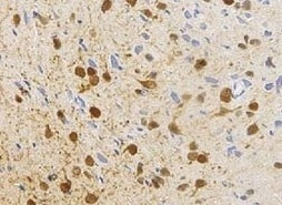
IL‑17/IL‑17A in Human Crohn's Disease Intestine.
IL-17/IL-17A was detected in immersion fixed paraffin-embedded sections of human Crohn's disease intestine using Mouse Anti-Human/Primate IL-17/IL-17A Monoclonal Antibody (Catalog # MAB317) at 5 µg/mL for 1 hour at room temperature followed by incubation with the Anti-Mouse IgG VisUCyte™ HRP Polymer Antibody (Catalog # VC001). Tissue was stained using DAB (brown) and counterstained with hematoxylin (blue). Specific staining was localized to lymphocytes. View our protocol for IHC Staining with VisUCyte HRP Polymer Detection Reagents.
Immunoprecipitation of Human IL‑17.
Human IL-17 was immunoprecipitated from 100 µg of human primary differentiated Th17 cell lysate following incubation with 3 µg Mouse Anti-Human/Primate IL-17 Monoclonal Antibody (Catalog # MAB317) or isotype control antibody (Catalog # MAB004) overnight at 4 °C. IL-17-antibody complexes were absorbed using anti-mouse agarose beads. Immunoprecipitated IL-17 was detected by Western blot using 1 µg/mL Goat Anti-Human IL-17 Antigen Affinity-purified Polyclonal Antibody (Catalog # AF-317-NA). View our recommended buffer recipes for immunoprecipitation.
IL‑6 Secretion Induced by IL‑17 and Neutralization by Human IL‑17 Antibody.
Recombinant Human IL-17 (Catalog # 317-ILB) stimulates IL-6 secretion in the NIH-3T3 mouse embryonic fibroblast cell line in a dose-dependent manner (orange line), as measured by the Mouse IL-6 Quantikine ELISA Kit (Catalog # M6000B). IL-6 secretion elicited by Recombinant Human IL-17 (15 ng/mL) is neutralized (green line) by increasing concentrations of Mouse Anti-Human/Primate IL-17 Monoclonal Antibody (Catalog # MAB317). The ND50 is typically 1-3 µg/mL.
Detection of IL‑17/IL‑17A in Human Crohn's Intestine.
Formalin-fixed paraffin-embedded tissue sections of human Crohn’s Disease Intestine were probed for IL-17A mRNA (ACD RNAScope Probe, catalog #310931; Fast Red chromogen, ACD catalog # 322750). Adjacent tissue section was processed for immunohistochemistry using mouse anti-human IL-17A monoclonal antibody (R&D Systems catalog # MAB317) at 5ug/mL with 1 hour incubation at room temperature followed by incubation with anti-mouse IgG VisUCyte HRP Polymer Antibody (Catalog # VC001) and DAB chromogen (yellow-brown). Tissue was counterstained with hematoxylin (blue). Specific staining was localized to lymphocytes.
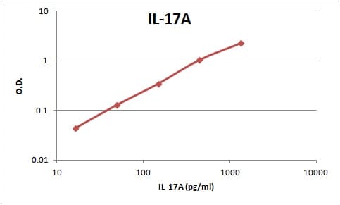
Human IL-17 / IL-17A ELISA Standard Curve
Recombinant Human IL‑17/IL‑17A (Catalog # 317-ILB) was serially diluted and captured by Mouse Anti-Human/Primate IL‑17/IL‑17A Monoclonal Antibody (Catalog # MAB317) coated on a Clear Polystyrene Microplate (Catalog # DY990). Goat Anti-Human IL‑17/IL‑17A Antigen Affinity-purified Polyclonal Antibody (Catalog # AF-317-NA) was biotinylated and incubated with the protein captured on the plate. Detection of the standard curve was achieved by incubating Streptavidin-HRP (Catalog # DY998)Applications for IL-17/IL-17A Antibody (41809)
Application
Recommended Usage
Dual RNAscope ISH-IHC Compatible
3-25 µg/mL
Sample: Immersion fixed paraffin-embedded sections of human crohn's intestine
Sample: Immersion fixed paraffin-embedded sections of human crohn's intestine
Immunohistochemistry
5-25 µg/mL
Sample: Immersion fixed paraffin-embedded sections of human Crohn's disease intestine
Sample: Immersion fixed paraffin-embedded sections of human Crohn's disease intestine
Immunoprecipitation
3 µg/100 µg cell lysate
Sample: Human primary differentiated Th17 cells
Sample: Human primary differentiated Th17 cells
Neutralization
Measured by its ability to neutralize IL‑17-induced IL‑6 secretion in the NIH-3T3 mouse embryonic fibroblast cell line. Yao, Z. et al. (1995) Immunity 3:811. The Neutralization Dose (ND50) is typically 1-3 µg/mL in the presence of 15 ng/mL Recombinant Human IL‑17.
Human/Primate IL-17 Sandwich Immunoassay
Please Note: Optimal dilutions of this antibody should be experimentally determined.
Reviewed Applications
Read 3 reviews rated 5 using MAB317 in the following applications:
Formulation, Preparation, and Storage
Purification
Protein A or G purified from ascites
Reconstitution
Reconstitute at 0.5 mg/mL in sterile PBS. For liquid material, refer to CoA for concentration.
Loading...
Formulation
Lyophilized from a 0.2 μm filtered solution in PBS with Trehalose. See Certificate of Analysis for details.
*Small pack size (-SP) is supplied either lyophilized or as a 0.2 µm filtered solution in PBS.
*Small pack size (-SP) is supplied either lyophilized or as a 0.2 µm filtered solution in PBS.
Shipping
Lyophilized product is shipped at ambient temperature. Liquid small pack size (-SP) is shipped with polar packs. Upon receipt, store immediately at the temperature recommended below.
Stability & Storage
Use a manual defrost freezer and avoid repeated freeze-thaw cycles.
- 12 months from date of receipt, -20 to -70 °C as supplied.
- 1 month, 2 to 8 °C under sterile conditions after reconstitution.
- 6 months, -20 to -70 °C under sterile conditions after reconstitution.
Calculators
Background: IL-17/IL-17A
Long Name
Interleukin 17
Alternate Names
CTLA-8, CTLA8, IL-17A, IL17, IL17A
Entrez Gene IDs
Gene Symbol
IL17A
UniProt
Additional IL-17/IL-17A Products
Product Documents for IL-17/IL-17A Antibody (41809)
Certificate of Analysis
To download a Certificate of Analysis, please enter a lot or batch number in the search box below.
Note: Certificate of Analysis not available for kit components.
Product Specific Notices for IL-17/IL-17A Antibody (41809)
For research use only
Citations for IL-17/IL-17A Antibody (41809)
 Powered by Bioz
Customer Reviews for IL-17/IL-17A Antibody (41809) (3)
5 out of 5
3 Customer Ratings
Have you used IL-17/IL-17A Antibody (41809)?
Submit a review and receive an Amazon gift card!
$25/€18/£15/$25CAN/¥2500 Yen for a review with an image
$10/€7/£6/$10CAN/¥1110 Yen for a review without an image
Submit a review
Customer Images
Showing
1
-
3 of
3 reviews
Showing All
Filter By:
-
Application: ImmunohistochemistrySample Tested: Ischemic brain tissueSpecies: HumanVerified Customer | Posted 08/19/2021

-
Application: ELISASample Tested: Cell culture supernatantSpecies: HumanVerified Customer | Posted 08/22/2018Use the antibody to coat the wells in an immunoassay.
-
Application: ELISASample Tested: Serum and PlasmaSpecies: HumanVerified Customer | Posted 01/18/2018
There are no reviews that match your criteria.
Protocols
Find general support by application which include: protocols, troubleshooting, illustrated assays, videos and webinars.
- Antigen Retrieval Protocol (PIER)
- Antigen Retrieval for Frozen Sections Protocol
- Appropriate Fixation of IHC/ICC Samples
- Cellular Response to Hypoxia Protocols
- Chromogenic IHC Staining of Formalin-Fixed Paraffin-Embedded (FFPE) Tissue Protocol
- Chromogenic Immunohistochemistry Staining of Frozen Tissue
- ClariTSA™ Fluorophore Kits
- Detection & Visualization of Antibody Binding
- Fluorescent IHC Staining of Frozen Tissue Protocol
- Graphic Protocol for Heat-induced Epitope Retrieval
- Graphic Protocol for the Preparation and Fluorescent IHC Staining of Frozen Tissue Sections
- Graphic Protocol for the Preparation and Fluorescent IHC Staining of Paraffin-embedded Tissue Sections
- Graphic Protocol for the Preparation of Gelatin-coated Slides for Histological Tissue Sections
- IHC Sample Preparation (Frozen sections vs Paraffin)
- ISH-IHC Protocol for Chromogenic Detection on Formalin Fixed Paraffin Embedded (FFPE) Tissue
- Immunofluorescent IHC Staining of Formalin-Fixed Paraffin-Embedded (FFPE) Tissue Protocol
- Immunohistochemistry (IHC) and Immunocytochemistry (ICC) Protocols
- Immunohistochemistry Frozen Troubleshooting
- Immunohistochemistry Paraffin Troubleshooting
- Immunoprecipitation Protocol
- Preparing Samples for IHC/ICC Experiments
- Preventing Non-Specific Staining (Non-Specific Binding)
- Primary Antibody Selection & Optimization
- Protocol for Heat-Induced Epitope Retrieval (HIER)
- Protocol for Making a 4% Formaldehyde Solution in PBS
- Protocol for VisUCyte™ HRP Polymer Detection Reagent
- Protocol for the Preparation & Fixation of Cells on Coverslips
- Protocol for the Preparation and Chromogenic IHC Staining of Frozen Tissue Sections
- Protocol for the Preparation and Chromogenic IHC Staining of Frozen Tissue Sections - Graphic
- Protocol for the Preparation and Chromogenic IHC Staining of Paraffin-embedded Tissue Sections
- Protocol for the Preparation and Chromogenic IHC Staining of Paraffin-embedded Tissue Sections - Graphic
- Protocol for the Preparation and Fluorescent IHC Staining of Frozen Tissue Sections
- Protocol for the Preparation and Fluorescent IHC Staining of Paraffin-embedded Tissue Sections
- Protocol for the Preparation of Gelatin-coated Slides for Histological Tissue Sections
- TUNEL and Active Caspase-3 Detection by IHC/ICC Protocol
- The Importance of IHC/ICC Controls
- Troubleshooting Guide: Immunohistochemistry
- View all Protocols, Troubleshooting, Illustrated assays and Webinars
Loading...
Associated Pathways


