Human CCR7 Antibody (150503)
R&D Systems | Catalog # MAB197
Clone 150503 was used by HLDA to establish CD designation

Key Product Details
Validated by
Biological Validation
Species Reactivity
Validated:
Human
Cited:
Human, Mouse, Canine, Complex Species Category, Primate, Primate - Callithrix jacchus (Common Marmoset), Primate - Macaca fascicularis (Crab-eating Monkey or Cynomolgus Macaque), Primate - Macaca mulatta (Rhesus Macaque), Rabbit
Applications
Validated:
Immunohistochemistry, Neutralization, Flow Cytometry, Dual RNAscope ISH-IHC Compatible, Immunocytochemistry, CyTOF-reported
Cited:
Immunohistochemistry, Immunohistochemistry-Paraffin, Western Blot, Neutralization, Flow Cytometry, Immunocytochemistry, Assay Development, Blocking, CyTof, Flow Cytomery, Functional Assay, Mass Cytometry
Label
Unconjugated
Antibody Source
Monoclonal Mouse IgG2A Clone # 150503
Loading...
Product Specifications
Immunogen
Human CCR7 transfectants
Met1-Pro378
Accession # AAA58615
Met1-Pro378
Accession # AAA58615
Specificity
Detects human CCR7.
Clonality
Monoclonal
Host
Mouse
Isotype
IgG2A
Endotoxin Level
<0.10 EU per 1 μg of the antibody by the LAL method.
Scientific Data Images for Human CCR7 Antibody (150503)
Detection of CCR7 in Human PBMCs by Flow Cytometry.
Human peripheral blood mononuclear cells (PBMCs) were stained with Mouse Anti-Human CD4 PE-conjugated Monoclonal Antibody (Catalog # FAB3791P) and either (A) Mouse Anti-Human CCR7 Monoclonal Antibody (Catalog # MAB197) or (B) Mouse IgG2AIsotype Control (Catalog # MAB003) followed by Allophycocyanin-conjugated Anti-Mouse IgG Secondary Antibody (Catalog # F0101B). View our protocol for Staining Membrane-associated Proteins.
CCR7 in Human PBMCs.
CCR7 was detected in immersion fixed human peripheral blood mononuclear cells (PBMCs) using 25 µg/mL Mouse Anti-Human CCR7 Monoclonal Antibody (Catalog # MAB197) for 3 hours at room temperature. Cells were stained (red) and counterstained (green). View our protocol for Fluorescent ICC Staining of Non-adherent Cells.
CCR7 in Human Lung.
CCR7 was detected in immersion fixed paraffin-embedded sections of human lung using Mouse Anti-Human CCR7 Monoclonal Antibody (Catalog # MAB197) at 5 µg/mL for 1 hour at room temperature followed by incubation with the Anti-Mouse IgG VisUCyte™ HRP Polymer Antibody (Catalog # VC001). Tissue was stained using DAB (brown) and counterstained with hematoxylin (blue). Specific staining was localized to cytoplasm in macrophages. View our protocol for IHC Staining with VisUCyte HRP Polymer Detection Reagents.
Chemotaxis Induced by CCL19/MIP‑3 beta and Neutral-ization by Human CCR7 Antibody.
Recombinant Human CCL19/MIP-3 beta (Catalog # 361-MI) chemoattracts the BaF3 mouse pro-B cell line transfected with human CCR7 in a dose-dependent manner (orange line). The amount of cells that migrated through to the lower chemotaxis chamber was measured by Resazurin (Catalog # AR002). Chemotaxis elicited by Recombinant Human CCL19/MIP-3 beta (50 ng/mL) is neutralized (green line) by increasing concentrations of Mouse Anti-Human CCR7 Monoclonal Antibody (Catalog # MAB197). The ND50 is typically 1-5 µg/mL.
Detection of Human CCR7 by Flow Cytometry
HCMV strain-dependent variability of UL4015-23 sequences and HCMV strain-specific HLA-EUL40 T-cell response in hosts.(A, B) Genomic DNAs isolated from HCMV positive blood samples in HCMV+ transplant recipients (n = 25) were sequenced for the identification of UL40 protein (amino acids 1–221) provided by the circulating HCMV strains. (A) Amino acid variability, expressed as a number of amino acid variants, within the HLA-E-binding peptide (UL4015-23, shown in red) among the sequence for HCMV UL40 signal peptide (UL401-37, shown in grey). A total of 32 UL40 sequences from 25 hosts were analysed. UL40 protein sequence from the Merlin HCMV clinical strain was used as reference. Positions 1 to 9 of residues in the HLA-E-binding peptide (UL4015-23) are indicated. (B) Sequence LOGO of the UL4015-23 HLA-E-binding peptide from 25 transplanted hosts. The height of the letter is proportional to the frequency of each amino acid in a given position (P1 to P9). Major anchor residues for binding in the HLA-E peptide groove are indicated in blue. Red letters highlight the important variability observed in position 8 of the HLA-E-binding peptide. Grey boxes correspond to a constitutive deletion of the corresponding amino acid in the UL40 sequence from the infecting viral strain. (C) Representative dot plot analyses showing the detection of strain-specific anti-UL40 HLA-E-restricted CD8 T-cell responses in 4 KTRs (KTR#026, #105, #108 and #109). Frequencies (%) of the HLA-EUL40-specific T cells among total circulating alpha beta CD8 T cells are indicated. Image collected and cropped by CiteAb from the following publication (https://dx.plos.org/10.1371/journal.ppat.1007041), licensed under a CC-BY license. Not internally tested by R&D Systems.
Detection of Human CCR7 by Proximity Ligation Assay
CXCR4 ligand binding facilitates CXCR4/CCR7 hetero-oligomer formation.(A) CCR7 homo-oligomer formation in H9 cells after treatment with or without 1 μg/ml recombinant gp120 was examined by in situ PLA. The number of in situ PLA signals per cell was counted by using the Duolink Image Tool software. The result shown is a representative result from three independent experiments showing the mean number of the signals plotted on the vertical axis. *, p < 0.05 by Mann-Whitney’s U test. (B) CCR7 homo-oligomer formation in H9 cells after treatment with 100 ng/ml CXCL12 was examined, as described in (A). Quantification of CCR7 homo-oligomers after CXCL12 pretreatment in the presence or absence of 1 or 5 μg/ml AMD3100. The result shown is a representative result from five independent experiments. *, p < 0.05 by Mann-Whitney’s U test. (C) CXCR4/CCR7 or CXCR4/CCR1 hetero-oligomer formation in H9 cells by in situ PLA using the indicated combinations of antibodies. The z-stack images derived from sections covering 10 μm with a 0.5-μm step are shown. Scale bar represents 50 μm. (D) Quantification of the detected in situ PLA signals per cell was performed. The result shown is a representative result from three independent experiments. *, p < 0.05 by Mann-Whitney’s U test. (E) CXCR4/CCR7 hetero-oligomer formation in H9 cells after treatment with 1 μg/ml gp120 in a native form (N) or control heat-denatured form (D) was examined by the PLA using the indicated combinations of antibodies. (F) CXCR4/CCR7 hetero-oligomer formation was examined with 100 ng/ml CXCL12 as described in (E). Image collected and cropped by CiteAb from the following publication (https://dx.plos.org/10.1371/journal.pone.0117454), licensed under a CC-BY license. Not internally tested by R&D Systems.
Detection of Rhesus Macaque CCR7 by Flow Cytometry
Dynamics of CCR7 expression on CD1c+ mDC and pDC during primary SIVmac239 infection.(A) The dynamics of CCR7 expression on CD1c+ mDC; (B) The dynamics of CCR7 expression on pDC. Image collected and cropped by CiteAb from the following publication (https://dx.plos.org/10.1371/journal.pone.0029036), licensed under a CC-BY license. Not internally tested by R&D Systems.
Detection of Human CCR7 by Immunohistochemistry
CCR7 homodimers are dissociated by the CCR7 TM4-derived peptide. (A) The levels of bioluminescence signals are presented for HEK293T cells transfected with the indicated combinations of CCR7-CGLuc, CCR7-NGLuc, CGLuc and NGLuc plasmids (1.5 μg each). A representative experiment from at least three independent experiments is shown. Data represent mean ± SD of triplicate samples. *p < 0.05 by one-way ANOVA. (B) The levels of bioluminescence signals are shown for cells transfected with combinations of CCR7-CGLuc and CCR7-NGLuc in the presence of the CCR7 TM4 peptide or the shuffled peptide (15 μg/ml). Relative luminescence was obtained by normalizing the values against untransfected cells. A representative experiment from at least three independent experiments is shown. Data represent mean ± SD of triplicate samples. *p < 0.05 by Student’s t test. (C) The levels of CCR7 homodimer in parental H9 cells were measured by in situ PLA. CCR7-CCR1 heterodimer formation detected by anti-CCR7 mAb-PLAPlus and anti-CCR1 mAb -PLAMinus (left), and CCR7 homodimer formation by anti-CCR7 mAb-PLAPlus and -PLAMinus (right). A representative experiment from at least three independent experiments is shown with the mean number of PLA signals plotted on the vertical axis. *p < 0.05 by Mann-Whitney’s U test. (D) CCR7 (top) or CCR1 (bottom) homodimer formation after treatment with the CCR7 TM4 peptide (right panel) or the shuffled peptide (left panel). The number of PLA signals per cell was counted using the Duolink Image Tool software. A representative experiment from three independent experiments is shown with the mean number of PLA signals plotted on the vertical axis. *p < 0.05 by Mann-Whitney’s U test; NS, not significant. (E) CCR7 expression levels were evaluated in the presence (red open histogram) or absence (blue open histogram) of the CCR7 TM4 peptide (15 μg/ml) by flow cytometry using anti-human CCR7 antibody or isotype control antibody (gray-filled histogram). MFI is indicated on th
Detection of Rhesus Macaque CCR7 by Flow Cytometry
Two-color strategy for identifying CD1c+ mDCs and pDCs.(A) PBMCs (R1) were first gated on Forward-scatter/side-scatter (FSC/SSC) scattergram. Then, CD1c+ mDCs (CD1c+CD14−CD20−) were selected in the R2 region and pDCs (CD123brightHLA-DR+) were selected in the R3 region. (B) The expressions of immunophenotypes, including CD4, CCR5, CD80, CD83, CD86, and CCR7 on DCs subsets were analyzed. Open histograms correspond to isotype controls and filled gray histograms correspond to specific mAbs staining. Image collected and cropped by CiteAb from the following publication (https://dx.plos.org/10.1371/journal.pone.0029036), licensed under a CC-BY license. Not internally tested by R&D Systems.
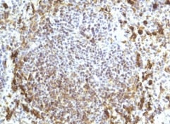
Detection of CCR7 in Human Tonsil.
Formalin-fixed paraffin-embedded tissue sections of human tonsil were probed for CCR7 mRNA (ACD RNAScope Probe, catalog # 410728; Fast Red chromogen, ACD catalog # 322750). Adjacent tissue section was processed for immunohistochemistry using mouse anti-human CCR7 monoclonal antibody (R&D Systems catalog # MAB197) at 15ug/mL with overnight incubation at 4 degrees Celsius followed by incubation with anti-mouse IgG VisUCyte HRP Polymer Antibody (Catalog # VC001) and DAB chromogen (yellow-brown). Tissue was counterstained with hematoxylin (blue). Specific staining was localized to lymphocytes.Applications for Human CCR7 Antibody (150503)
Application
Recommended Usage
CyTOF-reported
Mei, H.E. et al. (2015) J. Immunol. 194: 2022. Ready to be labeled using established conjugation methods. No BSA or other carrier proteins that could interfere with conjugation.
Dual RNAscope ISH-IHC Compatible
3-25 µg/mL
Sample: Immersion fixed paraffin-embedded sections of human tonsil
Sample: Immersion fixed paraffin-embedded sections of human tonsil
Flow Cytometry
0.25 µg/106 cells
Sample: Human peripheral blood mononuclear cells (PBMCs)
Sample: Human peripheral blood mononuclear cells (PBMCs)
Immunocytochemistry
8-25 µg/mL
Sample: Immersion fixed human peripheral blood mononuclear cells (PBMCs)
Sample: Immersion fixed human peripheral blood mononuclear cells (PBMCs)
Immunohistochemistry
5-25 µg/mL
Sample: Immersion fixed paraffin-embedded sections of human lung
Sample: Immersion fixed paraffin-embedded sections of human lung
Neutralization
Measured by its ability to neutralize CCL19/MIP‑3 beta -induced chemotaxis in the BaF3 mouse pro‑B cell line transfected with human CCR7. The Neutralization Dose (ND50) is typically 1-5 µg/mL in the presence of 50 ng/mL Recombinant Human CCL19/MIP‑3 beta.
Reviewed Applications
Read 3 reviews rated 5 using MAB197 in the following applications:
Flow Cytometry Panel Builder
Bio-Techne Knows Flow Cytometry
Save time and reduce costly mistakes by quickly finding compatible reagents using the Panel Builder Tool.
Advanced Features
- Spectra Viewer - Custom analysis of spectra from multiple fluorochromes
- Spillover Popups - Visualize the spectra of individual fluorochromes
- Antigen Density Selector - Match fluorochrome brightness with antigen density

Formulation, Preparation, and Storage
Purification
Protein A or G purified from hybridoma culture supernatant
Reconstitution
Reconstitute at 0.5 mg/mL in sterile PBS. For liquid material, refer to CoA for concentration.
Loading...
Formulation
Lyophilized from a 0.2 μm filtered solution in PBS with Trehalose. See Certificate of Analysis for details.
*Small pack size (-SP) is supplied either lyophilized or as a 0.2 µm filtered solution in PBS.
*Small pack size (-SP) is supplied either lyophilized or as a 0.2 µm filtered solution in PBS.
Shipping
Lyophilized product is shipped at ambient temperature. Liquid small pack size (-SP) is shipped with polar packs. Upon receipt, store immediately at the temperature recommended below.
Stability & Storage
Use a manual defrost freezer and avoid repeated freeze-thaw cycles.
- 12 months from date of receipt, -20 to -70 °C as supplied.
- 1 month, 2 to 8 °C under sterile conditions after reconstitution.
- 6 months, -20 to -70 °C under sterile conditions after reconstitution.
Calculators
Background: CCR7
Alternate Names
BLR2, CC-CKR-7, CCR7, CD197, CDw197, CMKBR7, EBI1
Gene Symbol
CCR7
UniProt
Additional CCR7 Products
Product Documents for Human CCR7 Antibody (150503)
Certificate of Analysis
To download a Certificate of Analysis, please enter a lot or batch number in the search box below.
Note: Certificate of Analysis not available for kit components.
Product Specific Notices for Human CCR7 Antibody (150503)
For research use only
Citations for Human CCR7 Antibody (150503)
 Powered by Bioz
Customer Reviews for Human CCR7 Antibody (150503) (3)
5 out of 5
3 Customer Ratings
Have you used Human CCR7 Antibody (150503)?
Submit a review and receive an Amazon gift card!
$25/€18/£15/$25CAN/¥2500 Yen for a review with an image
$10/€7/£6/$10CAN/¥1110 Yen for a review without an image
Submit a review
Customer Images
Showing
1
-
3 of
3 reviews
Showing All
Filter By:
-
Application: ImmunohistochemistrySample Tested: Spleen tissueSpecies: HumanVerified Customer | Posted 08/03/2021

-
Application: Flow CytometrySample Tested: Human T cells from Cord and Maternal BloodSpecies: HumanVerified Customer | Posted 02/22/2016
-
Application: ImmunofluorescenceSample Tested: See PMID 23731727Species: HumanVerified Customer | Posted 02/12/2015
There are no reviews that match your criteria.
Protocols
Find general support by application which include: protocols, troubleshooting, illustrated assays, videos and webinars.
- 7-Amino Actinomycin D (7-AAD) Cell Viability Flow Cytometry Protocol
- Antigen Retrieval Protocol (PIER)
- Antigen Retrieval for Frozen Sections Protocol
- Appropriate Fixation of IHC/ICC Samples
- Cellular Response to Hypoxia Protocols
- Chromogenic IHC Staining of Formalin-Fixed Paraffin-Embedded (FFPE) Tissue Protocol
- Chromogenic Immunohistochemistry Staining of Frozen Tissue
- ClariTSA™ Fluorophore Kits
- Detection & Visualization of Antibody Binding
- Extracellular Membrane Flow Cytometry Protocol
- Flow Cytometry Protocol for Cell Surface Markers
- Flow Cytometry Protocol for Staining Membrane Associated Proteins
- Flow Cytometry Staining Protocols
- Flow Cytometry Troubleshooting Guide
- Fluorescent IHC Staining of Frozen Tissue Protocol
- Graphic Protocol for Heat-induced Epitope Retrieval
- Graphic Protocol for the Preparation and Fluorescent IHC Staining of Frozen Tissue Sections
- Graphic Protocol for the Preparation and Fluorescent IHC Staining of Paraffin-embedded Tissue Sections
- Graphic Protocol for the Preparation of Gelatin-coated Slides for Histological Tissue Sections
- ICC Cell Smear Protocol for Suspension Cells
- ICC Immunocytochemistry Protocol Videos
- ICC for Adherent Cells
- IHC Sample Preparation (Frozen sections vs Paraffin)
- ISH-IHC Protocol for Chromogenic Detection on Formalin Fixed Paraffin Embedded (FFPE) Tissue
- Immunocytochemistry (ICC) Protocol
- Immunocytochemistry Troubleshooting
- Immunofluorescence of Organoids Embedded in Cultrex Basement Membrane Extract
- Immunofluorescent IHC Staining of Formalin-Fixed Paraffin-Embedded (FFPE) Tissue Protocol
- Immunohistochemistry (IHC) and Immunocytochemistry (ICC) Protocols
- Immunohistochemistry Frozen Troubleshooting
- Immunohistochemistry Paraffin Troubleshooting
- Intracellular Flow Cytometry Protocol Using Alcohol (Methanol)
- Intracellular Flow Cytometry Protocol Using Detergents
- Intracellular Nuclear Staining Flow Cytometry Protocol Using Detergents
- Intracellular Staining Flow Cytometry Protocol Using Alcohol Permeabilization
- Intracellular Staining Flow Cytometry Protocol Using Detergents to Permeabilize Cells
- Preparing Samples for IHC/ICC Experiments
- Preventing Non-Specific Staining (Non-Specific Binding)
- Primary Antibody Selection & Optimization
- Propidium Iodide Cell Viability Flow Cytometry Protocol
- Protocol for Heat-Induced Epitope Retrieval (HIER)
- Protocol for Liperfluo
- Protocol for Making a 4% Formaldehyde Solution in PBS
- Protocol for VisUCyte™ HRP Polymer Detection Reagent
- Protocol for the Characterization of Human Th22 Cells
- Protocol for the Characterization of Human Th9 Cells
- Protocol for the Fluorescent ICC Staining of Cell Smears - Graphic
- Protocol for the Fluorescent ICC Staining of Cultured Cells on Coverslips - Graphic
- Protocol for the Preparation & Fixation of Cells on Coverslips
- Protocol for the Preparation and Chromogenic IHC Staining of Frozen Tissue Sections
- Protocol for the Preparation and Chromogenic IHC Staining of Frozen Tissue Sections - Graphic
- Protocol for the Preparation and Chromogenic IHC Staining of Paraffin-embedded Tissue Sections
- Protocol for the Preparation and Chromogenic IHC Staining of Paraffin-embedded Tissue Sections - Graphic
- Protocol for the Preparation and Fluorescent ICC Staining of Cells on Coverslips
- Protocol for the Preparation and Fluorescent ICC Staining of Non-adherent Cells
- Protocol for the Preparation and Fluorescent ICC Staining of Stem Cells on Coverslips
- Protocol for the Preparation and Fluorescent IHC Staining of Frozen Tissue Sections
- Protocol for the Preparation and Fluorescent IHC Staining of Paraffin-embedded Tissue Sections
- Protocol for the Preparation of Gelatin-coated Slides for Histological Tissue Sections
- Protocol for the Preparation of a Cell Smear for Non-adherent Cell ICC - Graphic
- Protocol: Annexin V and PI Staining by Flow Cytometry
- Protocol: Annexin V and PI Staining for Apoptosis by Flow Cytometry
- TUNEL and Active Caspase-3 Detection by IHC/ICC Protocol
- The Importance of IHC/ICC Controls
- Troubleshooting Guide: Fluorokine Flow Cytometry Kits
- Troubleshooting Guide: Immunohistochemistry
- View all Protocols, Troubleshooting, Illustrated assays and Webinars


