
Key Product Details
Validated by
Biological Validation
Species Reactivity
Validated:
Human
Cited:
Human, Mouse, Rat
Applications
Validated:
Immunohistochemistry, Neutralization, Intracellular Staining by Flow Cytometry, Immunocytochemistry, CyTOF-ready
Cited:
Immunohistochemistry, Immunohistochemistry-Paraffin, Western Blot, Neutralization, Flow Cytometry, Immunocytochemistry, Bioassay, Cell Culture, Immunoassay Development
Label
Unconjugated
Antibody Source
Monoclonal Mouse IgG2B Clone # 1936
Loading...
Product Specifications
Immunogen
E. coli-derived recombinant human IL‑6
Val30-Met212
Accession # P05231
Val30-Met212
Accession # P05231
Specificity
Detects human IL-6 in direct ELISAs. Does not cross-react with recombinant IL-6 from mouse, rat, or pig.
Clonality
Monoclonal
Host
Mouse
Isotype
IgG2B
Endotoxin Level
<0.20 EU per 1 μg of the antibody by the LAL method.
Scientific Data Images for Human IL-6 Antibody (1936)
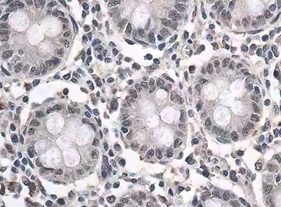
IL‑6 in Human Appendix.
IL-6 was detected in immersion fixed paraffin-embedded sections of human appendix using Mouse Anti-Human IL-6 Monoclonal Antibody (Catalog # MAB2061) at 1.7 µg/mL for 1 hour at room temperature followed by incubation with the Anti-Mouse IgG VisUCyte™ HRP Polymer Antibody (VC001). Before incubation with the primary antibody, tissue was subjected to heat-induced epitope retrieval using Antigen Retrieval Reagent-Basic (CTS013). Tissue was stained using DAB (brown) and counterstained with hematoxylin (blue). Specific staining was localized to cytoplasm in lymphocytes. View our protocol for IHC Staining with VisUCyte HRP Polymer Detection Reagents.
Detection of IL‑6 in Human PBMCs by Flow Cytometry.
Human peripheral blood mononuclear cells (PBMCs) treated with 100 ng/mL LPS for 24 hours were stained with Mouse Anti-Human IL-6 Monoclonal Antibody (Catalog # MAB2061, filled histogram) or isotype control antibody (MAB004, open histogram), followed by Phycoerythrin-conjugated Anti-Mouse IgG Secondary Antibody (F0102B). To facilitate intracellular staining, cells were fixed with Flow Cytometry Fixation Buffer (FC004) and permeabilized with Flow Cytometry Permeabilization/Wash Buffer I (FC005). View our protocol for Staining Intracellular Molecules.
Cell Proliferation Induced by IL‑6 and Neutralization by Human IL‑6 Antibody.
Recombinant Human IL-6 (206-IL) stimulates proliferation in the T1165.85.2.1 mouse plasmacytoma cell line in a dose-dependent manner (orange line). Proliferation elicited by Recombinant Human IL-6 (2.5 ng/mL) is neutralized (green line) by increasing concentrations of Mouse Anti-Human IL-6 Monoclonal Antibody (Catalog # MAB2061). The ND50 is typically 8.00-80.0 ng/mL.
Detection of IL‑6 in Human PBMCs.
IL‑6 was detected in immersion fixed Human PBMCs stimulated with LPS (positive control) and absent in untreated human PBMCs (negative control) using Mouse Anti-Human IL‑6 Monoclonal Antibody (Catalog # MAB2061) at 3 µg/mL for 3 hours at room temperature. Cells were stained using the NorthernLights™ 557-conjugated Anti-Mouse IgG Secondary Antibody (red; Catalog # NL007) and counterstained with DAPI (blue). Specific staining was localized to cytoplasm. View our protocol for Fluorescent ICC Staining of Non-adherent Cells.
Detection of IL‑6 in PBMC monocytes by Flow Cytometry.
PBMCs stimulated with 100 ng/ml LPS and Brefeldin A for 24 hrs (A) or unstimulated (B) were stained with Mouse Anti-Human IL‑6 Monoclonal Antibody (Catalog # MAB2061) and Mouse Anti-Human CD14 PE‑conjugated Monoclonal Antibody (Catalog # FAB3832P). To facilitate intracellular staining, cells were fixed with Flow Cytometry Fixation Buffer (1X) (Catalog # FC004) and permeabilized with Saponin. View our protocol for Staining Intracellular Molecules.
Detection of IL-6 by Immunocytochemistry/ Immunofluorescence
Effects of topo I-reactive B cells on the differentiation of CD4+ T cells in systemic sclerosis (SSc) patients.B cells with low affinity for topo I and those with high affinity for topo I as well as topo I-non-reactive B cells were obtained from anti-topo I antibody-positive SSc patients (n = 111). These B cells were co-cultured with CD4+ T cells. After 48 hr of co-culture, mRNA was extracted from these cells and FoxP3 and ROR gamma t expression levels were examined by real-time RT-PCR. The results were presented when 96-well plates were used as a co-culture site (A) and when microculture plates (B) were used (C). These cells were further co-cultured on microculture plates, and the protein expression of CD4, CD20, FoxP3, and ROR gamma t was confirmed by fluorescent cell staining and signal intensity was determined by ImageJ (D). Similarly, co-culture in microculture plates in the presence of anti-IL-10 (10 μg/ml), anti-IL-35 (5 μg/ml), anti-IL-6 (1 μg/ml), or anti-IL-23 (5 μg/ml) antibodies (Abs) was conducted, followed by fluorescent cell staining (E). These results represented seven experiments. The bar graphs show the mean + SD. Original magnification, ×1000. *p<0.05.Figure 3—source data 1.Source file for the effects of topo I-reactive B cells on the differentiation of CD4+ T cells in systemic sclerosis (SSc) patients.This archive contains all data used for the quantitative analysis shown in Figure 3.Source file for the effects of topo I-reactive B cells on the differentiation of CD4+ T cells in systemic sclerosis (SSc) patients.This archive contains all data used for the quantitative analysis shown in Figure 3. Image collected and cropped by CiteAb from the following open publication (https://pubmed.ncbi.nlm.nih.gov/34854378), licensed under a CC-BY license. Not internally tested by R&D Systems.Applications for Human IL-6 Antibody (1936)
Application
Recommended Usage
CyTOF-ready
Ready to be labeled using established conjugation methods. No BSA or other carrier proteins that could interfere with conjugation.
Immunocytochemistry
3-15 µg/mL
Sample: Immersion fixed Human PBMCs stimulated with LPS (positive control) and untreated human PBMCs (negative control)
Sample: Immersion fixed Human PBMCs stimulated with LPS (positive control) and untreated human PBMCs (negative control)
Immunohistochemistry
1-25 µg/mL
Sample: Immersion fixed paraffin-embedded sections of human appendix
Sample: Immersion fixed paraffin-embedded sections of human appendix
Intracellular Staining by Flow Cytometry
0.25 µg/106 cells
Sample: PBMCs treated +/- LPS (see details below)
Sample: PBMCs treated +/- LPS (see details below)
Neutralization
Measured by its ability to neutralize IL‑6-induced proliferation in the T1165.85.2.1 mouse plasmacytoma cell line. The Neutralization Dose (ND50) is typically 8.00-80.0 ng/mL in the presence of 2.5 ng/mL Recombinant Human IL‑6.
Reviewed Applications
Read 2 reviews rated 4.5 using MAB2061 in the following applications:
Flow Cytometry Panel Builder
Bio-Techne Knows Flow Cytometry
Save time and reduce costly mistakes by quickly finding compatible reagents using the Panel Builder Tool.
Advanced Features
- Spectra Viewer - Custom analysis of spectra from multiple fluorochromes
- Spillover Popups - Visualize the spectra of individual fluorochromes
- Antigen Density Selector - Match fluorochrome brightness with antigen density

Formulation, Preparation, and Storage
Purification
Protein A or G purified from hybridoma culture supernatant
Reconstitution
Reconstitute at 0.5 mg/mL in sterile PBS. For liquid material, refer to CoA for concentration.
Loading...
Formulation
Lyophilized from a 0.2 μm filtered solution in PBS and NaCl with Trehalose. See Certificate of Analysis for details.
*Small pack size (-SP) is supplied either lyophilized or as a 0.2 µm filtered solution in PBS.
*Small pack size (-SP) is supplied either lyophilized or as a 0.2 µm filtered solution in PBS.
Shipping
Lyophilized product is shipped at ambient temperature. Liquid small pack size (-SP) is shipped with polar packs. Upon receipt, store immediately at the temperature recommended below.
Stability & Storage
Use a manual defrost freezer and avoid repeated freeze-thaw cycles.
- 12 months from date of receipt, -20 to -70 °C as supplied.
- 1 month, 2 to 8 °C under sterile conditions after reconstitution.
- 6 months, -20 to -70 °C under sterile conditions after reconstitution.
Calculators
Background: IL-6
Long Name
Interleukin 6
Alternate Names
BSF-2, BSF2, IFNB2, IL6, MGI-2A
Entrez Gene IDs
Gene Symbol
IL6
UniProt
Additional IL-6 Products
Product Documents for Human IL-6 Antibody (1936)
Certificate of Analysis
To download a Certificate of Analysis, please enter a lot or batch number in the search box below.
Note: Certificate of Analysis not available for kit components.
Product Specific Notices for Human IL-6 Antibody (1936)
For research use only
Related Research Areas
Citations for Human IL-6 Antibody (1936)
 Powered by Bioz
Customer Reviews for Human IL-6 Antibody (1936) (2)
4.5 out of 5
2 Customer Ratings
Have you used Human IL-6 Antibody (1936)?
Submit a review and receive an Amazon gift card!
$25/€18/£15/$25CAN/¥2500 Yen for a review with an image
$10/€7/£6/$10CAN/¥1110 Yen for a review without an image
Submit a review
Customer Images
Showing
1
-
2 of
2 reviews
Showing All
Filter By:
-
Application: ImmunohistochemistrySample Tested: Colon tissueSpecies: HumanVerified Customer | Posted 06/18/2022

-
Application: ELISASample Tested: Mouse ear fibroblastSpecies: HumanVerified Customer | Posted 04/29/2016
There are no reviews that match your criteria.
Protocols
Find general support by application which include: protocols, troubleshooting, illustrated assays, videos and webinars.
- 7-Amino Actinomycin D (7-AAD) Cell Viability Flow Cytometry Protocol
- Antigen Retrieval Protocol (PIER)
- Antigen Retrieval for Frozen Sections Protocol
- Appropriate Fixation of IHC/ICC Samples
- Cellular Response to Hypoxia Protocols
- Chromogenic IHC Staining of Formalin-Fixed Paraffin-Embedded (FFPE) Tissue Protocol
- Chromogenic Immunohistochemistry Staining of Frozen Tissue
- ClariTSA™ Fluorophore Kits
- Detection & Visualization of Antibody Binding
- Extracellular Membrane Flow Cytometry Protocol
- Flow Cytometry Protocol for Cell Surface Markers
- Flow Cytometry Protocol for Staining Membrane Associated Proteins
- Flow Cytometry Staining Protocols
- Flow Cytometry Troubleshooting Guide
- Fluorescent IHC Staining of Frozen Tissue Protocol
- Graphic Protocol for Heat-induced Epitope Retrieval
- Graphic Protocol for the Preparation and Fluorescent IHC Staining of Frozen Tissue Sections
- Graphic Protocol for the Preparation and Fluorescent IHC Staining of Paraffin-embedded Tissue Sections
- Graphic Protocol for the Preparation of Gelatin-coated Slides for Histological Tissue Sections
- ICC Cell Smear Protocol for Suspension Cells
- ICC Immunocytochemistry Protocol Videos
- ICC for Adherent Cells
- IHC Sample Preparation (Frozen sections vs Paraffin)
- Immunocytochemistry (ICC) Protocol
- Immunocytochemistry Troubleshooting
- Immunofluorescence of Organoids Embedded in Cultrex Basement Membrane Extract
- Immunofluorescent IHC Staining of Formalin-Fixed Paraffin-Embedded (FFPE) Tissue Protocol
- Immunohistochemistry (IHC) and Immunocytochemistry (ICC) Protocols
- Immunohistochemistry Frozen Troubleshooting
- Immunohistochemistry Paraffin Troubleshooting
- Intracellular Flow Cytometry Protocol Using Alcohol (Methanol)
- Intracellular Flow Cytometry Protocol Using Detergents
- Intracellular Nuclear Staining Flow Cytometry Protocol Using Detergents
- Intracellular Staining Flow Cytometry Protocol Using Alcohol Permeabilization
- Intracellular Staining Flow Cytometry Protocol Using Detergents to Permeabilize Cells
- Preparing Samples for IHC/ICC Experiments
- Preventing Non-Specific Staining (Non-Specific Binding)
- Primary Antibody Selection & Optimization
- Propidium Iodide Cell Viability Flow Cytometry Protocol
- Protocol for Heat-Induced Epitope Retrieval (HIER)
- Protocol for Liperfluo
- Protocol for Making a 4% Formaldehyde Solution in PBS
- Protocol for VisUCyte™ HRP Polymer Detection Reagent
- Protocol for the Characterization of Human Th22 Cells
- Protocol for the Characterization of Human Th9 Cells
- Protocol for the Fluorescent ICC Staining of Cell Smears - Graphic
- Protocol for the Fluorescent ICC Staining of Cultured Cells on Coverslips - Graphic
- Protocol for the Preparation & Fixation of Cells on Coverslips
- Protocol for the Preparation and Chromogenic IHC Staining of Frozen Tissue Sections
- Protocol for the Preparation and Chromogenic IHC Staining of Frozen Tissue Sections - Graphic
- Protocol for the Preparation and Chromogenic IHC Staining of Paraffin-embedded Tissue Sections
- Protocol for the Preparation and Chromogenic IHC Staining of Paraffin-embedded Tissue Sections - Graphic
- Protocol for the Preparation and Fluorescent ICC Staining of Cells on Coverslips
- Protocol for the Preparation and Fluorescent ICC Staining of Non-adherent Cells
- Protocol for the Preparation and Fluorescent ICC Staining of Stem Cells on Coverslips
- Protocol for the Preparation and Fluorescent IHC Staining of Frozen Tissue Sections
- Protocol for the Preparation and Fluorescent IHC Staining of Paraffin-embedded Tissue Sections
- Protocol for the Preparation of Gelatin-coated Slides for Histological Tissue Sections
- Protocol for the Preparation of a Cell Smear for Non-adherent Cell ICC - Graphic
- Protocol: Annexin V and PI Staining by Flow Cytometry
- Protocol: Annexin V and PI Staining for Apoptosis by Flow Cytometry
- TUNEL and Active Caspase-3 Detection by IHC/ICC Protocol
- The Importance of IHC/ICC Controls
- Troubleshooting Guide: Fluorokine Flow Cytometry Kits
- Troubleshooting Guide: Immunohistochemistry
- View all Protocols, Troubleshooting, Illustrated assays and Webinars
Loading...
Associated Pathways


IL-21 Signaling Pathways and their Primary Biological Effects in Different Immune Cell Types
 Jak/STAT Signaling Pathway
 Mesenchymal Stem Cell Differentiation Pathways & Lineage-specific Markers
 NOD-like Receptor Signaling Pathways
 Th17 Differentiation Pathway
 Toll-Like Receptor Signaling Pathways
