Receptor Tyrosine Kinases (RTKs) are widely expressed transmembrane proteins that act as receptors for growth factors, neurotrophic factors, and other extracellular signaling molecules. Upon ligand binding, they undergo tyrosine phosphorylation at specific residues in the cytoplasmic tail. This leads to the binding of protein substrates and/or the establishment docking sites for adaptor proteins involved in RTK-mediated signal transduction. RTKs have critical functions in several developmental processes including regulating cell survival, proliferation, and motility. When unregulated, they play prominent roles in cancer formation. R&D Systems offers several different multiplex assay formats that allow the user to assess the activity of many RTKs in a single sample. Available RTK multiplex kits include bead-based multiplex kits for the Luminex platform, membrane-based antibody arrays, and microplate-based antibody arrays.
Proteome Profiler Human Phospho-RTK Array Kit
R&D Systems | Catalog # ARY001B

Key Product Details
Species
Product Summary for Proteome Profiler Human Phospho-RTK Array Kit
Kit Summary
A membrane-based antibody array for the parallel determination of the relative phosphorylation of human receptor tyrosine kinases. Validated for analyte detection in cell lysates.
Key Benefits
- Detects phosphorylation of 49 human receptor tyrosine kinases simultaneously
- Requires no specialized equipment
Principle of the Assay
The Proteome Profiler Human Phospho-RTK Array Kit is a membrane-based sandwich immunoassay. Capture antibodies spotted in duplicate on nitrocellulose membranes bind to specific target proteins present in the sample (Step 1). Tyrosine phosphorylation of the captured proteins is detected with an HRP-conjugated pan phospho-tyrosine antibody (Step 2) and then visualized using chemiluminescent detection reagents (Step 3). The signal produced is proportional to the amount phosphorylation in the bound analyte.
Why Use an Antibody Array to Detect Receptor Phosphorylation?
Determining the phosphorylation of multiple receptors in a single sample can be expensive, time consuming and can require specialized equipment. Performing multiple immunoprecipitations and Western blots requires time, labor, and reagents. The use of a multiplex antibody array to detect multiple phosphorylations in a single sample can be cost-effective and also save time and sample.
- 4 Array Membranes
- 4-Well Multi-dish
- Array Buffers
- Lysis Buffer
- Wash Buffer
- Anti-Phospho-Tyrosine-HRP Detection Antibody
- Chemiluminescent Detection Reagents
- Transparency Overlay Template
- Detailed Protocol
For a complete list of the kit contents and necessary materials, please see the Materials Provided/Other Supplies Required sections of the product datasheet.
Stability and Storage
Store the unopened kit at 2 °C to 8 °C. Do not use past kit expiration date.
| Simultaneously detect the relative phosphorylation of these RTKs in a single sample | ||
|---|---|---|
| ALK/CD246 | EphB4 | MuSK |
| Axl | EphB6 | PDGF R alpha |
| DDR1 | ErbB2 | PDGF R beta |
| DDR2 | ErbB3 | c-Ret |
| Dtk | ErbB4 | ROR1 |
| EGF R | FGF R1 | ROR2 |
| EphA1 | FGF R2 alpha | Ryk |
| EphA2 | FGF R3 | SCF R/c-kit |
| EphA3 | FGF R4 | Tie-1 |
| EphA4 | Flt-3/Flk-2 | Tie-2 |
| EphA5 | HGF R/c-MET | TrkA |
| EphA6 | IGF-I R | TrkB |
| EphA7 | Insulin R/CD220 | TrkC |
| EphA10 | M-CSF R | VEGF R1/Flt-1 |
| EphB1 | Mer | VEGF R2/KDR |
| EphB2 | MSP R/Ron | VEGF R3/Flt-4 |
| EphB3 | ||
Assays for Analytes represented in the Proteome Profiler Human Phospho-RTK Array Kit
| Analyte | Quantikine® ELISA Kits | DuoSet® ELISA Development System | DuoSet® IC ELISA Development Systems (Total) | DuoSet® IC ELISA Development Systems (Phospho) | Cell-based ELISA Kits |
|---|---|---|---|---|---|
| ALK/CD246 | |||||
| Axl | DY154 | DYC1643 | |||
| DDR1 | DYC2396 | DYC5859 | |||
| DDR2 | DYC2538 | DYC6170 | |||
| Dtk | DY859 | ||||
| EGF R | DEGFR0 | DY231 | DYC1854 | DYC1095B | |
| EphA1 | DYC638 | ||||
| EphA2 | DYC3035 | DYC4056 | |||
| EphA3 | |||||
| EphA4 | DYC3038 | DYC4057 | |||
| EphA5 | |||||
| EphA6 | |||||
| EphA7 | |||||
| EphA10 | |||||
| EphB1 | |||||
| EphB2 | |||||
| EphB3 | |||||
| EphB4 | DYC3038 | DYC4057 | |||
| ErbB2 | DY1129 | DYC1129 | DYC1768 | ||
| ErbB3 | DY348 | DYC234 | DYC1769 | KCB5677 | |
| ErbB4 | DYC1133 | DYC2115 | |||
| ErbB6 | |||||
| FGF R1 | DYC5079 | ||||
| FGF R2 alpha | DYC665 | DYC684 | |||
| FGF R3 | DYC766 | DYC2719 | |||
| FGF R4 | DYC685 | DYC5516 | |||
| Flt-3/Flk | DYC912 | DYC368 | |||
| HGF R/c-MET | DY358 | DYC358 | DYC2480 | ||
| IGF-I R | DY391 | DYC305 | DYC1770 | ||
| Insulin R/CD220 | DYC1544 | DYC2718 | |||
| M-CSF R | DY329 | DYC329 | DYC3268 | ||
| Mer | DY6488 | DYC891 | DYC2579 | ||
| MSP R/Ron | DYC691 | DYC1947 | |||
| MuSK | |||||
| PDGF R alpha | DYC322 | DYC2114 | |||
| Ret | |||||
| ROR1 | |||||
| ROR2 | |||||
| Ryk | |||||
| PDGF R beta | DYC385 | DYC1767 | |||
| DYC3096 | |||||
| SCF R/c-kit | DSCR00 | DY332 | DYC332 | DYC3527 | |
| Tie-1 | DY5907 | ||||
| Tie-2 | DTE200 | DY5159 | DCY2720 | ||
| TrkA | DYC175 | DYC2578 | |||
| TrkB | DYC397 | DYC688 | |||
| TrkC | DYC373 | ||||
| VEGF R1/Flt-1 | DVR100C | DY321B | DYC4170 | ||
| VEGF R2/KDR | DVR200 | DY357 | DYC1780 | DYC1766 | |
| VEGF R3/Flt-4 | DY349 | DYC3491 | DYC2724 |
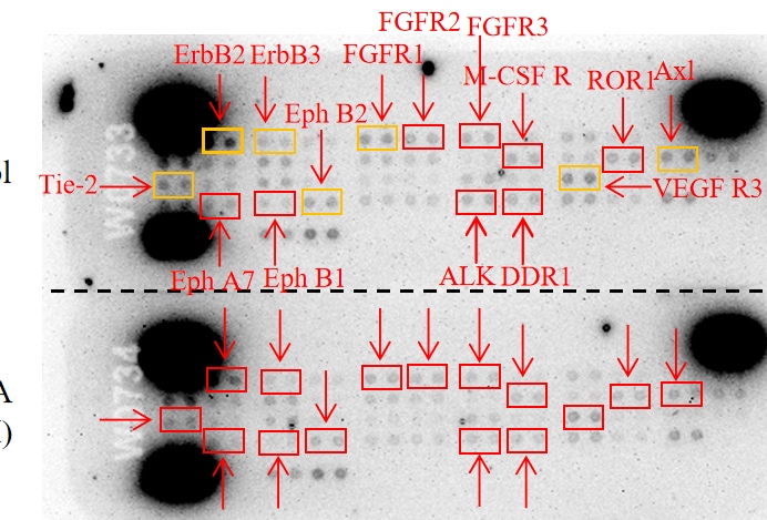
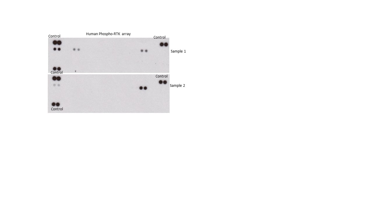
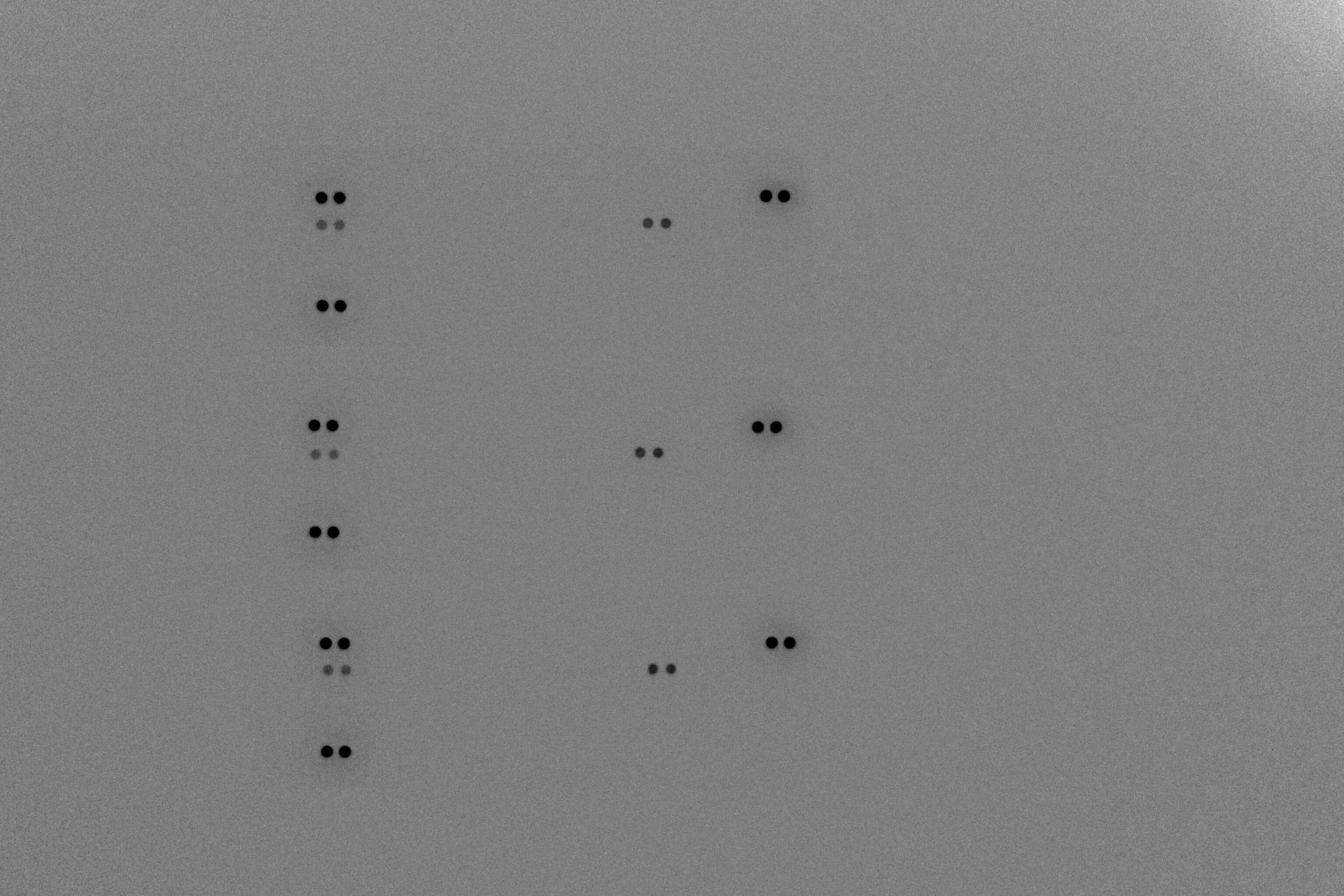
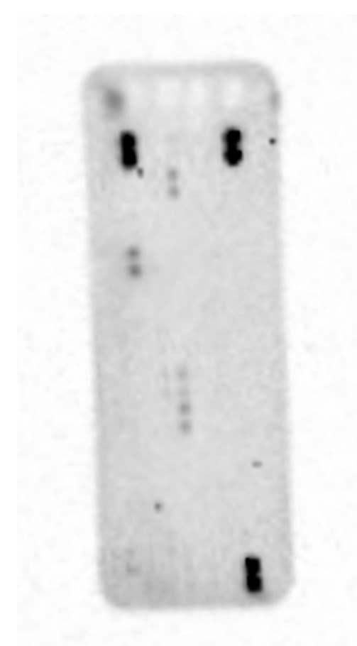
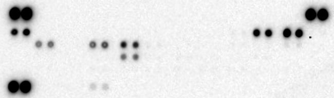
Scientific Data Images for Proteome Profiler Human Phospho-RTK Array Kit
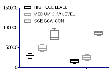
Detection of Human Receptor Tyrosine Kinase Phosphorylation in KATO-III Cell Line.
KATO-III human gastric carcinoma cells were either untreated or treated with 100 ng/mL rhFGF acidic and 1 μg/mL heparin for 15 minutes. Cells for rhFGF acidic/Heparin treatment either received pre-treatment with the reversible FGF R and VEGF R inhibitor PD 173074 (Tocris, Catalog # 3044), or with the FGF R-specific tyrosine kinase inhibitor SU 5402 (Tocris, Catalog # 3300), or no pre-treatment.
The Human Phospho-RTK Array is specific for ErbB2, ErbB3, and ErbB4 as shown by receptor competition.
MDA-MB-453 human breast cancer cells were treated with 100 ng/mL of rhNRG1-beta1/HRG1-beta1 (R&D Systems, Catalog # 396-HB) for 5 minutes. 5 µg of rhErbB2 (R&D Systems, Catalog # 1129-ER), rhErbB3 (R&D Systems, Catalog # 348-RB), or rhErbB4 (R&D Systems, Catalog # 1131-ER) extracellular domains were added to 50 µg of lysate and analyzed using the Human Phospho-RTK Array. Competition of a particular ErbB receptor was observed only with the corresponding recombinant soluble receptor.Formulation, Preparation, and Storage
Shipping
Storage
Background: RTK Assay Kits
Additional RTK Assay Kits Products
Product Documents for Proteome Profiler Human Phospho-RTK Array Kit
Certificate of Analysis
To download a Certificate of Analysis, please enter a lot or batch number in the search box below.
Note: Certificate of Analysis not available for kit components.
Product Specific Notices for Proteome Profiler Human Phospho-RTK Array Kit
For research use only
Related Research Areas
Citations for Proteome Profiler Human Phospho-RTK Array Kit
 Powered by Bioz
Customer Reviews for Proteome Profiler Human Phospho-RTK Array Kit (8)
Have you used Proteome Profiler Human Phospho-RTK Array Kit?
Submit a review and receive an Amazon gift card!
$25/€18/£15/$25CAN/¥2500 Yen for a review with an image
$10/€7/£6/$10CAN/¥1110 Yen for a review without an image
Submit a review
Customer Images

-
Verified Customer | Posted 10/31/2023The reagent kit testing remains as sensitive and stable as ever. We have repeated it multiple times in tissue samples, and the results are reliable.

-
Verified Customer | Posted 08/31/2021Great, excellent, can't wait to start my experiences right away.
-
Verified Customer | Posted 07/15/2021Purchased to screen changes induced by RTK inhibitors of cancer cell lines. Easy protocol, good clear results which could be independently validated by western blot. The only issue is that R&D are unable to supply information regarding the phospho-site detected, the antibody used in the array or the receptor chain recognised, so you just have to make an educated guess as to which antibody is most appropriate for an independent validation by western blotting etc. Excellent as an initial screen though.
-
Verified Customer | Posted 11/18/2019I would recommend to my colleagues, nicely performed. (for image acquiring, I used ImageQuant LAS 4000)
-
Verified Customer | Posted 03/13/2019
-
Verified Customer | Posted 10/29/2018
-
Verified Customer | Posted 08/28/2018The kit is excellent for p-RTK array in human cell lines. I recommended it to my colleague.
-
Verified Customer | Posted 10/20/2017
There are no reviews that match your criteria.
FAQs for Proteome Profiler Human Phospho-RTK Array Kit
-
Q: Can the Human Phospho-RTK Array be used for measuring the relative levels of total RTKs present in a sample?
A: No. Although the array kit uses capture antibodies that recognize both phosphorylated and unphosphorylated RTKs, the detection antibody is a pan anti-phospho-tyrosine antibody which only detects phosphorylated tyrosines on activated RTKs.
-
Q: To confirm positive signal for an RTK by IP-Western blot, are the capture antibodies from the Human Phospho-RTK Array Kit offered separately?
A: The identities of the capture antibodies on the Human Phospho-RTK Array are considered proprietary. Antibodies for each RTK analyte in the kit are available as well as Phospho-Tyrosine HRP-conjugated Antibody (Catalog #HAM1676). It can be challenging to directly compare Western Blot results of the linearized and reduced protein to Array detection of the native form. In ELISA, the protein will also be in the native form. ELISA kits are offered for many of the analytes targeted by the Array. These options are listed in a table on the Array product webpage.
-
Q: What are the phosphorylation site(s) of the 49 different tyrosine residues on the RTKs?
A: This kit uses a pan anti-phospho-tyrosine antibody as the detection antibody, which means it is capable of detecting phosphorylation at any available tyrosine. It is not designed to be specific for one phosphorylation site on each molecule. For instance, PDGF R alpha has multiple sites of tyrosine phosphorylation (see the “PTM/Processing” section of http://www.uniprot.org/uniprot/P16234 for a list of the currently identified sites).
-
Q: Which lysis buffers can be used for sample preparation?
A: Lysis Buffer 17 (Part #895943), provided in the kit, has been validated for optimal sample performance. Use of other lysis buffer requires independent evaluation.
-
Q: Can the Human Phospho-RTK Array be used for measuring the relative levels of total RTKs present in a sample?
A: No. Although the array kit uses capture antibodies that recognize both phosphorylated and unphosphorylated RTKs, the detection antibody is a pan anti-phospho-tyrosine antibody which only detects phosphorylated tyrosines on activated RTKs.
-
Q: To confirm positive signal for an RTK by IP-Western blot, are the capture antibodies from the Human Phospho-RTK Array Kit offered separately?
A: The identities of the capture antibodies on the Human Phospho-RTK Array are considered proprietary. Antibodies for each RTK analyte in the kit are available as well as Phospho-Tyrosine HRP-conjugated Antibody (Catalog #HAM1676). It can be challenging to directly compare Western Blot results of the linearized and reduced protein to Array detection of the native form. In ELISA, the protein will also be in the native form. ELISA kits are offered for many of the analytes targeted by the Array. These options are listed in a table on the Array product webpage.
-
Q: What are the phosphorylation site(s) of the 49 different tyrosine residues on the RTKs?
A: This kit uses a pan anti-phospho-tyrosine antibody as the detection antibody, which means it is capable of detecting phosphorylation at any available tyrosine. It is not designed to be specific for one phosphorylation site on each molecule. For instance, PDGF R alpha has multiple sites of tyrosine phosphorylation (see the “PTM/Processing” section of http://www.uniprot.org/uniprot/P16234 for a list of the currently identified sites).
-
Q: Which lysis buffers can be used for sample preparation?
A: Lysis Buffer 17 (Part #895943), provided in the kit, has been validated for optimal sample performance. Use of other lysis buffer requires independent evaluation.