
Alzheimer's Disease Research
Alzheimer's disease (AD) is a devastating progressive neurodegenerative disease characterized by synaptic dysfunction, neuronal loss, brain atrophy, and cognitive decline. As the most common form of dementia, AD accounts for approximately 60-80% of cases worldwide. Neurodegeneration occurs primarily in the hippocampus and cerebral cortex, where progressive brain changes contribute to memory loss, impaired learning, and declining cognitive function, which are hallmark features of the disease. These neurodegenerative changes are accompanied by brain atrophy, leading to a reduction in overall brain volume.
The pathological hallmarks of Alzheimer's disease are extracellular amyloid beta (Aβ) plaques and intracellular neurofibrillary tangles composed of hyperphosphorylated tau. However, growing evidence indicates that AD is a multifactorial disorder driven by interconnected mechanisms, including amyloid and tau pathology, neuroinflammation, complement-mediated synaptic dysfunction, impaired protein clearance, altered lipid metabolism, mitochondrial dysfunction, and blood-brain barrier and neurovascular dysfunction.
R&D Systems portfolio supports Alzheimer's disease research with proteins, antibodies, immunoassays, and small molecules for investigating disease mechanisms, validating targets, and advancing biomarker discovery and translational research.
Organoid and 3D Cell Culture Products
Brain organoids provide physiologically relevant 3D models that recapitulate key aspects of human brain biology, enabling the study of Alzheimer's disease-associated mechanisms, including amyloid pathology, neuroinflammation, and neurodegenerative processes. Explore our Cultrex™ matrices, proteins, and small molecules for robust, reproducible organoid cultures.
Human Amyloid β42 and β40 Quantikine ELISAs
Quantify key Alzheimer's disease biomarkers with Amyloid β42 and Amyloid β40 Quantikine™ ELISAs. These sensitive assays enable accurate measurement of amyloid β species across a range of sample types, supporting studies of Alzheimer's disease pathogenesis, biomarker discovery, and therapeutic development.
Fluorescent Amyloid β Probes
Track Aβ aggregation in real time with fluorescent probes and dyes that provide insight into amyloid assembly and support the screening of aggregation inhibitors. Available probes include Thioflavin T, Thioflavin X, Methoxy-X04, and K 114.
Understanding how Alzheimer's disease develops requires investigation of the molecular and cellular pathways that contribute to neuronal dysfunction and neurodegeneration. Explore the major biological mechanisms implicated in Alzheimer's disease pathogenesis and the potential therapeutic targets being studied to better understand disease progression and inform new treatment strategies.
The sections below highlight the key pathways and targets involved in Alzheimer's disease research.
Amyloid beta (Aβ) plaques are one of the defining pathological hallmarks of Alzheimer's disease (AD). According to the amyloid cascade hypothesis, an imbalance between Aβ production and clearance leads to the accumulation of pathogenic amyloid beta species in the brain. Soluble Aβ oligomers are widely regarded as highly neurotoxic and are increasingly thought to contribute to synaptic dysfunction prior to the appearance of extensive plaque pathology, ultimately promoting oxidative stress, neurodegeneration, and cognitive decline. As disease progresses, Aβ oligomers aggregate into amyloid fibrils and eventually form insoluble extracellular plaques that accumulate throughout affected brain regions.
Aβ peptides are generated through proteolytic processing of amyloid precursor protein (APP). APP can undergo either:
- Non-amyloidogenic processing: Cleavage of APP by α-secretase within the Aβ region, followed by γ-secretase processing, produces soluble APPα (sAPPα) and other non-amyloidogenic fragments while preventing generation of amyloid-β (Aβ) peptides.
- Amyloidogenic processing: Sequential cleavage of APP by β-secretase (BACE1) and γ-secretase produces amyloid-β (Aβ) peptides, including Aβ40 and Aβ42. Aβ42 is particularly prone to aggregation.
Due to its increased tendency to self-associate, Aβ42 readily forms toxic oligomers, amyloid fibrils, and extracellular plaques that are strongly associated with Alzheimer's disease pathology.
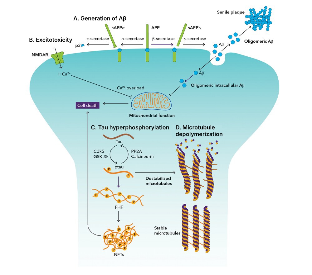
Figure 1. (A) Amyloid precursor protein (APP) is processed by two pathways. In the non-amyloidogenic pathway: α-secretase cleaves APP within the amyloid beta (Aβ) domain, preventing Aβ formation. In the amyloidogenic pathway: β-secretase and γ-secretase release Aβ intraneuronally before it is exported from the neuron and aggregates to form senile plaques. (B) Excessive activation of NMDA receptors leads to elevated intracellular calcium, which overloads the mitochondria, leading to cell death by excitotoxicity. (C) Hyperphosphorylation of tau generates p-tau, which forms paired helical filaments (PHF) and further aggregates into neurofibrillary tangles (NFTs). (D) In taupathies, hyperphosphorylation of tau leads to dissociation from microtubules causing them to depolymerize and breakdown, disrupting neuronal function.
Key Targets in Amyloid Biology and APP Processing
Small Molecule Inhibitors and Modulators
| Amyloid beta Peptides | Fluorescent Aβ Probes | γ-secretase Modulators |
| Amyloid beta Peptide Inhibitors | Secretase Inhibitors | |
| Amyloid beta Peptide Modulators | γ-Secretase Inhibitors |
Additional Resources
APP Processing and Amyloid Plaque Formation Research Area page
Molecular Balance in the Brain Technical Information page
A-beta Pathways: Plaque Formation & APP Metabolism
The intracellular accumulation of neurofibrillary tangles (NFTs) composed of misfolded, hyperphosphorylated tau (pTau) is closely associated with neuronal dysfunction in Alzheimer's disease (AD). Under normal conditions, tau binds to and stabilizes microtubules, helping maintain neuronal structure and intracellular transport. In AD, abnormal tau hyperphosphorylation, regulated by kinases including GSK-3β and CDK5, reduces tau's affinity for microtubules, promoting the accumulation of misfolded tau and disrupting normal neuronal function.
Hyperphosphorylated tau undergoes conformational changes that favor misfolding and self-aggregation. As pathological tau accumulates, it contributes to synaptic dysfunction and neurodegeneration while assembling into paired helical filaments and neurofibrillary tangles that closely correlate with cognitive decline and disease progression.
Increasing evidence indicates that pathological tau can spread throughout the brain through a prion-like seeding mechanism, transferring between neurons and potentially other neural cell types and driving the progressive expansion of tau pathology across interconnected neural networks. Because the extent of tau pathology closely correlates with cognitive impairment and disease progression, tau remains a major focus of Alzheimer's disease research and disease-modifying therapeutic development.
Key Targets in Tau Pathology and Neurofibrillary Tangles
| CDK5 | GSK-3β | p38γ |
| DYRK1A | MARK1 | PP2A |
| ERK1 | MARK2 | Tau |
| ERK2 | MARK3 | Tau 441 |
| ERK1/2 | MARK4 | |
| Fyn | p38α |
Small Molecule Inhibitors and Degraders
| CDK Inhibitors | GSK-3 Inhibitors | Microtubule Binding Compounds |
| DYRK Inhibitors | GSK-3 Degraders |
Neuroinflammation is a key feature of Alzheimer's disease (AD) and contributes to multiple processes associated with disease progression. In response to amyloid beta (Aβ), tau pathology, and other disease-associated stressors, microglia shift into activated states that can become chronically dysregulated over the course of the disease. Activated microglia and reactive astrocytes often accumulate around amyloid plaques, where they amplify inflammatory and complement-mediated responses. While these pathways normally support tissue maintenance and synaptic remodeling, their sustained activation may drive excessive synapse elimination and neuronal dysfunction in Alzheimer's disease. These synaptic changes are accompanied by altered neurotransmitter signaling, including dysregulation of glutamatergic and cholinergic pathways that further impair neuronal communication. As synaptic integrity and neural connectivity are progressively lost, neuronal communication becomes impaired, contributing to the cognitive decline characteristic of Alzheimer's disease.
Key Targets in Neuroinflammation
| CCL2 | IDO | P2X7/P2RX7 |
| CD33 | IL-1β | TDO2 |
| CX3CR1 | M-CSF R/CD115 | TNF-α |
| DAP12/TYROBP | NLRP3 | TREM2 |
Key Targets in Complement Signaling
| C1q | C4 | Factor H |
| C3 | CR1/CD35 | |
| C3aR | C5aR1 |
Key Targets in Synaptic Dysfunction
| Acetylcholinesterase/ACHE | GluR1 | PSD-95 |
| BDNF | GluR2 | SNAP25 |
| CHRNA4 | GluR3 | Synaptophysin (SYP) |
| CHRNA7 | NMDAR1 | TrkB |
| CHRNB2 | NMDAR2A | |
| Dynamin | NMDAR2B |
Small Molecule Inhibitors, Modulators, Agonists, and Antagonists
Additional Resources
Microglia Activation During Neuroinflammation: Overview
Microglia Activation During Neuroinflammation: Microglia Activation
Microglia Activation During Neuroinflammation: Microglia Polarization
Neuroinflammation Research Area page
Neuroinflammatory Factors Research Area page
ApoE-mediated lipid transport and cholesterol homeostasis influence multiple aspects of Alzheimer's disease pathogenesis. ApoE4 is the strongest common genetic risk factor for late-onset Alzheimer's disease and is associated with disrupted cholesterol metabolism, impaired amyloid-β clearance, and increased neuroinflammation. As a key regulator of lipid transport and homeostasis in the central nervous system, ApoE-associated pathways are thought to influence both disease susceptibility and progression in Alzheimer's disease.
Key Targets in ApoE and Lipid Metabolism
| ABCA1 | ApoJ/Clusterin | SORL1 (SorLA) |
| ABCG1 | LDL R | TREM2 |
| ApoE | Lipoprotein Lipase/LPL | |
| ApoE4 | LRP-1 |
Small Molecule Agonists
| LXR-like Receptor Agonists | PPAR Receptor Agonists | RXR Agonists |
Neurons rely heavily on mitochondrial activity to meet their energy demands, making them particularly vulnerable to metabolic disturbances that accumulate during aging and disease. As mitochondrial function declines, reactive oxygen species (ROS) can accumulate, promoting oxidative damage that contributes to synaptic dysfunction and neurodegeneration. Ferroptosis, an iron-dependent form of regulated cell death driven by lipid peroxidation, has recently emerged as another potential mechanism linking metabolic stress to neuronal loss. Growing evidence suggests that iron accumulation and increased lipid peroxidation in the Alzheimer's disease brain may promote ferroptotic signaling, potentially contributing to neuronal vulnerability and disease progression. These interconnected pathways may help explain how age-related metabolic dysfunction contributes to neuronal loss and cognitive decline in Alzheimer's disease.
Key Targets in Mitochondrial Dysfunction, Oxidative Stress, and Ferroptosis
| ACO1 (IRP1) | HMOX1 (HO-1) | Nrf2 |
| ACSL4/FACL4 | IRP2 | PGC-1α |
| AMPKα2 | KEAP1 | SOD2 |
| Frataxin (FXN) | NQO-1 | TFAM/mtTFA |
| GPX4 | Nrf1 | xCT/SLC7A11 |
Small Molecule Activators and Inhibitors
| AMPK Activators | Ferroptosis Activators | Ferroptosis Inhibitors |
| Antioxidant Compounds | Ferroptosis Controls | Nrf2 Activators |
Additional Resources
Regulated Cell Death Research Area page
Autophagy-lysosomal pathways play a fundamental role in maintaining cellular homeostasis within the nervous system. By facilitating the clearance and recycling of damaged cellular components and potentially harmful protein aggregates, these pathways help preserve neuronal health and function. When these pathways become impaired, toxic proteins can accumulate and contribute to the pathological changes associated with Alzheimer's disease. As a result, disruptions in autophagy-lysosomal function may promote neuronal stress, synaptic dysfunction, and neurodegeneration.
Key Targets in Lysosomal Dysfunction, Autophagy, and Protein Clearance
| ATG5 | LAMP-1/CD107a | SMPD1 |
| ATG7 | LAMP-2/CD107b | SMPD3 |
| ATG16L1 | LC3B | TFEB |
| Beclin 1 | mTOR | ULK1 |
| Cathepsin B | p62/SQSTM1 | VCP |
| Cathepsin D | Rab7a |
Small Molecule Activators, Inhibitors, and Modulators
| Autophagy Activators | Autophagy-Related Compounds | ULK Agonist |
| Autophagy-Inducing Peptides | Cathepsin Inhibitors | ULK Inhibitors |
| Autophagy Inhibitors | mTOR Inhibitors | VCP Inhibitors |
Additional Resources
Autophagy Signaling Research Area page
Autophagy-Inducing Peptides Research Area page
Webinar: Autophagy and Neurodegeneration with David Rubinsztein
The neurovascular unit, which coordinates communication between endothelial cells, pericytes, glia, and neurons, plays a critical role in maintaining brain homeostasis and supporting adequate cerebral blood flow. Growing evidence suggests that dysfunction of the neurovascular unit can disrupt this tightly regulated environment, compromise blood-brain barrier (BBB) integrity, and contribute to the pathological processes that drive cognitive decline. Rather than being a secondary consequence of neurodegeneration, neurovascular dysfunction is increasingly recognized as a contributor to disease progression.
Key Targets for Blood-Brain Barrier and Neurovascular Dysfunction
| ABCB1 | ICAM-1/CD54 | PECAM-1/CD31 |
| ANGPT1 | LRP-1 | RAGE/AGER |
| ANGPT2 | MMP-2 | Tight Junction Protein 1 (TJP1)/ZO-1 |
| Aquaporin-4/AQP4 | MMP-9 | VCAM-1/CD106 |
| Caveolin-1 | Occludin | VEGF |
| Claudin-5 | PDGF Rβ |
Small Molecule Activators, Inhibitors, and Modulators
| Multidrug Transport Inhibitors | RAGE Antagonists | VEGF Receptor Inhibitors |
Additional Resources
Blood-Brain Barrier Permeability Research Area page
Endothelial Cell Markers Research Area page
Biomarkers play an increasingly important role in Alzheimer's disease research, supporting early disease detection, patient stratification, target validation, and therapeutic development. Fluid biomarkers including Aβ42, Aβ40, phosphorylated tau species, neurofilament light chain (NfL), GFAP, and YKL-40 provide insight into the amyloid, tau, neurodegenerative, and neuroinflammatory processes that contribute to disease progression. As a result, biomarker discovery and validation have become essential components of both preclinical and clinical Alzheimer's disease research.
Key Biomarkers for Translational Research
| Aβ40 | Neurogranin | phospho-Tau (T231) |
| Aβ42 | NF-L | Tau |
| Chitinase 3-like 1/YKL-40 | phospho-Tau (T181) | TREM2 |
| GFAP | phospho-Tau (T217) |
Alzheimer's Disease Research Product Guide
This product guide provides a background to Alzheimer's disease research and lists around 100 products for the study of:
- Amyloid and tau proteins
- Excitotoxicity
- Mitochondrial dysfunction
- Oxidative stress
- Emerging targets
Alzheimer's Disease Neuropathology and Epidemiology
Alzheimer's disease (AD) is a debilitating and progressive neurodegenerative disease and the most common cause of dementia, affecting approximately 30% of individuals aged over 85 years. This poster summarizes the cellular and molecular mechanisms of AD.
Alzheimer's disease (AD) is a complex, multifactorial neurodegenerative disorder that arises from the interaction of genetic, molecular, cellular, and environmental factors. While the exact cause remains incompletely understood, growing evidence suggests that disease progression is driven by interconnected mechanisms including amyloid and tau pathology, neuroinflammation, microglial activation, complement signaling, synaptic dysfunction, impaired protein clearance, blood-brain barrier dysfunction, neurovascular impairment, and altered lipid metabolism. Genetic risk factors, particularly ApoE4, can further influence disease susceptibility by affecting pathways involved in amyloid clearance, inflammation, and brain lipid homeostasis. Researchers continue to investigate how these processes interact to promote neurodegeneration, cognitive decline, and Alzheimer's disease progression.
Alzheimer's disease is the most common cause of dementia, but it is one of several neurodegenerative disorders that can lead to cognitive impairment. Other forms of dementia include Lewy body dementia, frontotemporal dementia, vascular dementia, and mixed dementias, in which multiple disease processes contribute to cognitive decline. Although these disorders share some clinical features, they are associated with distinct pathological mechanisms, biomarkers, and patterns of neurodegeneration. For example, Alzheimer's disease is characterized by Aβ plaques and tau neurofibrillary tangles, whereas Lewy body dementia is associated with the accumulation of α-synuclein-containing Lewy bodies.
The defining pathological hallmarks of Alzheimer's disease are extracellular amyloid beta (Aβ) plaques and intracellular neurofibrillary tangles composed of hyperphosphorylated tau protein. Amyloid plaques are extracellular deposits formed by the aggregation of Aβ peptides, whereas neurofibrillary tangles are intracellular aggregates of abnormal tau that accumulate within neurons.
Amyloid beta aggregation is thought to occur early in the disease process and is associated with the formation of toxic Aβ oligomers and plaques that can disrupt neuronal function and trigger neuroinflammatory responses. Tau pathology develops when tau becomes abnormally phosphorylated, causing it to detach from microtubules, misfold, and aggregate into neurofibrillary tangles. Both amyloid pathology and tau pathology contribute to synaptic dysfunction, neurodegeneration, and cognitive decline, although the extent of tau pathology is more closely correlated with disease severity and progression.
The amyloid cascade hypothesis proposes, an imbalance between Aβ production and clearance initiates a series of pathological events that contribute to Alzheimer's disease progression. Support for this hypothesis comes from genetic studies showing that mutations in APP, PSEN1, and PSEN2 can alter amyloid beta production and processing, promoting the accumulation of pathogenic Aβ species and causing familial Alzheimer's disease.
While amyloid and tau remain the defining pathological hallmarks of Alzheimer's disease, growing evidence suggests that disease progression is also influenced by additional mechanisms including neuroinflammation, impaired protein clearance, altered lipid metabolism, and neurovascular dysfunction.
Phosphorylated tau (pTau) refers to tau protein that has been modified by the addition of phosphate groups at specific amino acid residues. Although tau phosphorylation normally helps regulate neuronal function, excessive phosphorylation can promote tau misfolding, aggregation, and neurofibrillary tangle formation in Alzheimer's disease.
Beyond its role in disease pathology, pTau has become one of the most important biomarker classes in Alzheimer's disease research. Biomarkers such as pTau181, pTau217, and pTau231 can be measured in cerebrospinal fluid and blood and are increasingly used to detect and characterize Alzheimer's disease-associated tau pathology, support patient stratification, monitor disease progression, and assess responses to emerging therapies. Because changes in pTau can occur before significant cognitive impairment develops, these biomarkers have become important tools for Alzheimer's disease research and clinical trial design.
Protein clearance and autophagy play essential roles in maintaining neuronal health by removing damaged organelles, misfolded proteins, and toxic protein aggregates. Autophagy is a highly regulated cellular process that delivers unwanted cellular components to lysosomes for degradation and recycling, helping preserve cellular homeostasis and protein quality control.
Evidence from human studies and experimental models suggests that autophagy-lysosomal pathways become progressively impaired during Alzheimer's disease. These defects may reduce the ability of neurons to clear amyloid beta (Aβ), tau, and other potentially toxic protein species, promoting the accumulation of pathogenic protein aggregates and cellular dysfunction. Researchers are therefore investigating key regulators of the autophagy-lysosomal pathway, including TFEB, mTOR, Beclin 1, and ULK1, as potential targets for enhancing cellular clearance mechanisms and facilitating the removal of disease-associated protein aggregates.
Neuroinflammation is increasingly recognized as an important contributor to Alzheimer's disease progression, with microglia playing a central role in this process. Microglia are the resident immune cells of the central nervous system and normally help maintain brain health by clearing cellular debris, removing damaged proteins, supporting synaptic remodeling, and responding to injury. In Alzheimer's disease, microglia become activated in response to amyloid beta (Aβ) plaques, tau pathology, and other disease-associated signals. While this response may initially help limit damage and promote the clearance of pathological proteins, chronic microglial activation can become dysregulated and contribute to sustained neuroinflammation.
As Alzheimer's disease progresses, activated microglia and reactive astrocytes produce inflammatory mediators and complement proteins that can alter protein clearance, disrupt synaptic function, and promote excessive synapse elimination. Persistent neuroinflammation has been linked to neuronal dysfunction, neurodegeneration, and cognitive decline. Genetic studies identifying Alzheimer's disease risk genes such as TREM2, CD33, and CR1/CD35 have further highlighted the importance of neuroimmune pathways in disease pathogenesis. TREM2 regulates microglial activation, phagocytosis, and lipid sensing; CD33 influences microglial activation and has been implicated in the regulation of amyloid beta clearance; and CR1/CD35 participates in complement-mediated immune processes that may influence amyloid clearance, synaptic remodeling, and neuroinflammation. Together, these pathways represent important areas of Alzheimer's disease research and therapeutic development.
Genetics plays an important role in Alzheimer's disease risk and pathogenesis. Rare variants of APP, PSEN1, and PSEN2 are associated with familial forms of Alzheimer's disease, while common genetic variants in APOE, TREM2, CD33, BIN1, ABCA7, CLU, and SORL1 influence susceptibility to late-onset Alzheimer's disease.
Among these, ApoE4 is the strongest common genetic risk factor for late-onset Alzheimer's disease. APOE encodes apolipoprotein E, a protein involved in lipid transport, cholesterol homeostasis, and amyloid beta clearance within the central nervous system. Compared with other APOE variants, ApoE4 is associated with reduced clearance and increased accumulation of amyloid beta in the brain. ApoE4 has also been linked to altered lipid metabolism, increased neuroinflammation, impaired synaptic function, and blood-brain barrier dysfunction. Although carrying ApoE4 increases the risk of developing Alzheimer's disease, it is not a direct cause of the disease, and many ApoE4 carriers never develop dementia.
Additional genetic risk factors such as TREM2, CD33, BIN1, ABCA7, CLU, and SORL1 are associated with biological pathways involved in neuroinflammation, lipid metabolism, protein clearance, endosomal trafficking, and amyloid processing. Together, genetic studies continue to provide important insights into Alzheimer's disease biology, disease mechanisms, and potential therapeutic targets.
The blood-brain barrier (BBB) helps maintain a stable environment for the brain by regulating the movement of molecules between the circulation and the central nervous system. Increasing evidence suggests that BBB dysfunction may occur relatively early in Alzheimer's disease and could contribute to disease progression before extensive neurodegeneration is apparent.
Disruption of BBB integrity may alter cerebral blood flow, reduce amyloid beta clearance, promote neuroinflammatory responses, and expose the brain to circulating factors that are normally excluded from neural tissue. Because vascular dysfunction and neurodegeneration appear to influence one another throughout the course of disease, the BBB and the broader neurovascular unit have become major areas of Alzheimer's disease research and therapeutic development.
Biomarkers play a critical role in Alzheimer's disease research, supporting early disease detection, patient stratification, target validation, and therapeutic development. The most widely studied Alzheimer's disease biomarkers reflect amyloid pathology, tau pathology, neurodegeneration, and neuroinflammatory processes.
Common Alzheimer's disease biomarkers include amyloid beta 42 (Aβ42), amyloid beta 40 (Aβ40), the Aβ42/Aβ40 ratio, phosphorylated tau species including pTau181, pTau217, and pTau231, total tau, neurofilament light chain (NF-L), glial fibrillary acidic protein (GFAP), soluble TREM2 (sTREM2), neurogranin, and YKL-40 (CHI3L1). These biomarkers can be measured in cerebrospinal fluid, blood, and other research samples using immunoassays such as ELISA, multiplex assays, and other protein quantification technologies. In addition to fluid biomarkers, imaging biomarkers measured using amyloid and tau PET scans are widely used in Alzheimer's disease research to assess pathology, support patient stratification, and monitor disease progression. As biomarker-guided research continues to expand, these indicators have become essential tools for understanding disease biology and accelerating Alzheimer's disease drug development.
Common Alzheimer's disease research models include transgenic and knock-in mouse models expressing mutant APP, PSEN1, PSEN2, or tau genes, as well as induced pluripotent stem cell (iPSC)-derived neurons, brain organoids, primary neural cultures, and other experimental systems. Together, these models provide valuable platforms for studying Alzheimer's disease biology, investigating disease mechanisms, and evaluating potential therapeutic approaches.
Interactive Signaling Pathways
A-beta Pathways: Plaque Formation & APP Metabolism
A-beta Pathways: Uptake & Degradation
Microglia Activation During Neuroinflammation: Overview
Microglia Activation During Neuroinflammation: Microglia Activation
Microglia Activation During Neuroinflammation: Microglia Polarization