Angiogenesis is the tightly regulated process by which new blood vessels are formed from the existing vasculature. This process is physiologically important for development and wound healing, and is also a common driver in multiple diseases including rheumatoid arthritis, atherosclerosis, macular degeneration, and cancer. Angiogenesis occurs in response to a variety of molecular cues. Generally, the angiogenic process includes endothelial cell proliferation, chemotactic endothelial cell migration through the extracellular matrix barrier, and the formation of capillary tubes. Physiological and pathological angiogenesis utilize many of the same cellular processes and molecular signaling networks, however the structures that form during pathological angiogenesis are often functionally abnormal.
Proteome Profiler Human Angiogenesis Array Kit
R&D Systems | Catalog # ARY007

Key Product Details
Species
Product Summary for Proteome Profiler Human Angiogenesis Array Kit
Kit Summary
A membrane-based antibody array for the parallel determination of the relative levels of selected angiogenesis-related proteins. Validated for analyte detection in cell culture supernates, cell lysates, tissue lysates, serum, plasma and saliva.
Key Benefits
- Detects 55 human angiogenesis-related proteins simultaneously
- Requires no specialized equipment
- Compatible with LI-COR* and chemiluminescence detection
Principle of the Assay
The Proteome Profiler Human Angiogenesis Array Kit is a membrane-based sandwich immunoassay. Samples are mixed with a cocktail of biotinylated detection antibodies (Step 1) and then incubated with the array membrane which is spotted in duplicate with capture antibodies to specific target proteins (Step 2). Captured proteins are visualized using chemiluminescent detection reagents (Step 3). The signal produced is proportional to the amount of bound analyte. Analytes include soluble growth and differentiation factors, extracellular matrix components, proteases, membrane-bound receptors, and intracellular signaling molecules.
Why Use an Antibody Array to Detect Angiogenesis-Related Proteins?
Determining the expression of multiple proteins in a single sample can be expensive, time consuming and can require specialized equipment. Performing multiple immunoprecipitations and Western blots requires time, labor, and reagents. The use of a multiplex antibody array to detect multiple proteins in a single sample can be cost-effective and also save time and sample.
- 4 Array Membranes
- 4-Well Multi-dish
- Array Buffers
- Wash Buffer
- Antibody Detection Cocktail
- Streptavidin-HRP
- Chemiluminescent Detection Reagents
- Transparency Overlay Template
- Detailed Protocol
For a complete list of the kit contents and necessary materials, please see the Materials Provided/Other Supplies Required sections of the product datasheet.
Stability and Storage
Store the unopened kit at 2 °C to 8 °C. Do not use past kit expiration date.
| Simultaneously detect the relative levels of these angiogenesis-related proteins in a single sample. | ||
|---|---|---|
| Activin A | FGF-7/KGF | PD-ECGF |
| ADAMTS-1 | GDNF | PDGF-AA |
| Angiogenin | GM-CSF | PDGF-AB/PDGF-BB |
| Angiopoietin-1 | HB-EGF | Persephin |
| Angiopoietin-2 | HGF | CXCL4/PF4 |
| Angiostatin/Plasminogen | IGFBP-1 | PlGF |
| Amphiregulin | IGFBP-2 | Prolactin |
| Artemin | IGFBP-3 | Serpin B5/Maspin |
| Tissue Factor/Factor III | IL-1 beta | Serpin E1/PAI-1 |
| CXCL16 | CXCL8/IL-8 | Serpin F1/PEDF |
| DPPIV/CD26 | LAP (TGF-beta 1) | TIMP-1 |
| EGF | Leptin | TIMP-4 |
| EG-VEGF | CCL2/MCP-1 | Thrombospondin-1 |
| Endoglin/CD105 | CCL3/MIP-1 alpha | Thrombospondin-2 |
| Endostatin/Collagen XVIII | MMP-8 | uPA |
| Endothelin-1 | MMP-9 | Vasohibin |
| FGF acidic | NRG1-beta 1 | VEGF |
| FGF basic | Pentraxin 3 | VEGF-C |
| FGF-4 | ||
Assays for analytes represented in the Human Angiogenesis Antibody Array Kit
| Quantikine® ELISA Kits | DuoSet® ELISA Development Systems | Quantikine® HS (High Sensitivity) ELISA Kits | QuantiGlo® Chemiluminescent ELISA Kits | |
|---|---|---|---|---|
| Activin A | DAC00B | DY338 | ||
| ADAMTS-1 | DY2197 | |||
| Angiogenin | DAN00 | DY265 | ||
| Angiopoietin-1 | DANG10 | DY923 | ||
| Angiopoietin-2 | DANG20 | DY623 | ||
| Angiostatin/Plasminogen | ||||
| Amphiregulin | DY262 | |||
| Artemin | DY2589 | |||
| Tissue Factor/Factor III | DCF300 | DY2339 | ||
| CXCL16 | DCX160 | DY1164 | ||
| DPPIV/CD26 | DY1180 | |||
| EGF | DEG00 | DY236 | ||
| EG-VEGF | DY1209 | |||
| Endoglin | DNDG00 | DY1097 | ||
| Endostatin/Collagen XVII | DNST0 | DY1098 | ||
| Endothelin-1 | DET100 | QET00B | ||
| FGF acidic | DFA00B | DY232 | ||
| FGF basic | DFB50 | DY233 | HSFB00D | |
| FGF-4 | DY235 | |||
| FGF-7/KGF | DKG00 | DY251 | ||
| GDNF | DY212 | |||
| GM-CSF | DGM00 | DY215 | HSGM0 | |
| HB-EGF | DY259 | |||
| HGF | DHG00 | DY294 | ||
| IGFBP-1 | DY871 | |||
| IGFBP-2 | DGB200 | DY674 | ||
| IGFBP-3 | DGB300 | DY675 | ||
| IL-1 beta | DLB50 | DY201 | HSLB00D | QLB00B |
| CXCL8/IL-8 | D8000C | DY208 | HS800 | Q8000B |
| TGF-beta1 (LAP) | DB100B | DY240 | ||
| Leptin | DLP00 | DY398 | ||
| CCL2/MCP-1 | DCP00 | DY279 | ||
| CCL3/MIP-1 alpha | DMA00 | DY270 | ||
| MMP-8 | DMP800 | DY908 | ||
| MMP-9 | DMP900 | DY911 | ||
| NRG1-beta1 | DY377 | |||
| Pentraxin 3 | DPTX30 | DY1826 | ||
| PD-ECGF | ||||
| PDGF-AA | DAA00B | DY221 | ||
| PDGF-AB/PDGF-BB | DHD00C, DBB00 | DY222, DY220 | ||
| Persephin | ||||
| CXCL4/PF4 | DY795 | |||
| PlGF | DPG00 | DY264 | ||
| Prolactin | DPRL00 | DY682 | ||
| Serpin B5/Maspin | ||||
| Serpin E1/PAI-1 | DSE100 | DY1786 | ||
| Serpin F1/PEDF | ||||
| TIMP-1 | DTM100 | DY970 | ||
| TIMP-4 | DTM400 | DY974 | ||
| Thrombospondin-1 | DTSP10 | DY3074 | ||
| Thrombospondin-2 | DTSP20 | DY1635 | ||
| uPA | DUPA00 | DY1310 | ||
| Vasohibin | ||||
| VEGF | DVE00 | DY293B | QVE00B | |
| VEGF-C | DVEC00 | DY752B |
Scientific Data Images for Proteome Profiler Human Angiogenesis Array Kit
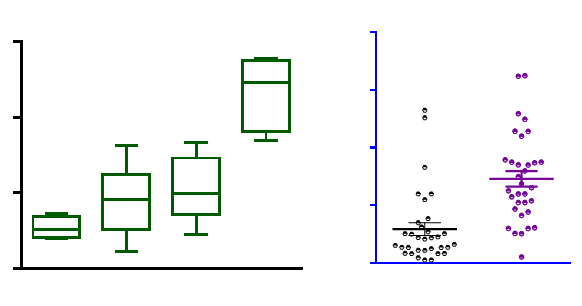
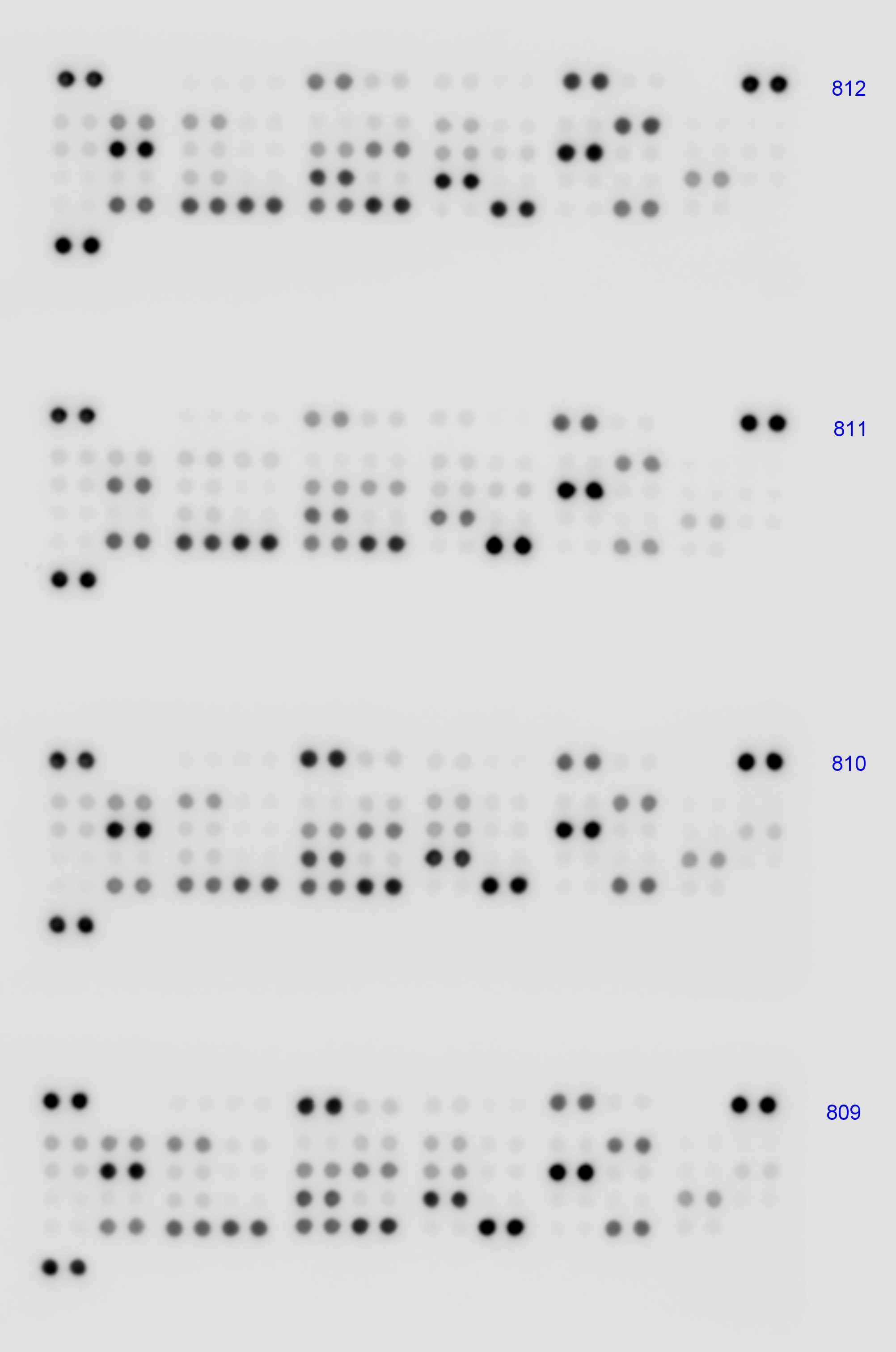
Profiling Angiogenesis-Related Proteins in Human Cancer Tissues.
A) The Proteome Profiler Human Angiogenesis Antibody Array (Catalog # ARY007) was used to profile angiogenesis-related proteins in tissues from human prostate, ovarian, and breast cancers. B) Histogram profiles for select analytes were generated by quantifying the mean spot pixel densities from the array membrane using image software.
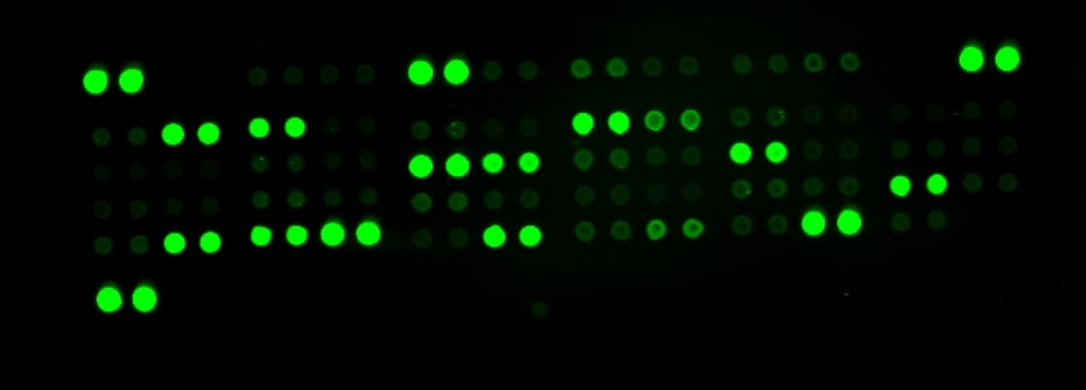
The Human Angiogenesis Array detects multiple analytes in cell culture supernates.
A. THP-1 human acute monocytic leukemia cell line treated with 1 µg/mL Recombinant Human IFN-gamma (Catalog # 285-IF) for 8 hours and 1 µg/mL lipopolysaccharide (LPS) for 16 hours. B. HUVEC human umbilical vein endothelial cells treated with 10 mg/ml of LPS for 24 hours. 500 µL of supernate was used for each array shown. Array images were collected and analyzed using the LI-COR Odyssey Infrared Imaging System.
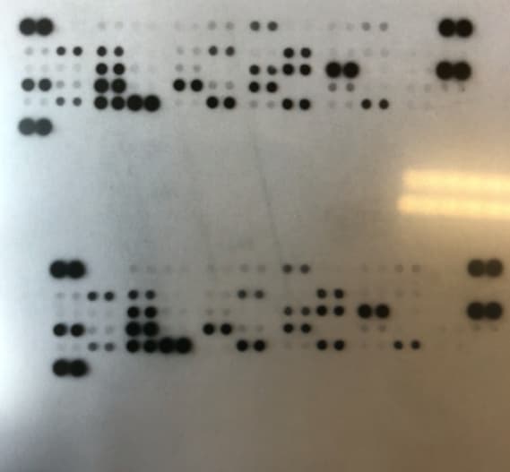
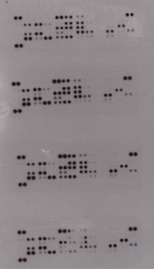
The Human Angiogenesis Array detects multiple analytes in tissue lysate, serum, and plasma.
Representative membranes and selective quantification are shown for human ovarian cancer tissue lysate (A., 200 µg), serum (B., 100 µL), and EDTA plasma (C., 100 µL). Array images were collected and analyzed using the LI-COR Odyssey Infrared Imaging System.Formulation, Preparation, and Storage
Shipping
Storage
Background: Angiogenesis Assay Kits
Additional Angiogenesis Assay Kits Products
Product Documents for Proteome Profiler Human Angiogenesis Array Kit
Certificate of Analysis
To download a Certificate of Analysis, please enter a lot or batch number in the search box below.
Note: Certificate of Analysis not available for kit components.
Product Specific Notices for Proteome Profiler Human Angiogenesis Array Kit
For research use only
Citations for Proteome Profiler Human Angiogenesis Array Kit
 Powered by Bioz
Customer Reviews for Proteome Profiler Human Angiogenesis Array Kit (10)
Have you used Proteome Profiler Human Angiogenesis Array Kit?
Submit a review and receive an Amazon gift card!
$25/€18/£15/$25CAN/¥2500 Yen for a review with an image
$10/€7/£6/$10CAN/¥1110 Yen for a review without an image
Submit a review
Customer Images

-
Verified Customer | Posted 10/31/2023We ordered the Angiogenesis Array Kit for both mice and humans, and it is highly sensitive for both solid tumor and serum samples.

-
Verified Customer | Posted 11/21/2022HepG2 lysate concentrated, DMEM FBS free, 300 ug loaded.
-
Verified Customer | Posted 02/07/2019
-
Verified Customer | Posted 02/06/2019Worked well to look at THP-1 angiogenesis factors.
-
Verified Customer | Posted 10/22/2018It worked well in cell culture supernatants using 125 ug of total protein per membrane.
-
Verified Customer | Posted 08/08/2018
-
Verified Customer | Posted 05/17/2018Used it on Conditioned medium collected from Breast Cancer Cells treated with Angiotensin II to look into its effect on Angiogenesis. Angiogenesis array worked really well and very easy to use. It provided some new promising gene targets which we did not know before.
-
Verified Customer | Posted 04/01/2018
-
Verified Customer | Posted 10/30/2017
-
Verified Customer | Posted 12/06/2016Human Angiogenesis Antibody Array was used to evaluate mesenchymal stem cells secretion profiles when seeded in various extracellular matrices. Great tool that ganarates lots of data!
There are no reviews that match your criteria.
FAQs for Proteome Profiler Human Angiogenesis Array Kit
-
Q: Does the Human Angiogenesis Proteome Profiler Array cross-react with other species like equines?
A: We have not tested this kit with equine samples. However, when looking at the cross reactivity with our Proteome Profiler Arrays, there are a few things to keep in mind. When the arrays are being designed, we look for the best antibodies that will work for the species stated. This does not mean there will not be any cross reactivity between species but it does suggest that the assay would not perform equally between species. While it is possible that some of the antibodies may detect the equine protein, different antibodies cross-react at different levels.
It is also important to note that the Proteome Profiler Arrays are designed to be used as a screening tool. This may be a good place to start if you are trying determine what may be having an increase or a decrease in signal. We recommend running one untreated sample and comparing them to your treated samples. Even though you may not have the exact same cross reactivity with each analyte, you may be able to detect the increase or decrease from your samples.
We have searched for citations that may have referenced using this kit with equine samples and were able to find 1. Here is the link to it: https://www.ncbi.nlm.nih.gov/pubmed/25313202. -
Q: What are the associated pathways each factor is involved in?
A: We do not have specific angiogenesis-related categorization of the target for this array, but we do have some informative material available in our website:
- https://www.rndsystems.com/research-area/angiogenesis
- https://www.novusbio.com/research-areas/cancer/angiogenesis -
Q: Which gene of the VEGF family is detected on E19 and E20
A: The VEGF spots are detecting VEGF-A.
-
Q: Will the Array identification number stamped on the Array membrane interfere with detection if it is not cut-off before the membrane is blocked?
A: The dye used for printing the Array identification number on the membranes will fluoresce and interfere with the LI-COR detection. It is critical that the number is cut off before beginning the experiment.
-
Q: Does the Human Angiogenesis Proteome Profiler Array cross-react with other species like equines?
A: We have not tested this kit with equine samples. However, when looking at the cross reactivity with our Proteome Profiler Arrays, there are a few things to keep in mind. When the arrays are being designed, we look for the best antibodies that will work for the species stated. This does not mean there will not be any cross reactivity between species but it does suggest that the assay would not perform equally between species. While it is possible that some of the antibodies may detect the equine protein, different antibodies cross-react at different levels.
It is also important to note that the Proteome Profiler Arrays are designed to be used as a screening tool. This may be a good place to start if you are trying determine what may be having an increase or a decrease in signal. We recommend running one untreated sample and comparing them to your treated samples. Even though you may not have the exact same cross reactivity with each analyte, you may be able to detect the increase or decrease from your samples.
We have searched for citations that may have referenced using this kit with equine samples and were able to find 1. Here is the link to it: https://www.ncbi.nlm.nih.gov/pubmed/25313202. -
Q: What are the associated pathways each factor is involved in?
A: We do not have specific angiogenesis-related categorization of the target for this array, but we do have some informative material available in our website:
- https://www.rndsystems.com/research-area/angiogenesis
- https://www.novusbio.com/research-areas/cancer/angiogenesis -
Q: Which gene of the VEGF family is detected on E19 and E20
A: The VEGF spots are detecting VEGF-A.
-
Q: Will the Array identification number stamped on the Array membrane interfere with detection if it is not cut-off before the membrane is blocked?
A: The dye used for printing the Array identification number on the membranes will fluoresce and interfere with the LI-COR detection. It is critical that the number is cut off before beginning the experiment.