Antibodies for Multiplex Immunofluorescence (mIF)
Multiplexed immunofluorescence (mIF) is a powerful technique for investigating complex biological systems and cellular interactions. It allows researchers to simultaneously detect multiple targets within a single tissue, providing a deeper understanding of cellular dynamics, spatial relationships, and biomarker expression patterns.
Table of Contents
- Featured COMET Qualified Antibodies
- Qualified Antibodies Table
- Qualification Process on COMET
- Other Spatial Tested Antibodies
Multiplex Immunofluorescence (mIF) on COMET™
The COMET platform from Lunaphore, is a fully-automated, high-throughput, spatial biology platform that performs sequential immunofluorescence (seqIF™) through staining, imaging, and elution cycles. It offers the unique flexibility to work with off-the-shelf, non-conjugated primary antibodies.
Featured Multiplex IF Antibodies Qualified on COMET
Figure 1: Detection of beta-Actin in Mouse Stomach via seqIF™ staining on COMET using Rat Anti-Mouse beta-Actin Monoclonal Antibody (Catalog # MAB11702) Tissue was stained using the Alexa Fluor™ 555 Goat anti-Rat IgG Secondary Antibody (yellow;
Bio-Techne Spatial Catalog # DR555RT) and counterstained with DAPI (blue; Bio-Techne Spatial Catalog # DR100). Specific staining was localized to the cell membrane.
Figure 2: Detection of CD21 in Human Lymph Node via seqIF™ staining on COMET using Rabbit Anti-Human CD21 Monoclonal Antibody (Catalog # NBP3-20354). Tissue was stained using the Alexa Fluor™ Plus 555 Goat anti-Rabbit IgG Secondary Antibody (yellow;
Bio-Techne Spatial Catalog # DR555RB) and counterstained with DAPI (blue; Bio-Techne Spatial Catalog # DR100). Specific staining was localized to the membrane.
Figure 3: Detection of CD31 in Mouse Thymus via seqIF™ staining on COMET using Rat Anti-Mouse CD31 Monoclonal Antibody (Catalog # MAB11714). Tissue was stained using the Alexa Fluor™ 647 Goat anti-Rat IgG Secondary Antibody (yellow; Bio-Techne Spatial Catalog # DR647RT) and counterstained with DAPI (blue; Bio-Techne Spatial Catalog # DR100). Specific staining was localized to the membrane.
COMET Qualified mIF Antibodies
Table 1: List of COMET Qualified Antibodies.
Antibody Testing Process on COMET
The antibody validation process on COMET includes the antibody qualification process for protocol optimization, with a focus on assessing specificity, sensitivity, and elution efficiency using positive control tissue. R&D Systems™, by Bio-Techne, IHC validated antibodies have already undergone a rigorous process to be added to the catalog. Antibody qualification process for COMET involves several key aspects as shown in Figure 4.
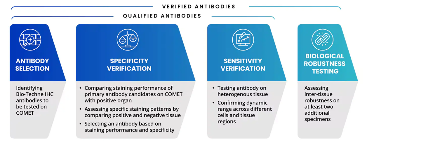
Figure 4. Steps of Antibody Testing Process on COMET.
Multiplex IF (mIF) validated antibodies with a COMET application validation are taken through the qualification or verification process listed above. This rigorous testing process R&D Systems and Novus antibodies deliver high-quality and specific staining on the COMET platform. R&D Systems COMET tested antibodies are also in the Panel Builder and added to the expanding COMET marker database.
Explore our curated selection of antibodies and take your spatial biology studies to new heights. All R&D Systems antibodies are provided in conjugation-ready format, free from BSA and Azide. Find our growing list of spatial biology tested antibodies from Novus Biologicals and R&D Systems below.
Have you used one of our antibodies in a spatial biology platform? Submit a review and receive a reward!
| Target | Platform | Catalog Number | External Data Link | OMAP_ID |
| ABCA3 | Cell DIVE™ | NBP1-89310 | Citation | OMAP-7 |
| AGER | Cell DIVE™ | AF1145 | Citation | OMAP-7 |
| APP / β Amyloid | Nanostring GeoMx® DSP | NBP2-13075AF647 | Data Image | |
| BST2 / CD317 | IMC (Hyperion™ and Hyperion Xti™) | DDX0390P-100 | Citation | |
| CD11b | Nanostring GeoMx® DSP | NB110-89474AF647 | Data Image | |
| CD161 / NK1.1 | IMC (Hyperion™ and Hyperion Xti™) | NB100-77528 | Citation | |
| CD20 | Nanostring GeoMx® DSP | NBP2-47840AF647 | Data Image | |
| CD20 | Nanostring GeoMx® DSP | NBP2-47840AF594 | Data Image | |
| CD20 | Nanostring GeoMx® DSP | NBP2-47840AF488 | Data Image | |
| CD3 | Nanostring GeoMx® DSP | NBP2-54392AF647 | Data Image | |
| CD3 | Nanostring GeoMx® DSP | NBP2-54392AF594 | Data Image | |
| CD3 | Nanostring GeoMx® DSP | NBP2-54392AF532 | Data Image | |
| CD3 | Nanostring GeoMx® DSP | NBP2-54392AF488 | Data Image | |
| CD4 | SIMS | NBP2-25199 | Citation | |
| CD4 | Canopy CellScape | FAB8165G | Citation | |
| CD68 | Nanostring GeoMx® DSP | NBP2-34587AF488 | Data Image | |
| CD68 | Nanostring GeoMx® DSP | NBP2-34587AF594 | Data Image | |
| CD68 | Nanostring GeoMx® DSP | NBP2-34587AF647 | Data Image | |
| CD68 | Nanostring GeoMx® DSP | NBP2-34587AF532 | Data Image | |
| CD69 | IMC (Hyperion™ and Hyperion Xti™) | NBP1-51607 | Data Image | |
| CD7 | IMC (Hyperion™ and Hyperion Xti™) | NBP2-37368 | Data Image | |
| CHGA | CODEX | NBP2-34674 | Citation 1, Citation 2 | OMAP-2, OMAP-6, OMAP-13 |
| C-Kit / CD117 | Nanostring GeoMx® DSP | NBP2-90037AF647 | Data Image | |
| CPS1 | SIMS | NBP3-08970 | Citation | OMAP-5 |
| CTSL | Cell DIVE™ | NB100-1775 | Citation | OMAP-7 |
| CXCL13 | IBEX | AF801 | Citation 1, Citation 2 | OMAP-1 |
| CXCR6 | IBEX | FAB2145R | Citation | |
| ELA2 / Neutrophil Elastase | Nanostring GeoMx® DSP | MAB9167AF594 | Data Image | |
| ELA2 / Neutrophil Elastase | Nanostring GeoMx® DSP | MAB9167AF488 | Data Image | |
| Fibronectin | IBEX | NBP2-22113AF532 | Citation | |
| GCG | CODEX | MAB12491 | Citation | OMAP-6 |
| Gfap | Nanostring GeoMx® DSP | NBP1-05197AF647 | Data Image | |
| Gfap | Nanostring GeoMx® DSP | NBP1-05197AF594 | Data Image | |
| Gfap | Nanostring GeoMx® DSP | NBP1-05197AF532 | Data Image | |
| Gfap | Nanostring GeoMx® DSP | NBP1-05197AF488 | Data Image | |
| Gfap | Nanostring GeoMx® DSP | NBP2-33184AF647 | Data Image | |
| Gfap | Nanostring GeoMx® DSP | NBP2-33184DL594 | Data Image | |
| Gfap | Nanostring GeoMx® DSP | NBP2-33184AF532 | Data Image | |
| Gfap | Nanostring GeoMx® DSP | NBP2-33184AF488 | Data Image | |
| Gfap | Nanostring GeoMx® DSP | NBP2-34413AF647 | Data Image | |
| GHRL | CODEX | MAB8200 | Citation | OMAP-13 |
| GP2 | CODEX | NBP2-75781 | Citation | OMAP-13 |
| HAVCR1 | CODEX | MAB1750 | Citation | OMAP-9 |
| HLA-G | IMC (Hyperion™ and Hyperion Xti™) | NB110-55297 | OMAP-8 | |
| ITLN1 | CODEX | AF4254 | Citation | OMAP-2 |
| KRT (Pan-Cytokeratin) | Nanostring GeoMx® DSP | NBP2-33200AF594 | Data Image | |
| KRT (Pan-Cytokeratin) | Nanostring GeoMx® DSP | NBP2-33200AF488 | Data Image | |
| KRT8 | CODEX | NBP2-34501 | Citation | OMAP-9 |
| KRT8/18 (Cytokeratin) | Nanostring GeoMx® DSP | NBP2-34655AF594 | Data Image | |
| Laminin (Pan-Laminin) | Nanostring GeoMx® DSP | NB300-144AF532 | Data Image | |
| Laminin (Pan-Laminin) | Nanostring GeoMx® DSP | NB300-144AF488 | Data Image | |
| Laminin (Pan-Laminin) | Nanostring GeoMx® DSP | NB300-144AF594 | Data Image | |
| Laminin (Pan-Laminin) | Nanostring GeoMx® DSP | NB300-144AF647 | Data Image | |
| Lgr5 | Nanostring GeoMx® DSP | NBP1-28904AF594 | Data Image | |
| LRP2 | CODEX | MAB9578 | Citation | OMAP-9 |
| LUM | IBEX | AF2846 | Citation 1, Citation 2 | OMAP-1, OMAP-11 |
| LYVE1 | IBEX, Cell DIVE™, IMC (Hyperion™ and Hyperion Xti™), CODEX | AF2089 | Citation 1, Citation 2, Citation 3, Citation 4, Citation 5 | OMAP-1, OMAP-4, OMAP-7, OMAP-8, OMAP-13 |
| MMR/CD206 | IMC (Hyperion™ and Hyperion Xti™) | AF2535 | Citation | |
| MRC1 | CODEX | AF2534 | Citation | OMAP-9 |
| MUC1 | Nanostring GeoMx® DSP | NBP2-47888AF594 | Data Image | |
| MUC2 | CODEX | NB120-11197 | Citation | OMAP-2 |
| Nefh | Nanostring GeoMx® DSP | NB500-416AF594 | Data Image | |
| NEFH | Nanostring GeoMx® DSP | NB500-416AF647 | Data Image | |
| Nefh | Nanostring GeoMx® DSP | NB500-416AF488 | Data Image | |
| Nefh | Nanostring GeoMx® DSP | NB500-416AF532 | Data Image | |
| NOS2 | Cell DIVE™ | NBP2-22119 | Citation | OMAP-7 |
| OCLN | Nanostring GeoMx® DSP | NBP3-08879AF594 | Data Image | |
| OCLN | Nanostring GeoMx® DSP | NBP3-08879AF647 | Data Image | |
| p53 | IBEX | NB200-103PE | Citation | |
| PECAM1 | IMC (Hyperion™ and Hyperion Xti™) | NB600-562 | OMAP-8 | |
| Perilipin | IMC (Hyperion™ and Hyperion Xti™) | NB110-40760 | Data Image | |
| PODXL | CODEX | AF1658 | Citation | OMAP-9 |
| PPY | CODEX | MAB62971 | Citation | OMAP-13 |
| SPARC | IBEX | AF941 | Citation 1, Citation 2 | OMAP-1, OMAP-11 |
| SST | CODEX | NBP2-37447 | Citation 1, Citation 2 | OMAP-6, OMAP-13 |
| SYP | CODEX | NBP1-47483 | Citation | OMAP-2 |
| TGF-beta 1 | IMC (Hyperion™ and Hyperion Xti™) | NBP1-80289 | Data Image | |
| UMOD | CODEX | AF5144 | Citation | OMAP-9 |
*NanoString and GeoMx are registered trademarks of NanoString Technologies, Inc.
*Hyperion™ and Hyperion Xti™ are registered trademarks of Standard BioTools.